Acute Osteomyelitis
•Als PPTX, PDF herunterladen•
84 gefällt mir•31,828 views

Student Seminar Presentation under supervision of orthopedic specialist. Reference as mentioned in the slides.

Empfohlen
Weitere ähnliche Inhalte
Was ist angesagt?
Was ist angesagt? (20)
Ähnlich wie Acute Osteomyelitis
Ähnlich wie Acute Osteomyelitis (20)
Mehr von yuyuricci
Mehr von yuyuricci (20)
Kürzlich hochgeladen
Kürzlich hochgeladen (20)
Acute Osteomyelitis
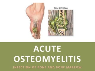
- 1. ACUTE OSTEOMYELITIS INFECTION OF BONE AND BONE MARROW
- 2. INTRODUCTION Infection of bone and bone marrow Can progress to osteonecrosis, bone destruction and septic arthritis Bimodal age distribution: Children under 5 years old Adults over 50 years of age Risk factors: Recent trauma or surgery Immunocompromised patients Illicit IV drug use Poor vascular supply Systemic conditions such as diabetes and sickle cells Peripheral neuropathy
- 3. CLASSIFICATION I. Acute within 2 weeks II. Subacute 2 – 6 weeks III. Chronic after 6 weeks Incidence of infection increases with increase in grade of open fracture: Type I – II : 2% Type III : 10 – 50% based on duration, mechanism, and host response DURAT I ON
- 4. CLASSIFICATION I. Hematogenous Originated or transported by blood Etiology of 20% of osteomyelitis Vertebrae most common site; and metaphysis of long bone S. aureus is most common organism II. Contiguous factor Associated with previous surgery, trauma, wounds or poor vascularity Can be bacterial (most common), mycobacterial or fungal in nature III. Direct inoculation Penetrating injuries Surgical contamination based on duration, mechanism, and host response MECHANI SM
- 5. CLASSIFICATION based on duration, mechanism, and host response HOST RESPONSE SY S T E M I C LO C A L Malnutrition Vascular compromise Renal failure, hepatic failure Chronic lymphodema Diabetes Extensive scarring Autoimmune disease Radiation fibrosis Malignancy Neuropathy Extreme age Immunosuppression drugs Immunodeficiency Smoking
- 6. CIERNY-MADER STAGING Medullary OM Infection confined to medullary cavity Superficial OM Contiguous type of infection. Confined to surface of bone Localized OM Full-thickness cortical sequestration which can easily be removed surgically Diffuse OM Loss of bone stability, even after surgical debridement A N AT O M I C P H Y S I O L O G I C Stage I: Medullary A. Host, good systemic defense and local vascularity Stage II: Superficial B. Host, good systemic and local compromise Stage III: Localized C. Host, non-candidate for surgery with treatment more problematic than disease process Stage IV: Diffuse
- 7. ORGANISM OST EOMYEL I T I S ORGANI SM TAB L E A G E G R O U P M O S T C O M M O N O R G A N I S M S Newborns (younger than 4 mo) S. Aureus Enterobacter species Group A & B streptococcus species Children (aged 4 mo to 4 y) S. Aureus Group A streptococcus species Kingella kingae Enterobacter species Children, adolescents (aged 4 y to adult) S. Aureus (80%) Group A streptococcus species H. Influenzae Enterobacter species Adult S. Aureus Occasionally Enterobacter or Streptococcus species Sickle Cell Anemia Patients S. Aureus is typically most common But Salmonella species is pathognomonic
- 8. ACUTE OSTEOMYELITIS Mainly a disease of children Occurs after an episode of bacteremia Trauma may determine the site of infection Possibly by causing small haematoma of fluid collection in the bone Most common organism in both children and adult is Staphyloccocus aureus (70%)
- 9. PATHOPHYSIOLOGY Affects metaphyseal region of long bones; femur (27%), tibia (22%), fibula (5%) more than upper extremity Bacteremia is the common event in childhood, consequence of other infections such as otitis media, pharyngitis and sinusitis Its presumed that bacteria gain access to metaphyseal location via nutrient arteries.
- 10. PATHOPHYSIOLOGY Staphylococcus aureus: Surface antigen plays key role in bacterial adherence to type 1 collagen and endotoxins that suppress local immune response Glycocalyx: may form around bacteria and enhance adherence to other bacteria and metallic implants
- 11. PATHOPHYSIOLOGY CAUSES OF METAPHYSEAL OST EOMYL I T I S Hair pin bend vessels Increased vascularity causes pooling of blood also called as “lake of blood” Immature cells in metaphysis due to high cell turnover Relative lack of phagocytosis Presence of degenerative cartilage cells End arteries in metaphysis Prone for trauma Single endothelial lining in metaphyseal arteries
- 12. PATHOPHYSIOLOGY CASCADE OF EV ENTS Infective embolus enters nutrient artery trapped in vessels of small caliber Blocks vessels, area of bone become necrotic Active hyperemia in vicinity with infiltration of PMN cells which is poured as exudate Increased intraosseous pressure due to exudate and debris (intense pain)
- 13. PATHOPHYSIOLOGY CASCADE OF EV ENTS Vessels compressed, further necrosis occurs Exudate follow path of least resistance Gets accumulated in sub-periosteal spaces to form abscess and damage the blood supply If perforates, can spread to soft tissue
- 14. PATHOPHYSIOLOGY SECOND ROUT E OF SPREAD OF I NF EC T I ON Exudate into medullary cavity destroying marrow elements, blood supply In advanced stages cortex may be surrounded by pus, depriving blood supply Diaphyseal sequestration T HI RD ROUT E OF SPREAD OF I NF EC T I ON Through the physis into joints
- 15. PATHOPHYSIOLOGY
- 16. CLINICAL FEATURE infant, children and adult I NFANT Failure to thrive Metaphyseal tenderness, restricted joint movement CHI L DREN Fever (high grade) Child refuse to use limb (pseudoparalysis) Local redness, swelling, warmth, odema
- 17. CLINICAL FEATURE infant, children and adult ADULT Commonest site is thoracolumbar spine History of urological procedure Followed by mild fever and backache Local tenderness
- 18. PHYSICAL EXAMINATION Ill looking Increase temperature and pulse Limb is held still Local redness, swelling and warmth Tenderness at near one of the larger joint Restricted joint movement – pseudoparalysis Edema Lymphadenopathy
- 19. INVESTIGATIONS A physical examination may shows bone tenderness and possibly swelling and redness Tests may include: Blood cultures Bone biopsy Bone scan Bone x-ray Full blood count C-reactive protein (CRP) Erythrocyte sedimentation rate (ESR) MRI of the bone Needle aspiration of the area around affected bone
- 20. INVESTIGATION B LOOD & F LUI D Leukocytosis Elevated CRP & ESR Anti-staphylococcal antibody titre may be elevated Positive fluid or tissue aspiration Blood culture at the peak of fever may yield the causative organism X-RAY Earliest abnormality detected after first week of the onset of symptom Extra cortical outline (periosteal new bone formation at metaphysis)
- 21. INVESTIGATION ULT RASONOGRAPHY May detect a sub-periosteal collection of fluid in the early stages But it cannot distinguish between hematoma or pus B ONE SCAN (RADI ONUCL I DE SCANNI NG) Technetium – 99 Increased activity in both perfusion and bone phase Highly sensitive investigation even in the very early stages
- 22. INVESTIGATION MAGNET I C RESONANCE I MAGI NG (MRI ) Helpful in cases of doubtful diagnosis Best method of demonstrating bone marrow inflammation
- 23. MANAGEMENT
- 24. TREATMENT I. Supportive treatment for pain and dehydration Analgesic IV fluid II. Splintage for comfort and prevent joint contracture III. Antibiotic therapy Bactericidal drugs are important to: a. Stop the spread of infection to healthy bone b. Control acute flares Oral therapy followed by IV route for 10 – 14 days Antibiotics used in treating OM : a. Amoxicillin b. Ciprofloxacin plus clindamycin c. Levofloxacin plus clindamycin Antibiotics is continued for another 6 weeks (min) but usually more than 6 months
- 25. TREATMENT IV. Surgical drainage If antibiotics are given early (within first 48 hours) it is often unnecessary But if clinical features do not improve within 36 hours of starting treatment, or even earlier and there is sign of deep pus, drainage is required Debridement of infected tissues WHEN ANTIBIOTICS CAN BE STOPPED? CLINICALLY : Signs of healing, reduction of pain, no fever, can walk, no discharging sinus INFLAMMATORY PARAMETERS : normalizes SERIAL X-RAY : bone healing, no new OM changes
- 26. TREATMENT ALGORITHM OF CIERNY – MADER STAGES 3&4 LONG BONE OSTEOMYELITIS
- 27. DIFFERENTIAL DIAGNOSIS I. Rheumatic fever Gradual, poly-joint swelling II. Ewing’s sarcoma Radiological signs III. Acute suppurative arthritis Muscle spasm more marked, limited movements, effusions IV. Cellulitis No intense pain V. Erysipelas Raised red margin
- 28. COMPLICATIONS I. Epiphyseal damage and altered bone growth II. Suppurative arthritis III. Metastatic infection IV. Pathological fracture V. Chronic osteomyelitis