Clinical Examination of the Hip: Assessing Pain, Movement, and Alignment
•
689 gefällt mir•71,307 views

The document provides information on clinical examination of the hip joint. It begins with anatomy of the hip joint and associated muscles and ligaments. It then discusses elements of history taking including pain characteristics. The physical examination section covers inspection of gait, limb posture and length, palpation of bony landmarks and muscles, range of motion testing, and special tests like Trendelenburg test. Measurements of limb length discrepancies both apparent and true are also described.

Empfohlen
Weitere ähnliche Inhalte
Was ist angesagt?
Was ist angesagt? (20)
Andere mochten auch
Andere mochten auch (20)
Ähnlich wie Clinical Examination of the Hip: Assessing Pain, Movement, and Alignment
Ähnlich wie Clinical Examination of the Hip: Assessing Pain, Movement, and Alignment (20)
Kürzlich hochgeladen
Kürzlich hochgeladen (20)
Clinical Examination of the Hip: Assessing Pain, Movement, and Alignment
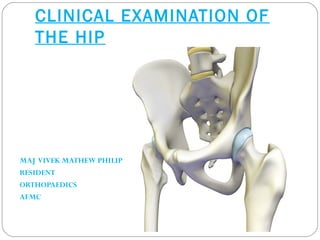
- 1. CLINICAL EXAMINATION OF THE HIP MAJ VIVEK MATHEW PHILIP RESIDENT ORTHOPAEDICS AFMC
- 2. CLINICAL EXAMINATION OF THE HIP Anatomy History Clinical Examination
- 3. ANATOMY. Ball and socket type of synovial joint. Connects the pelvic girdle to the lower limb Made up of femoral head and acetabulum Designed for stability and wide range of movement Covered with a thin layer of hyaline cartilage
- 4. Anatomy The articular surface of is horse-shoe shaped and is deficient inferiorly- acetabular notch Has a labrum -is a circular layer of cartilage which surrounds the outer part of the acetabulum making the socket deeper and so helping provide more stability Capsule
- 5. Iliofemoral Ligament This is a strong ligament which connects the pelvis to the femur at the front of the joint It resembles a Y in shape Stabilises the hip by limiting hyperextension
- 6. Ischiofemoral ligament: This is a ligament which reinforces the posterior aspect of the capsule attaches the ischium to the two trochanters of the femur.
- 7. Pubofemoral ligament The pubofemoral ligament attaches the pubis to the femur
- 8. Transverse acetabular Ligament: Bridges acetabular notch. Ligament of head of femur: flat and triangular in shape Lies within joint, ensheathed by synovium
- 10. Muscles Gluteals: Gluteus Maximus, Gluteus Minimus and Gluteus Medius Attach to the Ilium and travel laterally to insert into the greater trochanter of the femur Medius and Minimus abduct and medially rotate the hip joint, as well as stabilising the pelvis Gluteus maximus extends and laterally rotates the hip joint
- 11. More Muscles Quadriceps The four Quadricep muscles are Vastus lateralis, medialis, intermedius and Rectus femoris All attach inferiorly to the tibial tuberosity Rectus femoris originates at the Anterior Inferior Iliac Spine and acts to flex the hip The 3 other Quad muscles do not cross the hip joint, and attach around the greater trochanter and just below it.
- 12. : Iliopsoas: The is the primary hip flexor muscle which consists of 2 parts Attaches superiorly to the lower part of the spine and the inside of the ilium Cross the hip joint and insert to the lesser trochanter of the femur
- 13. Muscles: Hamstrings: The hamstrings are three muscles which form the back of the thigh Attach superiorly to the ischial tuberosity Cause hip extension
- 14. Functional Group of Muscles Acting on the Hip Flexors: Iliopsoas, sartorius, tensor fascia lata, rectus femorus, Extensors: - hamstrings, addcutor magnus, gluteus maximus Adductors: - adductor longus, brevis, and magnus, gracilis, pectineus Abductors: - gluteus medius, minimus, tensor fascia lata - gamelli, obturators, piriformis in sitting External rotators: - obturator externus, internus, piriformis, quadratus femoris, gluteus maximus Internal Rotators: - gluteus medius, minimus, tensor fascia lata.
- 15. Nerves Femoral (L2,3,4) Obturator (L2, 3, 4) Sciatic (L4,5, S1, 2,)
- 16. Blood Supply
- 17. EXAMINATION OF HIP History General examination Gait Inspection Palpation Movements Measurements Special tests
- 18. Clinical features of Hip Pathology: Pain. Swelling. Loss of function. Limp. Leg length discrepancy.
- 19. PAIN Most important reported symptom. Site Anterior hip pain : arthritis, hip flexor strain, iliopsoas bursitis, labral tear Lateral hip pain : greater trochanteric bursitis, gluteus medius tear, iliotibial band syndrome (athletes), meralgia paresthetica (an entrapment syndrome of the lateral femoral cutaneous nerve syndrome) Posterior hip pain DDx: hip extensor and external rotator pathology, degenerative disc disease, spinal stenosis REFERED PAIN: to knee. hip pathology can be referred to the knee
- 20. The Pain Continues... Onset: When did it start? Hours, days, weeks, years Character Sharp: muscle strain/tear, fracture Dull: OA, RA Achy: OA, RA, AVN Radiation Sciatica can run from the hip, down the back of the thigh, into the foot Radiates to the groin can imply inguinal hernia, groin strain, etc.
- 21. Pain: What were they doing when the pain came on? Did they fall? fractures, muscle tears, haematomas, etc Playing sports? Muscle sprain, labral tear, etc Prolonged exercise? OA Gradual vs sudden? RA,OA vs. trauma
- 22. Pain: Do they have any aggravating or relieving factors? OA gets worse as they day goes on and is relieved by rest Muscle tears/sprains may be exacerbated by movement RA is worse after prolonged periods of rest If analgesia works, find out what they take and how often!
- 23. How does the pain affect their daily life? How far can they walk? Difficulty walking up/down stairs? Are they still able to do their favourite hobbies? Has their partner noticed their pain limiting them? Are they taking regular analgesia?
- 24. SWELLING Site Onset Duration Association with pain Progression over time
- 25. LIMP Usually noted by kin Onset Duration Association with pain Progression Ambulatory status
- 26. PAST HISTORY Trauma Tuberculosis Surgery around hip Skin /hematological disorders Neurological disorders Connective tissue disorders Steroid intake Any other significant medical /surgical illness
- 27. PERSONAL HISTORY Occupation and work tolerance Diet Smoking/alcohol Sexual history Menopausal history
- 28. FAMILY HISTORY TB in close relative Dysplasia Metabolic storage disorders Inflammatory arthritis
- 29. GENERAL EXAMINATION • Ht/wt/BMI • Fever • Vital signs • Pallor • External iliac/inguinal lymph nodes • Stigmata of rheumatoid arthritis/TB • Chest expansion
- 30. LOCAL EXAMINATION Inspection Palpation Movements Measurements Special tests
- 31. GAIT Simplest of all definitions “mode of walking” Normal gait is rhythmical bipedal biphasic walking in which the lumbar spine, hip and legs move in unison Limping is the most common abnormality Can be defined as any abnormality of normal rhythmic biphasic walking
- 32. TYPES OF GAIT Antalgic gait in painful hip conditions pt walks with reduced stance phase on the affected side
- 33. Waddling gait Body sways from side to side on a wide base seen in b/l DDH,pregnancy
- 34. Trendelenberg gait In double stance forces distributed equally over two hips In single stance forces increases 6 fold Patient lurches on the affected siade and pelvis drops on to sound side
- 36. Cont’d… Short limb gait- When the affected limb becomes short Up and down movement of half of the body Pt lurches on the affected side with a pelvis drop on the same side
- 37. Circumduction gait- In fixed abduction deformity or in hemiparesis the pt moves his limbs while dragging his body along with limb in a semi circle
- 38. Gluteus maximus gait- In paralysis of gluteus maximus Pt lurches backward during stance phase
- 39. Quadriceps gait In quadriceps weakness body collapses-hence the trunk goes for anterior bending to shift the vertical vector anterior to the knee to balance
- 40. Toe ingait Pt walks with both feet turned inwards-seen in femoral anteversion
- 41. Toe outgait Pt walks with both feet turned outwards-seen in femoral retroversion
- 42. ATTITUDE OF THE LIMB Standing : position of the head level of scapulae and nipples curvature of the spine attitude of hip, knee & ankle position of the ASIS-square or oblique
- 43. ATTITUDE OF THE LIMB • Supine: Position of the upper limbs • lower limbs parallel/rotated • Patella facing up/in/out • exaggerated lumbar lordosis
- 44. INSPECTION FROM BACK Scoliosis Gluteal muscle wasting PSIS Back of iliac crest Scars and sinuses
- 45. LOOK FOR LIMB LENGTH DESCREPANCY
- 46. PALPATION “Confirms the findings of inspection” Local temperature Increased in acute arthritis Joint tenderness Anteriorly-2cms below and lateral to mid- inguinal point Posteriorly- junction of medial 2/3rd and lateral 1/3rd of a line joiningGT & PSIS
- 47. Tenderness ASIS GT PSIS pubic symphysis SI joint ischial tuberosity
- 48. PALPATION(Contd) Femoral artery pulsation at midinguinal pont Palpation of GT: smooth/irregular proximal migration Digital Bryant’s Test : supratrochanteric shortening
- 49. MEASUREMENT OF DEFORMITY Fixed Flexion Deformity unilateral -Thomas Test The examiner blocks the pelvis by bringing the contralateral sound hip into maximal flexion. This eliminates lumbar lordosis that can be used to compensate for the hip flexion contracture of the affected hip. The leg to be examined is then brought into maximal extension with the hip in neutral adduction and rotation.
- 50. BILATERAL FFD Patient in prone position with lower limbs hangigng out from the edge of the table Patient should be able to kep both thighs extended Measure the angle between thigh and bed for ffd
- 51. Fixed Abduction Deformity It is compensated by scoliosis with convexity towards the affected side & by the pelvis being tilted down causing apparent lengthening of limb
- 52. Fixed adduction deformity It is compensated by scoliosis with convexity towards the normal side & by the pelvis being tilted up causing apparent shortening of limb
- 53. Fixed external & internal rotation deformity Always remains revealed Determined by noting the direction of anterior surface of patella or the toes when the foot is held at right angle to the leg
- 54. Movements Flexion (135 deg):sitting • For ilio psoas contribution: Flex knee and move it towards the chest without moving the opposite leg when patient sits with the legs hanging on the edge of the examination couch • Active SLRT against resistance(supine)
- 55. Movements Extension ( 0 to 20 deg) • For gluteus maximus contribution: • Hamstring contribution
- 56. Movements Abduction ( 0 to 45 deg) Adduction(0 to 45 deg)
- 57. Movements External rotation 90 deg flexion(45 deg) full extension(45deg)
- 58. Movements Internal rotation Internal rotation in 90 deg flexion(45 deg) Internal Rotation in full extension(45 deg)
- 60. MEASUREMENT- Muscle bulk Muscle wasting
- 61. LIMB LENGTH:APPARENT • functional length • patient in straight line and limbs parellel, defromities not corrected • from the fixed midpoint to the medial malleolus • shows the compensation that the pt has developed to conceal any fixed deformity • here both limbs should be kept parallel to each other • measured from xiphisternum or umbilicus to medial malleolus
- 62. TRUE LENGTH •anatomical length •patient in straighat line and deformities corrected and the limbs are kept in identical position •measured from the ASIS to medial malleolus
- 63. BLOCK METHOD
- 64. MEASUREMENTS If True Shortening = Apparent Shortening: No compensation True Shortening >apparent shortening: only part of the deformity is compensated by tilting the pelvis(fixed abduction deformity) True Shortening<apparent Shortening:fixed adduction deformity with no compensation Every 10 degree of deformity : 01 cm
- 65. APPARENT SHORTENING & LENGTHENING ADDUCTION :APPARENT SHORTENING ABDUCTION :APPARENT LENGTHENING
- 66. SEGMENT OF TRUE SHORTENING
- 67. SEGMENTAL SHORTENING:SUPRATROCHANTERIC BRYANT’S TRINGLE NELATON’S LINE
- 68. MEASUREMENTS Chiene’s lines The lines joining the two ASIS and the two GTs are parallel to each other Disturbed in supratrochanteric shortening Shoemaker’s lines
- 69. MEASUREMENTS Morris’ Bistrochanteric Test: • it measues the distance between the GT and pubic symphysis on both sides • Reduced in hip dislocations
- 70. True shortening Supra trochanteric Coxa Vara Perthes SCFE Malunited basal # NOF Congenital Coxa Vara Arthritis Dislocation Infra trochanteric Malunion Fracture femur & tibia Growth arrest from polio Trauma and infective sequale
- 71. TELESCOPY Flex the hip to 90 deg •one hand with the thumb on asis and the remaining fingers over the soft tissue proximal to femur •other hand at the distal femur •push and pull the femur
- 72. VASCULAR SIGN OF NARATH
- 74. This test examine the strength of the abductor mechanism of the hip. Fulcrum: head of femur Load arm:weight of the body Power arm: abductors Lever: neck and trochanters of the femur Normally, in a one legged stance, the pelvis is raised up on the unsupported side. If the weight bearing hip is unstable, the pelvis drops on the unsupported side, to avoid falling the patient has to throw his or her body towards the loaded side. •In the classic test, the examiner stands behind the patient. If the patient stands on a healthy hip the gluteal fold on this side drops. •If the patient stands on a diseased leg the gluteal fold on the opposite side drops (the sound side sags). 1.. Weakness of the hip abductors e.g. poliomyelitis 2.. Shortening of femoral neck e.g. coxa vara. 3. Dislocation or subluxation of the hip
- 75. TESTS FOR DDH BARLOW’S MANOUVRE The maneuver is easily performed by adducting the hip while applying light pressure on the knee, directing the force posteriorly.[2] If the hip is dislocatable - that is, if the hip can be popped out of socket with this maneuver - the test is considered positive ORTOLANI TEST It is performed by an examiner first flexing the hips and knees of a supine infant to 90 degrees, then with the examiner's index fingers placing anterior pressure on the greater trochanters, gently and smoothly abducting the infant's legs using the examiner's thumbs. A positive sign is a distinctive 'clunk' which can be heard and felt as the femoral head relocates anteriorly into the acetabulum:[2] hip
- 77. TESTS FOR JOINT CONTRACTURES FLEXION:THOMAS TEST
- 78. TESTS FOR JOINT CONTRACTURES OBER’S TEST: Test for ileo-tibial tract contracture. In lateral decubitus position knee is flexed to 90 degree hip is abducted to 40 degree and pelvis is stabilised. limb is gently adducted towards the examining table normally the hip adducts and the limb crosses the midline
- 79. TESTS FOR JOINT CONTRACTURES ELY’S TEST for the contracture of the rectus femoris prone position with the knees extended passively flex one knee to be tested normally knee can be flexed fully in contracted rectus full flexion of the knee forces the hip into flexion causing the rise of buttocks
- 80. PHELP’S TEST: To detect the contracture of gracilis muscle Prone position with the knee extended Passive abduction to the maximum with the extended knee Knees are then flexed to relax gracilis Attempt to further abduct the hip with knee in flexion Further abduction is possible in gracilis contracture
- 81. TEST FOR FEMORAL ANTEVERSION:CRAIG’S TEST 1.Positioned prone 2.Knee flexed 90 deg 3.One hand over trochanter 4.Other hand is rotating the leg till the trocanter felt prominent 5.Angle subtended between the imaginary vertical to the long axis of the leg
- 82. PIRIFORMIS TEST(FADIR) Lateral decubitus position •hip is flexed to 45 degree •knee is flexed to 90 degree •one hand stabilises the pelvis •other hand pushes the knee to the floor causing the internal rotation •pain locally-piriformis tendinitis •pain radiates down-piriformis syndrome
- 83. PATRICK’S TEST(FABER) Tend to stress the ipsilateral s-i joint •pain is posterior in s-i arthritis •pain is anterior in hip arthritis
- 84. PELVIC STRESS TESTS LATERAL PELVIC COMPRESSION TEST ANTERIOR PELVIC COMPRESSION TEST
- 85. PELVIC STRESS TESTS PUBIC SYMPHYSIS STRESS TEST STINCHFIELD TEST
- 86. IMPINGEMENT TEST FLEXION ADDUCTION INTERNAL ROTATION
- 87. FULCRUM TEST It tests for the stress fractures of the shaft of femur
- 88. NOT TO FORGET •OTHER LOWER LIMB JOINTS •SPINE •PER RECTAL EXAMINATION
- 89. THANK YOU