SlideShare ist ein Scribd-Unternehmen logo
1 von 51
Downloaden Sie, um offline zu lesen
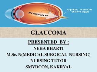
GLAUCOMA
PRESENTED BY :
NEHA BHARTI
M.Sc. N(MEDICAL SURGICAL NURSING)
NURSING TUTOR
SMVDCON, KAKRYAL
DEFINITION
• It is a group of disorders characterized by an
abnormally high intra ocular pressure (IOP), optic
nerve dystrophy (weakness) and peripheral visual
field loss (tunnel vision.)
• It is a symptomatic condition of the eye where
the IOP is more than normal (above 25mm Hg).
• Untreated of glaucoma leads to permanent
damage of the optic nerve and resultant visual
field loss, which can progress to blindness.
• Glaucoma is an eye disease where the eye’s
optic nerve is damaged. It is one of the
leading causes of blindness
www.opto.ca
EYE ANATOMY
• The optic nerve is a
bundle of nerve
fibers
• It carries visual
information from the
retina to the brain
www.opto.ca
Fluid Circulation
• The eye has an
internal fluid
circulation system
• Fluid is produced at
the base of the iris
www.opto.ca
Fluid Circulation
• The fluid flows
through the pupil to
the front of the iris
www.opto.ca
Fluid Circulation
• The fluid exits the
eye at the angle
between the iris and
the cornea where it
drains through a
spongy meshwork
CAUSES AND RISK FACTORS
• Genetics:- Family history of glaucoma
• Ageing
• Ocular hypertension is a condition where the
pressure in your eyes, or IOP, is too high. Continually
high pressure within the eye can eventually damage
the optic nerve and lead to glaucoma or permanent
vision loss.
• Severe myopia:- It is associated with an increased
risk of pathological ocular complications and
may lead to blinding disorders like glaucoma
• Eye trauma:- It is most commonly caused by
blunt trauma, which is an injury that doesn't penetrate
the eye, such as a blow to the head or an injury directly
on the eye. This can lead to an increase
in eye pressure, which can damage the optic nerve.
• Ocular surgery:- can cause a change in the eye's
pressure. Sharp increases in eye pressure are called
“pressure spikes” and sometimes occur in patients
after cataract surgery. Often these pressure spikes are
short-term and can be treated with medicines.
• Migraine:- Prolonged increased pressure
can lead to visual loss if not corrected.
• Black ethnicity:- African Americans are also
more likely to develop glaucoma at a younger
age and suffer blindness from the disease. The
genetic causes underlying glaucoma remain
unclear, but these ethnic disparities in the risk
of developing glaucoma suggest a genetic
basis that is ethnicity-specific
• Prolonged use of local or systemic
corticosteroids:- Long-term use of topical and
systemic steroids produces secondary open-
angle glaucoma similar
to chronic simple glaucoma. The increased
intraocular pressure
[IOP] caused by prolonged steroid therapy is
reversible but the damage produced by it is
irreversible. (edema glucocorticoid receptors
on trabecular meshwork cells.)
PATHOPHYSIOLOGY
•IOP is a function of production of liquid aqueous
humor by the ciliary processes of the eye and its
drainage through the trabecular meshwork.
•Aqueous humor is produced by the ciliary body
and flow into the posterior chamber behind the iris.
•The trabecular meshwork filters the aqueous
humor into Schlemm’s canal. Where is picked up by
the episcleral vessels and mixed with blood.
CLASSIFICATIONS
1. CONGENITAL GLAUCOMA
2. ACQUIRED GLAUCOMA
•PRIMARY GLAUCOMA
•SECONDARY GLAUCOMA
1. CONGENITAL GLAUCOMA
• It is rare disease, occurs when a congenital defect in
the angle of the anterior chamber obstructs the out
flow of aqueous humor. If untreated, causes damage
to the optic nerve and blindness. In most cases,
surgery is required.
1.TRUE CONGENITAL GLAUCOMA
2. INFANTILE GLAUCOMA
3. JUVENILE GLAUCOMA
A.TRUE CONGENITAL GLAUCOMA
• It is labeled when IOP is raised during
intrauterine life and child is born with ocular
enlargement. It occurrence is about 40% of
cases.
B. INFANTILE GLAUCOMA:-
• It is labeled when the disease manifests prior
to the child’s third birthday. It occurs in about
50% of cases.
C. JUVENILE GLAUCOMA
• It is labeled in the rest 10% of cases who
develop pressure rise between 3-6 years of
life.
2. ACQUIRED GLAUCOMA
A. PRIMARY GLAUCOMA
• 1. PRIMARY OPEN ANGLE GLAUCOMA
• 2. PRIMARY ANGLE CLOSURE GLAUCOMA
B. SECONDARY GLAUCOMA
A. PRIMARY GLAUCOMA
1. PRIMARY OPEN ANGLE GLAUCOMA
• POAG is the most common form of glaucoma
• It occurs when the fluid drainage is poor and
fluid builds up in the eye and the internal eye
pressure goes up
• This increased pressure can cause damage to
the optic nerve and vision loss
• The exact mechanism of damage is still
unknown
www.opto.ca
Symptoms of Primary Open
Angle Glaucoma
• POAG develops gradually and painlessly and has
no initial symptoms
Vision is normal in
the early stages
www.opto.ca
Symptoms of Primary Open
Angle Glaucoma
• If untreated, peripheral or side vision is slowly lost
Tunnel vision:-
Defective sight in
which objects cannot
be properly seen if
not close to the
centre of the field of
view.
www.opto.ca
Symptoms of Primary Open
Angle Glaucoma
• Eventually, all vision may be lost
B. SECONDARY GLAUCOMA
2. PRIMARY ANGLE CLOSURE GLAUCOMA
• This type of glaucoma is an emergency
situation
• It occurs when the iris itself blocks the
drainage angle and results in a sudden
increase in pressure
• Symptoms include severe eye pain, nausea,
eye redness and very blurred vision
• Immediate treatment is required
B. SECONDARY GLAUCOMA
• Glaucoma can develop as a complication from other
conditions including:
– Eye injuries
– Diabetes
– Steroid use
www.opto.ca
3. Low Tension Glaucoma
• Low Tension (or Normal Tension) Glaucoma is not
as common
• In these cases, the eye pressure is in the normal
range but the optic nerve still gets damaged
• The exact mechanism of damage is still unknown
DIAGNOSTIC EVALUATION
• Regular eye examinations by an optometrist
or ophthalmologist are vital to detecting
glaucoma
• A number of tests are performed
• A patient’s medical history, family history and
background are important to determine the
presence of risk factors
www.opto.ca
Glaucoma Tests:
Slit Lamp & Gonioscopy
• A special microscope called a slit lamp is used to
examine the structures of the eye
• A gonioscopy lens may be used to view the drainage
angle
SLIT- LAMP EXAM
• Once patient in the examination
chair, the doctor will place an
instrument in front of patient on
which to rest chin and forehead.
• This helps steady head for the
exam. Doctor may put drops in
eyes to make any abnormalities
on the surface of cornea more
visible.
• The drops contain a yellow dye
called fluorescein, which will
wash away the tears. Additional
drops may also be put in eyes to
allow pupils to dilate, or get
bigger.10/26/2018
• The doctor will use a low-powered microscope, along
with a slit lamp, which is a high-intensity light. They
will look closely at eyes. The slit lamp has different
filters to get different views of the eyes. Some
doctor’s offices may have devices that capture digital
images to track changes in the eyes over time.
• During the test, the doctor will examine all areas of
your eye, including the:- eyelids, conjunctiva, iris,
lens, sclera, cornea, retina and optic nerve.
10/26/2018
Glaucoma Tests:
Tonometry
• Eye pressure is measured with an instrument
called a tonometer
TONOMETERY
• Tonometry is the
procedure eye care
professionals perform
to determine the
intraocular pressure,
the fluid pressure inside
the eye. It is an
important test in the
evaluation of patients at
risk from glaucoma.
• (normal pressure range
is 12 to 22 mm Hg)
10/26/2018
www.opto.ca
Glaucoma Tests:
Ophthalmoscopy
• Eye drops may be placed in the eyes to dilate the pupils
• Special magnifying lenses are used to examine the
retina and optic nerve for damage
Normal Optic Nerve Suspicious Optic Nerve
www.opto.ca
Glaucoma Tests:
Ophthalmoscopy
• Advances are being made in digital imaging of the retina
COLOR FUNDUS PHOTOGRAPHY
• Fundus camera to record color images of the
condition of the interior surface of the eye, in order
to document the presence of disorders and monitor
their change over time.
• A fundus camera or retinal camera is a specialized
low power microscope with an attached camera
designed to photograph the interior surface of the
eye, including the retina, retinal vasculature, optic
disc, macula, and posterior pole (i.e. the fundus).
10/26/2018
MANAGEMENT
MEDICAL MANAGEMENT:-
• BETA ADRENERGIC BLOCKERS:- Timolol,
betaxolol are used to decreased aqueous
humor production.
• CHOLINERGIC (MIOTICS):- Pilocarpine,
carbacol are used to reduce IOP by facilitating
the outflow of aqueous humor
• CARBONIC ANHYDRASE INHIBITORS:-
Dorzolamide, methazolamide or
acetazolamide to decrease the formation and
secretion of aqueous humor.
• PROSTAGLANDIN ANALOGS:- Latanoprost to
reduce IOP by increasing uveoscleral outflow.
SURGICAL MANAGEMENT
ARGON LASER TRABECULOPLASTY:-
• It may be used to treat open angle glaucoma.
In this, thermal argon laser burns are applied
to the inner surface of the trabecular
meshwork to open the intra trabecular spaces
and widen the canal of Schlemm, thereby
increasing the outflow of aqueous humor and
decreasing IOP.
LASER IRIDOTOMY:-
• An opening is made by the laser bean in the
iris to eliminate the pupillary block. It relieves
pressure and preserves vision by promoting
outflow of the aqueous humor.
CYCLOCRYOTHERAPY:-
• Application of a freezing probe to the sclera
over the Cilliary body that destroy some of the
Cilliary processes, results in the reduction of
the amount of aqueous humor produced.
CYCLODIALYSIS:-
• Through a small incision in the sclera, a
spatula type instrument is passed into the
anterior chamber, creating an opening in the
angle.
• FILTERING PROCEDURES:- for chronic
glaucoma filtering procedure are used to
create an opening or fistula in the trabecular
meshwork to drain aqueous humor.
• TRABECULOTOMY:- A partial thickness
incision is made in the sclera and further
section of sclera is removed to produce an
opening for aqueous humor outflow under
the conjunctiva, creating a filtering bleb.
• SCLERECTOMY:- A partial thickness incision is
made in the sclera and one or more openings
are made with a punch. The top flap of sclera
is closed over the punched holes.
NURSING MANAGEMENT
ASSESSMENT:-
• History or presence of risk factor:- positive
family history, tumour of eye, haemorrhage,
uveitis, trauma etc.
• Physical examination based on those in
general assessment of the eye may indicate:-
blurred vision, decreased light perception
redness cloudy appearance etc.
DIAGNOSIS:-
• Acute pain related to increased IOP and
surgical complications as evidenced by patient
verbalization or facial expression of the
patient
GOAL:- The pain of patient will be reduced.
INTERVENTIONS:-
• Monitor vital signs of the patient
• Monitor the degree of eye pain very 30 min
during the acute phase.
• Monitor visual acuity at any time before hatching
ophthalmic agent for glaucoma.
• Maintain the bed rest in semi- fowler position
• Give analgesic prescription and evaluation of its
effectiveness.

Weitere ähnliche Inhalte

Was ist angesagt? (20)

Retinal detachment new
Retinal detachment newRetinal detachment new
Retinal detachment new
 
Glaucoma
GlaucomaGlaucoma
Glaucoma
 
Trachoma
TrachomaTrachoma
Trachoma
 
Refractive error
Refractive errorRefractive error
Refractive error
 
Stye or hordeolum
Stye  or hordeolumStye  or hordeolum
Stye or hordeolum
 
Blepharitis
BlepharitisBlepharitis
Blepharitis
 
Glaucoma
Glaucoma Glaucoma
Glaucoma
 
Keratitis
KeratitisKeratitis
Keratitis
 
Uveitis ppt
Uveitis pptUveitis ppt
Uveitis ppt
 
Trachoma
TrachomaTrachoma
Trachoma
 
Refractive errors (eye condions)
Refractive errors (eye condions)Refractive errors (eye condions)
Refractive errors (eye condions)
 
Glaucoma - symptomes and treatment
Glaucoma - symptomes and treatment Glaucoma - symptomes and treatment
Glaucoma - symptomes and treatment
 
Catract
Catract Catract
Catract
 
Retinal detachment
Retinal detachmentRetinal detachment
Retinal detachment
 
Glaucoma
GlaucomaGlaucoma
Glaucoma
 
Refractive errors
Refractive errorsRefractive errors
Refractive errors
 
Eyelid infections ppt
Eyelid infections pptEyelid infections ppt
Eyelid infections ppt
 
Dacryocystitis
DacryocystitisDacryocystitis
Dacryocystitis
 
Corneal Ulcer
Corneal Ulcer  Corneal Ulcer
Corneal Ulcer
 
Eye bank
Eye bankEye bank
Eye bank
 

Ähnlich wie Glaucoma

Ähnlich wie Glaucoma (20)

Presentation (8).pptx
Presentation (8).pptxPresentation (8).pptx
Presentation (8).pptx
 
glaucoma and causes sign symptoms treatment
glaucoma and causes sign symptoms treatmentglaucoma and causes sign symptoms treatment
glaucoma and causes sign symptoms treatment
 
Glaucoma.pptx
Glaucoma.pptxGlaucoma.pptx
Glaucoma.pptx
 
Glaucoma, a major cause of blindness.
Glaucoma, a major cause of blindness.Glaucoma, a major cause of blindness.
Glaucoma, a major cause of blindness.
 
GLAUCOMA of human eye for certificate nurses
GLAUCOMA of human eye for certificate nursesGLAUCOMA of human eye for certificate nurses
GLAUCOMA of human eye for certificate nurses
 
Glaucoma.pptx
 Glaucoma.pptx Glaucoma.pptx
Glaucoma.pptx
 
Glaucoma-1.pptx disorder of eye affects the vision
Glaucoma-1.pptx disorder of eye affects the visionGlaucoma-1.pptx disorder of eye affects the vision
Glaucoma-1.pptx disorder of eye affects the vision
 
POAG AND PACG
POAG AND PACGPOAG AND PACG
POAG AND PACG
 
Glaucoma
GlaucomaGlaucoma
Glaucoma
 
slide for glaucoma.pdf
slide for glaucoma.pdfslide for glaucoma.pdf
slide for glaucoma.pdf
 
Glaucoma - DISEASE PRESENTATION
Glaucoma - DISEASE PRESENTATIONGlaucoma - DISEASE PRESENTATION
Glaucoma - DISEASE PRESENTATION
 
Glaucoma,topic 4 presentation
Glaucoma,topic 4 presentationGlaucoma,topic 4 presentation
Glaucoma,topic 4 presentation
 
Glaucoma their diagnosis and treatment
Glaucoma their diagnosis and treatmentGlaucoma their diagnosis and treatment
Glaucoma their diagnosis and treatment
 
Glaucoma
GlaucomaGlaucoma
Glaucoma
 
Glaucoma
GlaucomaGlaucoma
Glaucoma
 
Pilocarpine use in glaucoma
Pilocarpine use in glaucomaPilocarpine use in glaucoma
Pilocarpine use in glaucoma
 
Glaucoma
GlaucomaGlaucoma
Glaucoma
 
Glaucoma
GlaucomaGlaucoma
Glaucoma
 
Glaucoma
GlaucomaGlaucoma
Glaucoma
 
Glaucoma diska
Glaucoma diskaGlaucoma diska
Glaucoma diska
 

Mehr von NEHA BHARTI

ANTI MALARIAL.pptx
ANTI MALARIAL.pptxANTI MALARIAL.pptx
ANTI MALARIAL.pptxNEHA BHARTI
 
sulfonamides.pptx
sulfonamides.pptxsulfonamides.pptx
sulfonamides.pptxNEHA BHARTI
 
4. macrolide antibiotics.pptx
4. macrolide antibiotics.pptx4. macrolide antibiotics.pptx
4. macrolide antibiotics.pptxNEHA BHARTI
 
3. aminoglycosides.pptx
3. aminoglycosides.pptx3. aminoglycosides.pptx
3. aminoglycosides.pptxNEHA BHARTI
 
2. cephalosporins.ppt
2. cephalosporins.ppt2. cephalosporins.ppt
2. cephalosporins.pptNEHA BHARTI
 
CHEMOTHERAPY- PENICILLIN.pptx
CHEMOTHERAPY- PENICILLIN.pptxCHEMOTHERAPY- PENICILLIN.pptx
CHEMOTHERAPY- PENICILLIN.pptxNEHA BHARTI
 
Polycystic kidney disease
Polycystic kidney diseasePolycystic kidney disease
Polycystic kidney diseaseNEHA BHARTI
 
Nephrotic syndrome
Nephrotic syndromeNephrotic syndrome
Nephrotic syndromeNEHA BHARTI
 
Acute renal failure and chronic renal failure
Acute renal failure and chronic renal failureAcute renal failure and chronic renal failure
Acute renal failure and chronic renal failureNEHA BHARTI
 
ABDELLAH’S THEORY
ABDELLAH’S THEORYABDELLAH’S THEORY
ABDELLAH’S THEORYNEHA BHARTI
 
Nursing assessment and assessment of eye
Nursing assessment and assessment of eyeNursing assessment and assessment of eye
Nursing assessment and assessment of eyeNEHA BHARTI
 
Bowel elimination
Bowel eliminationBowel elimination
Bowel eliminationNEHA BHARTI
 

Mehr von NEHA BHARTI (15)

ANTI MALARIAL.pptx
ANTI MALARIAL.pptxANTI MALARIAL.pptx
ANTI MALARIAL.pptx
 
QUINOLONES.pptx
QUINOLONES.pptxQUINOLONES.pptx
QUINOLONES.pptx
 
sulfonamides.pptx
sulfonamides.pptxsulfonamides.pptx
sulfonamides.pptx
 
4. macrolide antibiotics.pptx
4. macrolide antibiotics.pptx4. macrolide antibiotics.pptx
4. macrolide antibiotics.pptx
 
3. aminoglycosides.pptx
3. aminoglycosides.pptx3. aminoglycosides.pptx
3. aminoglycosides.pptx
 
2. cephalosporins.ppt
2. cephalosporins.ppt2. cephalosporins.ppt
2. cephalosporins.ppt
 
CHEMOTHERAPY- PENICILLIN.pptx
CHEMOTHERAPY- PENICILLIN.pptxCHEMOTHERAPY- PENICILLIN.pptx
CHEMOTHERAPY- PENICILLIN.pptx
 
Polycystic kidney disease
Polycystic kidney diseasePolycystic kidney disease
Polycystic kidney disease
 
Hydronephrosis
HydronephrosisHydronephrosis
Hydronephrosis
 
NEPHRITIS
NEPHRITISNEPHRITIS
NEPHRITIS
 
Nephrotic syndrome
Nephrotic syndromeNephrotic syndrome
Nephrotic syndrome
 
Acute renal failure and chronic renal failure
Acute renal failure and chronic renal failureAcute renal failure and chronic renal failure
Acute renal failure and chronic renal failure
 
ABDELLAH’S THEORY
ABDELLAH’S THEORYABDELLAH’S THEORY
ABDELLAH’S THEORY
 
Nursing assessment and assessment of eye
Nursing assessment and assessment of eyeNursing assessment and assessment of eye
Nursing assessment and assessment of eye
 
Bowel elimination
Bowel eliminationBowel elimination
Bowel elimination
 

Kürzlich hochgeladen

Influencing policy (training slides from Fast Track Impact)
Influencing policy (training slides from Fast Track Impact)Influencing policy (training slides from Fast Track Impact)
Influencing policy (training slides from Fast Track Impact)Mark Reed
 
4.16.24 21st Century Movements for Black Lives.pptx
4.16.24 21st Century Movements for Black Lives.pptx4.16.24 21st Century Movements for Black Lives.pptx
4.16.24 21st Century Movements for Black Lives.pptxmary850239
 
Expanded definition: technical and operational
Expanded definition: technical and operationalExpanded definition: technical and operational
Expanded definition: technical and operationalssuser3e220a
 
ENG 5 Q4 WEEk 1 DAY 1 Restate sentences heard in one’s own words. Use appropr...
ENG 5 Q4 WEEk 1 DAY 1 Restate sentences heard in one’s own words. Use appropr...ENG 5 Q4 WEEk 1 DAY 1 Restate sentences heard in one’s own words. Use appropr...
ENG 5 Q4 WEEk 1 DAY 1 Restate sentences heard in one’s own words. Use appropr...JojoEDelaCruz
 
Visit to a blind student's school🧑‍🦯🧑‍🦯(community medicine)
Visit to a blind student's school🧑‍🦯🧑‍🦯(community medicine)Visit to a blind student's school🧑‍🦯🧑‍🦯(community medicine)
Visit to a blind student's school🧑‍🦯🧑‍🦯(community medicine)lakshayb543
 
4.18.24 Movement Legacies, Reflection, and Review.pptx
4.18.24 Movement Legacies, Reflection, and Review.pptx4.18.24 Movement Legacies, Reflection, and Review.pptx
4.18.24 Movement Legacies, Reflection, and Review.pptxmary850239
 
Congestive Cardiac Failure..presentation
Congestive Cardiac Failure..presentationCongestive Cardiac Failure..presentation
Congestive Cardiac Failure..presentationdeepaannamalai16
 
EmpTech Lesson 18 - ICT Project for Website Traffic Statistics and Performanc...
EmpTech Lesson 18 - ICT Project for Website Traffic Statistics and Performanc...EmpTech Lesson 18 - ICT Project for Website Traffic Statistics and Performanc...
EmpTech Lesson 18 - ICT Project for Website Traffic Statistics and Performanc...liera silvan
 
How to Add Barcode on PDF Report in Odoo 17
How to Add Barcode on PDF Report in Odoo 17How to Add Barcode on PDF Report in Odoo 17
How to Add Barcode on PDF Report in Odoo 17Celine George
 
Incoming and Outgoing Shipments in 3 STEPS Using Odoo 17
Incoming and Outgoing Shipments in 3 STEPS Using Odoo 17Incoming and Outgoing Shipments in 3 STEPS Using Odoo 17
Incoming and Outgoing Shipments in 3 STEPS Using Odoo 17Celine George
 
AUDIENCE THEORY -CULTIVATION THEORY - GERBNER.pptx
AUDIENCE THEORY -CULTIVATION THEORY -  GERBNER.pptxAUDIENCE THEORY -CULTIVATION THEORY -  GERBNER.pptx
AUDIENCE THEORY -CULTIVATION THEORY - GERBNER.pptxiammrhaywood
 
TEACHER REFLECTION FORM (NEW SET........).docx
TEACHER REFLECTION FORM (NEW SET........).docxTEACHER REFLECTION FORM (NEW SET........).docx
TEACHER REFLECTION FORM (NEW SET........).docxruthvilladarez
 
Integumentary System SMP B. Pharm Sem I.ppt
Integumentary System SMP B. Pharm Sem I.pptIntegumentary System SMP B. Pharm Sem I.ppt
Integumentary System SMP B. Pharm Sem I.pptshraddhaparab530
 
4.16.24 Poverty and Precarity--Desmond.pptx
4.16.24 Poverty and Precarity--Desmond.pptx4.16.24 Poverty and Precarity--Desmond.pptx
4.16.24 Poverty and Precarity--Desmond.pptxmary850239
 
Field Attribute Index Feature in Odoo 17
Field Attribute Index Feature in Odoo 17Field Attribute Index Feature in Odoo 17
Field Attribute Index Feature in Odoo 17Celine George
 
Textual Evidence in Reading and Writing of SHS
Textual Evidence in Reading and Writing of SHSTextual Evidence in Reading and Writing of SHS
Textual Evidence in Reading and Writing of SHSMae Pangan
 
Oppenheimer Film Discussion for Philosophy and Film
Oppenheimer Film Discussion for Philosophy and FilmOppenheimer Film Discussion for Philosophy and Film
Oppenheimer Film Discussion for Philosophy and FilmStan Meyer
 
Q4-PPT-Music9_Lesson-1-Romantic-Opera.pptx
Q4-PPT-Music9_Lesson-1-Romantic-Opera.pptxQ4-PPT-Music9_Lesson-1-Romantic-Opera.pptx
Q4-PPT-Music9_Lesson-1-Romantic-Opera.pptxlancelewisportillo
 
HỌC TỐT TIẾNG ANH 11 THEO CHƯƠNG TRÌNH GLOBAL SUCCESS ĐÁP ÁN CHI TIẾT - CẢ NĂ...
HỌC TỐT TIẾNG ANH 11 THEO CHƯƠNG TRÌNH GLOBAL SUCCESS ĐÁP ÁN CHI TIẾT - CẢ NĂ...HỌC TỐT TIẾNG ANH 11 THEO CHƯƠNG TRÌNH GLOBAL SUCCESS ĐÁP ÁN CHI TIẾT - CẢ NĂ...
HỌC TỐT TIẾNG ANH 11 THEO CHƯƠNG TRÌNH GLOBAL SUCCESS ĐÁP ÁN CHI TIẾT - CẢ NĂ...Nguyen Thanh Tu Collection
 

Kürzlich hochgeladen (20)

Paradigm shift in nursing research by RS MEHTA
Paradigm shift in nursing research by RS MEHTAParadigm shift in nursing research by RS MEHTA
Paradigm shift in nursing research by RS MEHTA
 
Influencing policy (training slides from Fast Track Impact)
Influencing policy (training slides from Fast Track Impact)Influencing policy (training slides from Fast Track Impact)
Influencing policy (training slides from Fast Track Impact)
 
4.16.24 21st Century Movements for Black Lives.pptx
4.16.24 21st Century Movements for Black Lives.pptx4.16.24 21st Century Movements for Black Lives.pptx
4.16.24 21st Century Movements for Black Lives.pptx
 
Expanded definition: technical and operational
Expanded definition: technical and operationalExpanded definition: technical and operational
Expanded definition: technical and operational
 
ENG 5 Q4 WEEk 1 DAY 1 Restate sentences heard in one’s own words. Use appropr...
ENG 5 Q4 WEEk 1 DAY 1 Restate sentences heard in one’s own words. Use appropr...ENG 5 Q4 WEEk 1 DAY 1 Restate sentences heard in one’s own words. Use appropr...
ENG 5 Q4 WEEk 1 DAY 1 Restate sentences heard in one’s own words. Use appropr...
 
Visit to a blind student's school🧑‍🦯🧑‍🦯(community medicine)
Visit to a blind student's school🧑‍🦯🧑‍🦯(community medicine)Visit to a blind student's school🧑‍🦯🧑‍🦯(community medicine)
Visit to a blind student's school🧑‍🦯🧑‍🦯(community medicine)
 
4.18.24 Movement Legacies, Reflection, and Review.pptx
4.18.24 Movement Legacies, Reflection, and Review.pptx4.18.24 Movement Legacies, Reflection, and Review.pptx
4.18.24 Movement Legacies, Reflection, and Review.pptx
 
Congestive Cardiac Failure..presentation
Congestive Cardiac Failure..presentationCongestive Cardiac Failure..presentation
Congestive Cardiac Failure..presentation
 
EmpTech Lesson 18 - ICT Project for Website Traffic Statistics and Performanc...
EmpTech Lesson 18 - ICT Project for Website Traffic Statistics and Performanc...EmpTech Lesson 18 - ICT Project for Website Traffic Statistics and Performanc...
EmpTech Lesson 18 - ICT Project for Website Traffic Statistics and Performanc...
 
How to Add Barcode on PDF Report in Odoo 17
How to Add Barcode on PDF Report in Odoo 17How to Add Barcode on PDF Report in Odoo 17
How to Add Barcode on PDF Report in Odoo 17
 
Incoming and Outgoing Shipments in 3 STEPS Using Odoo 17
Incoming and Outgoing Shipments in 3 STEPS Using Odoo 17Incoming and Outgoing Shipments in 3 STEPS Using Odoo 17
Incoming and Outgoing Shipments in 3 STEPS Using Odoo 17
 
AUDIENCE THEORY -CULTIVATION THEORY - GERBNER.pptx
AUDIENCE THEORY -CULTIVATION THEORY -  GERBNER.pptxAUDIENCE THEORY -CULTIVATION THEORY -  GERBNER.pptx
AUDIENCE THEORY -CULTIVATION THEORY - GERBNER.pptx
 
TEACHER REFLECTION FORM (NEW SET........).docx
TEACHER REFLECTION FORM (NEW SET........).docxTEACHER REFLECTION FORM (NEW SET........).docx
TEACHER REFLECTION FORM (NEW SET........).docx
 
Integumentary System SMP B. Pharm Sem I.ppt
Integumentary System SMP B. Pharm Sem I.pptIntegumentary System SMP B. Pharm Sem I.ppt
Integumentary System SMP B. Pharm Sem I.ppt
 
4.16.24 Poverty and Precarity--Desmond.pptx
4.16.24 Poverty and Precarity--Desmond.pptx4.16.24 Poverty and Precarity--Desmond.pptx
4.16.24 Poverty and Precarity--Desmond.pptx
 
Field Attribute Index Feature in Odoo 17
Field Attribute Index Feature in Odoo 17Field Attribute Index Feature in Odoo 17
Field Attribute Index Feature in Odoo 17
 
Textual Evidence in Reading and Writing of SHS
Textual Evidence in Reading and Writing of SHSTextual Evidence in Reading and Writing of SHS
Textual Evidence in Reading and Writing of SHS
 
Oppenheimer Film Discussion for Philosophy and Film
Oppenheimer Film Discussion for Philosophy and FilmOppenheimer Film Discussion for Philosophy and Film
Oppenheimer Film Discussion for Philosophy and Film
 
Q4-PPT-Music9_Lesson-1-Romantic-Opera.pptx
Q4-PPT-Music9_Lesson-1-Romantic-Opera.pptxQ4-PPT-Music9_Lesson-1-Romantic-Opera.pptx
Q4-PPT-Music9_Lesson-1-Romantic-Opera.pptx
 
HỌC TỐT TIẾNG ANH 11 THEO CHƯƠNG TRÌNH GLOBAL SUCCESS ĐÁP ÁN CHI TIẾT - CẢ NĂ...
HỌC TỐT TIẾNG ANH 11 THEO CHƯƠNG TRÌNH GLOBAL SUCCESS ĐÁP ÁN CHI TIẾT - CẢ NĂ...HỌC TỐT TIẾNG ANH 11 THEO CHƯƠNG TRÌNH GLOBAL SUCCESS ĐÁP ÁN CHI TIẾT - CẢ NĂ...
HỌC TỐT TIẾNG ANH 11 THEO CHƯƠNG TRÌNH GLOBAL SUCCESS ĐÁP ÁN CHI TIẾT - CẢ NĂ...
 

Glaucoma

  • 1. GLAUCOMA PRESENTED BY : NEHA BHARTI M.Sc. N(MEDICAL SURGICAL NURSING) NURSING TUTOR SMVDCON, KAKRYAL
  • 2. DEFINITION • It is a group of disorders characterized by an abnormally high intra ocular pressure (IOP), optic nerve dystrophy (weakness) and peripheral visual field loss (tunnel vision.) • It is a symptomatic condition of the eye where the IOP is more than normal (above 25mm Hg). • Untreated of glaucoma leads to permanent damage of the optic nerve and resultant visual field loss, which can progress to blindness.
  • 3. • Glaucoma is an eye disease where the eye’s optic nerve is damaged. It is one of the leading causes of blindness
  • 4. www.opto.ca EYE ANATOMY • The optic nerve is a bundle of nerve fibers • It carries visual information from the retina to the brain
  • 5. www.opto.ca Fluid Circulation • The eye has an internal fluid circulation system • Fluid is produced at the base of the iris
  • 6. www.opto.ca Fluid Circulation • The fluid flows through the pupil to the front of the iris
  • 7. www.opto.ca Fluid Circulation • The fluid exits the eye at the angle between the iris and the cornea where it drains through a spongy meshwork
  • 8. CAUSES AND RISK FACTORS • Genetics:- Family history of glaucoma • Ageing • Ocular hypertension is a condition where the pressure in your eyes, or IOP, is too high. Continually high pressure within the eye can eventually damage the optic nerve and lead to glaucoma or permanent vision loss. • Severe myopia:- It is associated with an increased risk of pathological ocular complications and may lead to blinding disorders like glaucoma
  • 9. • Eye trauma:- It is most commonly caused by blunt trauma, which is an injury that doesn't penetrate the eye, such as a blow to the head or an injury directly on the eye. This can lead to an increase in eye pressure, which can damage the optic nerve. • Ocular surgery:- can cause a change in the eye's pressure. Sharp increases in eye pressure are called “pressure spikes” and sometimes occur in patients after cataract surgery. Often these pressure spikes are short-term and can be treated with medicines.
  • 10. • Migraine:- Prolonged increased pressure can lead to visual loss if not corrected. • Black ethnicity:- African Americans are also more likely to develop glaucoma at a younger age and suffer blindness from the disease. The genetic causes underlying glaucoma remain unclear, but these ethnic disparities in the risk of developing glaucoma suggest a genetic basis that is ethnicity-specific
  • 11. • Prolonged use of local or systemic corticosteroids:- Long-term use of topical and systemic steroids produces secondary open- angle glaucoma similar to chronic simple glaucoma. The increased intraocular pressure [IOP] caused by prolonged steroid therapy is reversible but the damage produced by it is irreversible. (edema glucocorticoid receptors on trabecular meshwork cells.)
  • 12.
  • 13. PATHOPHYSIOLOGY •IOP is a function of production of liquid aqueous humor by the ciliary processes of the eye and its drainage through the trabecular meshwork. •Aqueous humor is produced by the ciliary body and flow into the posterior chamber behind the iris. •The trabecular meshwork filters the aqueous humor into Schlemm’s canal. Where is picked up by the episcleral vessels and mixed with blood.
  • 14.
  • 15.
  • 16. CLASSIFICATIONS 1. CONGENITAL GLAUCOMA 2. ACQUIRED GLAUCOMA •PRIMARY GLAUCOMA •SECONDARY GLAUCOMA
  • 17. 1. CONGENITAL GLAUCOMA • It is rare disease, occurs when a congenital defect in the angle of the anterior chamber obstructs the out flow of aqueous humor. If untreated, causes damage to the optic nerve and blindness. In most cases, surgery is required. 1.TRUE CONGENITAL GLAUCOMA 2. INFANTILE GLAUCOMA 3. JUVENILE GLAUCOMA
  • 18. A.TRUE CONGENITAL GLAUCOMA • It is labeled when IOP is raised during intrauterine life and child is born with ocular enlargement. It occurrence is about 40% of cases. B. INFANTILE GLAUCOMA:- • It is labeled when the disease manifests prior to the child’s third birthday. It occurs in about 50% of cases.
  • 19. C. JUVENILE GLAUCOMA • It is labeled in the rest 10% of cases who develop pressure rise between 3-6 years of life.
  • 20. 2. ACQUIRED GLAUCOMA A. PRIMARY GLAUCOMA • 1. PRIMARY OPEN ANGLE GLAUCOMA • 2. PRIMARY ANGLE CLOSURE GLAUCOMA B. SECONDARY GLAUCOMA
  • 21. A. PRIMARY GLAUCOMA 1. PRIMARY OPEN ANGLE GLAUCOMA • POAG is the most common form of glaucoma • It occurs when the fluid drainage is poor and fluid builds up in the eye and the internal eye pressure goes up • This increased pressure can cause damage to the optic nerve and vision loss • The exact mechanism of damage is still unknown
  • 22. www.opto.ca Symptoms of Primary Open Angle Glaucoma • POAG develops gradually and painlessly and has no initial symptoms Vision is normal in the early stages
  • 23. www.opto.ca Symptoms of Primary Open Angle Glaucoma • If untreated, peripheral or side vision is slowly lost Tunnel vision:- Defective sight in which objects cannot be properly seen if not close to the centre of the field of view.
  • 24. www.opto.ca Symptoms of Primary Open Angle Glaucoma • Eventually, all vision may be lost
  • 25. B. SECONDARY GLAUCOMA 2. PRIMARY ANGLE CLOSURE GLAUCOMA • This type of glaucoma is an emergency situation • It occurs when the iris itself blocks the drainage angle and results in a sudden increase in pressure • Symptoms include severe eye pain, nausea, eye redness and very blurred vision • Immediate treatment is required
  • 26. B. SECONDARY GLAUCOMA • Glaucoma can develop as a complication from other conditions including: – Eye injuries – Diabetes – Steroid use
  • 27. www.opto.ca 3. Low Tension Glaucoma • Low Tension (or Normal Tension) Glaucoma is not as common • In these cases, the eye pressure is in the normal range but the optic nerve still gets damaged • The exact mechanism of damage is still unknown
  • 28. DIAGNOSTIC EVALUATION • Regular eye examinations by an optometrist or ophthalmologist are vital to detecting glaucoma • A number of tests are performed • A patient’s medical history, family history and background are important to determine the presence of risk factors
  • 29. www.opto.ca Glaucoma Tests: Slit Lamp & Gonioscopy • A special microscope called a slit lamp is used to examine the structures of the eye • A gonioscopy lens may be used to view the drainage angle
  • 30. SLIT- LAMP EXAM • Once patient in the examination chair, the doctor will place an instrument in front of patient on which to rest chin and forehead. • This helps steady head for the exam. Doctor may put drops in eyes to make any abnormalities on the surface of cornea more visible. • The drops contain a yellow dye called fluorescein, which will wash away the tears. Additional drops may also be put in eyes to allow pupils to dilate, or get bigger.10/26/2018
  • 31. • The doctor will use a low-powered microscope, along with a slit lamp, which is a high-intensity light. They will look closely at eyes. The slit lamp has different filters to get different views of the eyes. Some doctor’s offices may have devices that capture digital images to track changes in the eyes over time. • During the test, the doctor will examine all areas of your eye, including the:- eyelids, conjunctiva, iris, lens, sclera, cornea, retina and optic nerve. 10/26/2018
  • 32. Glaucoma Tests: Tonometry • Eye pressure is measured with an instrument called a tonometer
  • 33. TONOMETERY • Tonometry is the procedure eye care professionals perform to determine the intraocular pressure, the fluid pressure inside the eye. It is an important test in the evaluation of patients at risk from glaucoma. • (normal pressure range is 12 to 22 mm Hg) 10/26/2018
  • 34. www.opto.ca Glaucoma Tests: Ophthalmoscopy • Eye drops may be placed in the eyes to dilate the pupils • Special magnifying lenses are used to examine the retina and optic nerve for damage Normal Optic Nerve Suspicious Optic Nerve
  • 35. www.opto.ca Glaucoma Tests: Ophthalmoscopy • Advances are being made in digital imaging of the retina
  • 36. COLOR FUNDUS PHOTOGRAPHY • Fundus camera to record color images of the condition of the interior surface of the eye, in order to document the presence of disorders and monitor their change over time. • A fundus camera or retinal camera is a specialized low power microscope with an attached camera designed to photograph the interior surface of the eye, including the retina, retinal vasculature, optic disc, macula, and posterior pole (i.e. the fundus). 10/26/2018
  • 37. MANAGEMENT MEDICAL MANAGEMENT:- • BETA ADRENERGIC BLOCKERS:- Timolol, betaxolol are used to decreased aqueous humor production. • CHOLINERGIC (MIOTICS):- Pilocarpine, carbacol are used to reduce IOP by facilitating the outflow of aqueous humor
  • 38. • CARBONIC ANHYDRASE INHIBITORS:- Dorzolamide, methazolamide or acetazolamide to decrease the formation and secretion of aqueous humor. • PROSTAGLANDIN ANALOGS:- Latanoprost to reduce IOP by increasing uveoscleral outflow.
  • 39.
  • 40. SURGICAL MANAGEMENT ARGON LASER TRABECULOPLASTY:- • It may be used to treat open angle glaucoma. In this, thermal argon laser burns are applied to the inner surface of the trabecular meshwork to open the intra trabecular spaces and widen the canal of Schlemm, thereby increasing the outflow of aqueous humor and decreasing IOP.
  • 41.
  • 42. LASER IRIDOTOMY:- • An opening is made by the laser bean in the iris to eliminate the pupillary block. It relieves pressure and preserves vision by promoting outflow of the aqueous humor.
  • 43.
  • 44. CYCLOCRYOTHERAPY:- • Application of a freezing probe to the sclera over the Cilliary body that destroy some of the Cilliary processes, results in the reduction of the amount of aqueous humor produced.
  • 45. CYCLODIALYSIS:- • Through a small incision in the sclera, a spatula type instrument is passed into the anterior chamber, creating an opening in the angle.
  • 46. • FILTERING PROCEDURES:- for chronic glaucoma filtering procedure are used to create an opening or fistula in the trabecular meshwork to drain aqueous humor.
  • 47. • TRABECULOTOMY:- A partial thickness incision is made in the sclera and further section of sclera is removed to produce an opening for aqueous humor outflow under the conjunctiva, creating a filtering bleb.
  • 48. • SCLERECTOMY:- A partial thickness incision is made in the sclera and one or more openings are made with a punch. The top flap of sclera is closed over the punched holes.
  • 49. NURSING MANAGEMENT ASSESSMENT:- • History or presence of risk factor:- positive family history, tumour of eye, haemorrhage, uveitis, trauma etc. • Physical examination based on those in general assessment of the eye may indicate:- blurred vision, decreased light perception redness cloudy appearance etc.
  • 50. DIAGNOSIS:- • Acute pain related to increased IOP and surgical complications as evidenced by patient verbalization or facial expression of the patient GOAL:- The pain of patient will be reduced.
  • 51. INTERVENTIONS:- • Monitor vital signs of the patient • Monitor the degree of eye pain very 30 min during the acute phase. • Monitor visual acuity at any time before hatching ophthalmic agent for glaucoma. • Maintain the bed rest in semi- fowler position • Give analgesic prescription and evaluation of its effectiveness.