3. 2
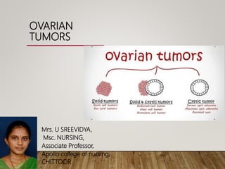
OVARIES
• The most important medical problems in
ovaries are the neoplasms
• Death from ovarian cancers is more than
that of cervix and uterus together
• Silent growth of ovarian tumors is the
rule ,which make them so dangerous
4. OVARY
THE HUMAN OVARY HAS A STRICKING PROPENSITY TO
DEVELOP A WIDE VARITY OF TUMORS MOST OF WHICH
ARE BENIGN.
80% OF ALL OVARIAN TUMORS ARE BENIGN,ALTHOUGH
THIS VARIES WITH AGE.
5.
6. BENIGN LESIONS OF OVARY
NON NEOPLASTIC NEOPLASTIC(BENIGN)
FUNCTIONAL PATHOLOGY
1.Follicular cyst 1.PCOS A. SURFACE EPITHELIUM
2.Corpus luteal cyst 2.Endometrial cyst 1. Serous
3.Theca lutein & (Chocolate cyst) 2. Mucinous
granulosa lutein cyst 3. Endometroid
4. Brenner
B.GERM CELL TUMOUR (BENIGN)
C. SEX CORD STROMAL TUMORS
1. Granulosa cell tumours
2. Thecoma/fibroma
3. Androblastoma
4. gynandro blastoma
5. unclassified tumours
D. Lipid cell tumors
E. Gonadoblastoma
7. FOLLICULAR CYST
(commonest)
CORPUS
LUTEUM CYST
THECA LUTEIN
CYST
AGE GROUP Adolescent,
reproductive age
groups ,can occur
in perimenopause
Reproductive age Reproductive age
CAUSE Hyper estronism Over activity of
corpus luteum
Excess chorionic
gonadotropin
secretion,
following to ovulation
induction drugs
Size
Laterality
Grow ≥3 & ≤8 cm
B/L or U/L
3 – 10 cm
U/L
Large upto 30 cm
B/L
8. FOLLICULAR CYST CORPUS LUTEUM
CYST
THECA LUTEIN CYST
GROSS Thin walled ,
unilocular , filled
with straw coloured
fluid
Pink or
haemorrhagic cyst,
cut section
yellowish orange,
filled with blood
clots
Multicystic, greyish
blue colour, filled
with straw colour
fluid or blood
Histology Lining epithelium
Granulosa cells
Luteinised
granulosa cell
Theca lutein
cells,granulosa
lutein cells
C/F Usually
asymptomatic ,
diagnosis incidental
-Dull pelvic pain with
U/L
-Rupture with
hemoperitoneum
more common
Small are
asymptomatic, large
-discomfort , pain
Rupture/torsion
(more common)
11. PCOS:
- 0.5-4%, infertile women, young reproductive age
-due to excess androgen , chronic anovulation
-Pathology: ovaries enlarged, stroma increased, capsule
thickened, pearly white appearance
-Gross appearance: multiple follicles in cortex
-Histology: thickened tunica albuginea, stromal hyperthecosis
-CF: amenorrhoea, hirsutism, obesity, enlarged PCO.
-Investigations: Hormonal investigations- Blood tests can be used to measure the
levels of FSH, LH and circulating male hormones.
•Ultrasound- TVS
•Laparoscopy
•Hysteroscopy.
-Management – Wt. reduction
COCP(Combined oral contraceptive pills)
Treatment Of hirsutism
Treatment Of infertility
12. Definition
• Ovarian tumors may arise at any age, but are
commonest between 30 and 60.
• 1.Ovarian tumors are particularly liable to
benign or to become malignant.
• 2.In their early stages they are asymptomatic
and painless.
• 3.They may grow to a large size and tend to
undergo mechanical complications such as
torsion and perforation.
OVARIAN TUMOURS
13. BENIGN OVARIAN TUMORS
These are the benign Ovarian neoplasms, may be divided
generally by cell type of origin into three types:
1.Epithelial
2.Stromal
3.Germcell
Incidence: It varies from 1-3 percent.
About 75% of these are benign.
14.
15.
16. ETIOLOGY
Etiology not well known
May be considered as,
• Repeated trauma to epithelium
• Hereditary or familial ovarian cancer
1) Gene mutations
2) Oncogenes
17. RISK MODIFIERS
• Nulliparity
• Infertility
• Early menarche
• Late menopause
• Endometriosis
• Family history
• Prolonged use of ovulation inducing
drugs
• HRT
• Oral contraceptive usage
• Multi parity
• Breast feeding
• Tubal –ligation
• Hysterectomy
• Prophylactic salpingo-
oophorectomy.
RISK FACTORS PROTECTIVE FACTORS
18. OVARIAN
TUMORS
Epithelial tumors: These tumors arise from a layer
of cells that surrounds the outside of the ovary called the
germinal epithelium.
• About 70-80% of all ovarian cancers are epithelial.
• These are most common in women who have been
through menopause (aged 45-70 years).
19. Stromal tumors: Stromal tumors develop from connective-
tissue cells that form the structure of the ovary and produce
hormones.
• These account for 5-10% of ovarian cancers and also these tumors
typically occur in women aged 40-60 years.
Germ cell tumors: Tumors that arise from germ cells (cells that
produce the egg) account for about 15% of all ovarian cancers.
• These tumors develop most often in young women (including teenaged
girls). Although 90% of women with this type of cancer are successfully
treated, many become permanently infertile.
20. 27
1. EPITHELIAL OVARIAN TUMORS
• 70 – 80 % of overall tumors
• Age 40+
• Traditionally divided into Benign, Malignant,
and Borderline in malignancy
• Can be strictly epithelial (serous, Mucinous)
• Only 25% diagnosed in Stage I
21. EPIDEMOLOGY:
-Incidence: 1-3% among outpatient , 75% -benign
-Racial factors: higher in white population, lowest in japan
-Economic status: higher in industrialised countries
PATHOLOGY:
-Origin: mesoepithelial cells on ovarian surface
-Incidence: epithelial tumours—80% of all ovarian tumours
serous cystadenoma– 50% of all epithelial tumours
mucinous cysts—12-15%
endometroid—10%
unspecified—25-27%
22. 26
CLASSIFICATION OF SURFACE EPITHELIAL TUMORS
-Serous Tumors : Benign ,Borderline and malignant
-Mucinous T. : Benign ,Borderline , and malignant
-Endometrioid T. : Benign, Borderline, and malignant
Clear cell carcinoma
-Transitional cell T. :Brenner tumors, Benign ,Borderline ,and
malignant
-Undifferentiated Carcinoma
23. SEROUS CYSTADENOMA
Generally benign
Bilateral – 10-25%
Risk of malignancy : 5 – 10 % borderline malignant
20 -25% malignant
GROSS : multilocular with papillary components.
MICRO : low columnar epithelium with cilia.
Associated fibrosis may lead to “cystadenofibroma”
24. 29
SEROUS TUMORS
• Most are large ,spherical to ovoid ,cystic
structures
• 5 – 10 cm and might be 30-40 cm
• 25% of benign tumors are bilateral
• The surface of the benign is smooth and
glistening .
• In contrast to the malignant forms, the
surface is nodular and irregular
27. MUCINOUS CYSTADENOMA
Have tendency to become huge masses
Round to ovoid masses with smooth capsules that are
usually translucent or bluish to whitish gray.
Interior divided by discrete septa into loculi containing
clear , viscid fluid.
Epithelium – tall, pale staining, secretary with basal nuclei
and goblet cells
5 – 10% are malignant
28.
29. 43
MUCINOUS TUMORS
• Epithelium consists of mucin-producing
cells
• Less likely to be malignant
• 10% of ovarian cancers
• 80% of them benign
• 10% Low malignant potential
• 10% malignant
31. SEROUS /MUCINOUS CYSTADENOMA
Gross appearance of a mucinous (A) and serous (B) cystadenoma of
the ovary. The mucinous type is generally multiloculated and can be
quite large.
32. ►Usually benign. occur in reproductive life
►They can be malignant.
►May be associated with endometrial
hyperplasia
►May coexist with mucinous cystadenoma
BRENNER TUMORS
35. BRENNER TUMOR
Uncommon tumor grossly identical to fibroma
Arise from Walthard cell nests ,also from surface epithelium, and
ovarian stroma
On microscopy – markedly hyperplastic fibromatous matrix
with nests of epitheloid cells showing coffee bean pattern
Considered uniformly benign. But scattered reports of malignant
Brenner's available.
Endocrinologically inert, but could spread with virilization and
endometrial hyperplasia
36.
37. ► Clear cell ovarian tumors are part of the surface
epithelial tumor group of ovarian cancers,
► Accounting for 6% of these cancers.
► Polypoid masses that protrude into the cyst.
► On microscopic examination, composed of cells
with clear cytoplasm (that contains glycogen)
► The pattern may be glandular, papillary or solid.
CLEAR CELL CARCINOMA
38.
39. ENDOMETROID TUMOUR:
-2% of all ovarian tumours
-Lined by glandular epithelium
-Moderate size, solid, with cystic areas
with haemorrhagic fluid.
40. 2. GERM CELL TUMORS
• Ovarian germ cell tumor is a disease in which
malignant (cancer) cells form in the germ (egg) cells
of the ovary.
• CAN OCCUR ATANY AGE
• 12-15% OF OVARIAN NEOPLASM
• 60% OF GCTS OCCURS IN CHILDREN
• MOST COMMON BENIGN TYPE IS ‘BENIGN CYSTIC TERATOMA’
41. GERM CELL TUMORS
► Dermoid cyst (benign cystic teratoma)
• 25% of all ovarian neoplasm
• Contain tissue derived from two or more germ cell
layers
• Unilocular cyst. May contain teeth, bone , cartilage,
nerves, hair, Tissues
• Almost always benign. Malignant changes may occur in
any component
• Occur at any age. peak is 20-30 years.
• Bilateral in 20%
42. DERMOID CYST
GROSS: thick, opaque , whitish wall.
CONTENTS: hair, bone, cartilage, and a large amount of greasy
sebaceous material.
MICROSCOPICALLY : all the three germ layers (ectoderm, mesoderm
and endoderm)
Malignant change occurs in 1-3%. Usually of a squamoustype.
Risk of torsion is 15%
An ovarian cystectomy is almost always possible, even if it appears that
only a small amount of ovarian tissue remains
43.
44.
45. Benign
ovarian
tumors
MUCINOUS
CYST
ADENOMA
SEROUS
CYST
ADENOMA
BRENNER BENIGN
CYSTIC
TERATOMA
INCIDENCE 12-15 % of
Epithelial
tumors
50 % of all
Epithelial
tumors
2 – 3 % of all
Epithelial
tumors
95 % of Germ
cell tumors
20-25% of all
OV.tumors
40% of all
ovarian tumors
1 -2% of all
ovarian tumors
15 – 25% of all
ovarian tumors
Bilateral 10% 40% 8 -10% 15 -20%
Malignant
chance
5 –10% 40% rare 1 -2%
20- 40 % of all
ov. Tumors in
pregnancy
46. TUMOR MUCINOUS SEROUS DERMOID
ORIGIN
Totipotent surface
epithelium of ovary
Totipotent surface
epithelium of
ovary
germ cells arrested
after 1st meoitic
division
PATHOLOGY
naked eye : huge size
& wt 5-10 kg
pedunculated, largest.
smooth, lobulated
with whitish or
bluish white ,
translucent tumor.
naked eye:
smooth, shiny,
greyish white
exuberant
papillary
projections .
naked eye:
moderate size,
capsule tense &
smooth
c/s: thick, visid
mucin (glycoprotein)
colourless
multiloculated with
papillary. honey
combed appearance
c/s:multilobulated
clear fluid (serum)
proteins (albumin
& globulin)
c/s: trabeculated
appearance ,
sebaceous material
with hair , clear
protruberance
47. -microscopy:
lined by 1 layer of
tall coloumnar
epithelium with
dark staining
basal nuclei
without any cilia.
Epithelium
resemble to those
of endocervix.
-complication:
rupture
pseudomyxoma
peritonei &
shows adhesions
with visera .
microscopy:-
lined by cubical
epithelium
- papillary
structures –
dense fibrous
stroma covered
by single or
multiple layers of
columnar
epithelium.
ciliated secretory
& peg cells.
Epithelium
resemble to those
of endosalpingeal
epithelium
micro: stratified
squamous
epithelium,granulat
ion tissue, may be
transitional/
columnar
.
48. 3. SEX CORD STROMAL OVARIAN
NEOPLASMS
• Hormone secreting tumors of the ovary.
• These tumors include fibromas,
Sertoli-Leydig cell tumors
(Arrheno–blastomas or androblastomas).
49. FIBROMA
Most common benign, solid neoplasms of the ovary.
Compose approx 5% of benign ovarian neoplasms and 20% of
all solid tumors of the ovary.
Frequently seen in middle-aged women.
Characterized by their firmness and resemblance to myomas
Misdiagnosed as exophytic fibroids or primary ovarian
malignancy
Not hormonally active
Fibromas may be associated with ascites or hydrothorax as a
result of increased capillary permeability.
51. FIBROMA:
-origin: stromal cells of ovarian cortex
-small nodule with long pedicle ,solid, smooth surfaced tumour
-microscopy: interlacing bundles of spindle shaped cells
-complication: torsion ,meig syndrome
ANDROBLASTOMA/SERTOLI-LEYDIG CELL TUMOR
-testicular adenoma
-androgen secreting tumour
-seen in women less than 30 years
-gynandroblastoma (granulosa + androblastoma cells)
-cf: amennorhoea, atrophy of breasts, enlargement of clitoris, body
hair growth, deepening of voice.
52.
53. THECOMA
Solid fibromatous lesions that show varying degrees of yellow or
orange discoloration.
Almost always confined to one ovary.
Usually >40 years, 65% after menopause
May be hormonally active and hence associated with estrogenic or
occasionally androgenic effects.
Luetinised thecoma – younger, sclerosing peritonitis and ascites
Leydeig cell thecoma – associated with Reinke crystals
Rarely malignant
54. 4. GONADOBLASTOMAS
Gonadoblastoma is a rare benign tumor that has the potential for malignant
transformation and affects a subset of patients with an intersex disorder or
disorder of sex development (DSD).
Contain both germ cells and sex cord stromalcells.
Arise in patients with dysgenetic gonads - 44 XXY / 45XO
Presents usually as phenotypic female <30 years with primary amenorrhea
and virilization.
Treatment – laparoscopy or laparotomy with removal of b/l dysgenetic
gonads.
Further treatment depends on malignant germ cell component
58. CLINICAL FEATURES
AGE:- late child bearing age
-dermoid, mucinous adenoma common in reproductive
period.
-dermoid common in pregnancy
symptoms: -asymptomatic
- detected accidently
-heaviness in lower abdomen, a gradually increasing
mass in lower abdomen
- dull aching pain,
- cardiorespiratory & gastrointestinal upset
(nausea, indigestion)
-menstrual pattern unaffected except in hormone
producing tumours
signs: cachetic due to protein loss, pitting edema of legs
59. ABDOMINAL EXAMINATION
Inspection -- bulging of lower abdomen
mass – central/ one side/ whole abdomen
visible veins , flanks – flat
Palpation -- cystic / tense cystic
• freely mobile from side to side but restricted from above down,
smooth surface, nontender
Percussion -- dull in center and resonant in flanks
fluid thrill +
Auscultation -- friction rub +
Bimanual pelvic examination --
uterus separated from mass
Groove is felt between uterus & mass
movement of mass p/a fails to move cx
lower pole of cyst felt through fornix
absence of pulsation of ut vessels through the fornices
69. • Second most common gynaecological
malignancy
• Major cause of death from gyn. Cancer
• 2% lifetime risk (1.4% in general
population.)
• Mean age – 64y
• Rare in young (3% <35y)
70. • Present with late stage (66% - stage 3 or more)
• Most common,
– Persistent pelvic/abdominal pain
– Abdominal distention/bloating
– Early satiety
– Constitutional ( fatigue,weight loss)
– Abnormal uterine bleeding
– Ascites , effusions
– Urinary symptoms (frequency, urgency)
71. • Benign ovarian tumour or cyst.
• Uterine or tubal mass.
• Endometriosis.
• Bowel mass or primary peritoneal carcinoma.
• Secondary carcinoma: breast, gastrointestinal
tract, lymphomas and pelvic-organ tumours.
72. • Fixed, Hard mass arising from pelvis
• Ascites
• Adenexal mass
– In premenopausal <20% malignant
– In postmenopausal 50% malignant
• Pleural effusions
• Lymphadenopathy (cervical, inguinal)
73. • Toconfirm the diagnosis
• Toidentify the extent of lesion
• Todetect the primary site
75. • USG for suggestive of malignancy
– Solid + cystic areas
– Ascites
– Multilocular/complex cyst
– Papillary projections
– Neovascularization
– Bilateral tumor
– Liver deposits
76. MANAGEMENT
• Once an ovarian tumor is diagnosed, the
patient should be admitted for operation,
sooner the better.
• The complication can occur at any time and
the nature of the tumor cannot be assessed
clinically.
• Differentiation between benign and malignant
ovarian tumours could be made by clinical
examination, ultrasonography, laparotomy and
finally biopsy.
79. • Primarily surgical
– Diagnosis
– Staging
– Treatment
• Ideally done by Gynaecological oncologist
• Objective is,
– Accurate staging
– Removing all visible tumor
• No residual disease after laparotomy
80. INDICATIONS FOR SURGERY
Any solid ovarian lesion
Any ovarian lesion with papillary vegetation on the cyst wall
Any adnexal mass >10cm in diameter
Palpable adnexal mass in a premenarcheal or
postmenopausal women
Torsion or rupture suspected
81. OVARIAN MASS IN REPRODUCTIVE
AGE GROUP
<5 cms. >/= 5 cms
USG USG
cystic
observation
Complex,
solid,
suspiciou
s
Persistence or progression
surgery
82. GUIDELINES FOR SURGERY IN
BENIGN TUMOR
• Incision should be vertical paramedian & sufficiently big
enough to deliver the cyst intact.
• Inspect the nature of the peritoneal fluid – clear, straw colour,
hemorrhagic or infective
• Deliver the tumor intact and note it carefully about its nature.
• Inspect and palpate the other ovary, pelvic organs, omentum
and liver.
• Proceed for the definitive surgery
• Cut the tumor and inspect the inner side for any evidence of
malignancy.
83. DEFINITIVE SURGERY:
• In young women- ovarian cystectomy
leaving behind the healthy ovarian tissue
• Ovariotomy (salpingo-oophorectomy) –
For a big tumor.
• In parous women Around
40 years - TAH+BSO
• Others (any age other than
above)- individualisation.
84. OPERATIVE MODALITIES
Laparoscopic cystectomy / ovariotomy laparoscopy/
USG guided aspiration of cyst/Laparotomy.
Laparoscopy vs laparotomy – decision based on suspicion of
malignancy and technical expertise
No recurrence rates following laparoscopy or laparotomy.
The objective is to try cystectomy if possible.
Laparoscopic surgery for benign ovarian tumours is associated with
less pain, shorter hospital stay, and fewer adverse events than with
laparotomy.
85. The standards for laparoscopy in benign tumours
1. careful examination of the external surface of the tumour and
sampling of the peritoneal cavity
2. avoidance of any tumoral rupture
3. protection of the ovarian tumour with an endoscopic bag
before removal
86. • Borderline malignant epithelial tumours have got
some features of malignancy but not all.
• Young females
– U/L: salpingo-oophorectomy
• Older females
– TAH + BSO
• If unfit for surgery
– Primary chemotherapy
– If responds, interval surgery after 3-6 cycles