
Empfohlen
Weitere ähnliche Inhalte
Ähnlich wie 268307418-Management-of-Mandibular-fractures-ppt.ppt
Ähnlich wie 268307418-Management-of-Mandibular-fractures-ppt.ppt (20)
Kürzlich hochgeladen
Kürzlich hochgeladen (20)
268307418-Management-of-Mandibular-fractures-ppt.ppt
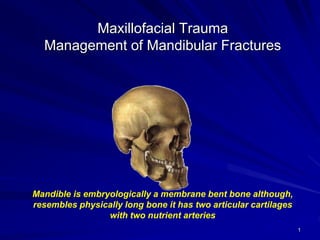
- 1. 1 Maxillofacial Trauma Management of Mandibular Fractures Mandible is embryologically a membrane bent bone although, resembles physically long bone it has two articular cartilages with two nutrient arteries
- 2. 2 Mandible in trauma Mandibular fracture is more common than middle third fracture (anatomical factor) It could be observed either alone or in combination with other facial fractures Minor mandibular fracture may be associated with head injury owing to the cranio-mandibular articulation Mandibular fracture may compromise the patency of the airway in particular with loss of consciousness Fracture of mandible occurred with frontal impact force as low as 425 lb (190 Kg) {Condylar fracture}
- 3. 3 Fracture of condyle regarded as a safety mechanism to the patient Frontal force of 800-900 lb (350-400 Kg) is required to cause symphesial fracture Mandible was more sensitive to lateral impact than frontal one Frontal impact is substantially cushioned by opening and retrusion of the jaw (Nahum 1975) Long canine tooth and partially erupted wisdoms represent line of relatively weakness
- 4. 4 Anatomical considerations Attached muscles: Masseter Temporalis Medial and lateral pterygoid Mylohyoid Geniohyoid and genioglosus anterior belly of digastrics
- 5. 5 Blood supply Endosteal supply via the ID artery and vein Periosteal supply, important in aging due to diminishes and disappearance of alveolar artery Bradley 1972 Nerve Damage of inferior dental nerve Facial palsy by direct trauma to ramus Damage of facial nerve in temporal bone fracture Goin 1980 Damage to mandibular division of facial nerve
- 6. 6 Factors influenced site of fracture and displacement Anatomy of the mandible and attached muscle (canine & wisdoms) Weakening areas of mandible (resorption and pathologyl) Direction of force of the blow Age of the patient
- 7. 7 Types of fracture Simple Greenstick fracture (rare, exclusively in children) Fracture with no displacement (Linear) Fracture with minimal displacement Displaced fracture Comminuted fracture Extensive breakage with possible bone and soft tissue loss Compound fracture Severe and tooth bearing area fractures Pathological fracture (osteomyelities, neoplasm and generalized skeletal disease)
- 8. 8 Sites of fractures Condyle fracture – Intracapsular fracture – Extracapsular fracture High condyle neck fracture Low condylar fracture Angle/ ramus fracture (body fracture) Canine region (parasymphesial fracture) Midline fracture (symphesis fracture) Coronoid fracture (rare)
- 9. Mandible Fracture Frequency Condyle 30% Ramus 3% Angle 25% Body 25% Parasymphyseal / Mental 15% Coronoid process 2%
- 10. 10 Favourable or unfavourable They can be vertically or horizontally in direction They are influenced by the medial pterygoid- masseter “sling” If the vertical direction of the fracture favours the unopposed action of medial pterygoid muscle, the posterior fragment will be pulled lingually If the horizontal direction of the fracture favours the unopposed action of messeter and pterygoid muscles in upward direction, the posterior fragment will be pulled lingually Favourable fracture line makes the reduced fragment easier to stabilize
- 11. 11 Effects of muscles on displacement Transverse midline fracture (symphesial) stabilizes by the action of mylohyoid and geniohyoid Oblique fracture (parasymphesial) tends to overlap under the influence of muscles action Bilateral parasymphesial fracture results in backward displacement associated with loss of tongue control when the level of consciousness is depressed
- 12. 12 Condylar fractures The most common mandibular fracture Unilateral or bilateral Intracapsular or extracapsular Antero-medial displacement is common but it may remain angulated with the ramus Dislocation of the glenoid fossa and fracture of petrous temporal bone which is very rare
- 13. 13 Sign and symptoms Swelling, pain, tenderness and restriction of movement Deviation of mandible towards the side of fracture Gagging of occlussion (premature contact on the posterior teeth) with bilateral condylar displaced or over-riding fractures Displacement of mandible toward the affected side Anterior open bite on opposite side of fracture Laceration of EAM**** Retroauricular ecchymosis**** Cerebrospinal leak and otorrhea in association with skull base fracture Condylar fractures
- 14. 14 Sequlae of TMJ injury Artheritic changes Haemartherosis, fibrosis and aknylosis Meniscal damage and detachment TMD Staph infection with condylar backward displacement and external auditory meatus injury Meningitis with petrous temporal bone fracture and intracranial involvement Condylar fractures
- 15. 15 Coronoid process fracture: Rare fracture caused by direct trauma to ramus and results from reflux contraction of temporalis Can be seen following operation of large ramus cyst Elicit tenderness over the anterior part of ramus Development of tell-tale haematoma
- 16. 16 Fracture of the ramus: Type I Single fracture Mimics low condylar fracture that runs below the sigmoid notch Type II comminuted fracture Common in missile injuries and appears to be with little displacement due to effects of messeter and medial pterygoid muscles
- 17. 17 Fracture of the angle and body Pain, tenderness and trismus Extra-oral swelling at the angle with obvious deformity Step deformity behind the molar teeth Movement and crepitus at the fracture site Derangement of occlussion Intra-oral buccal and lingula heamatoma Involvement of IDN Gingival tear if fracture in dentated area Tooth involvement and possible longitudinal split fracture
- 18. 18 Midline fracture The most common missed fracture (always fine crack) Can be symphesial or parasymphesial fracture Commonly associated with one or both condyles fracture Unilateral fracture leads to over-riding of the fragments and bilateral may contribute in loss of voluntery tongue control Long canine tooth represent a weak area and contributes to parasymphesial fracture Rarely runs across mental foramen
- 19. 19 Signs and symptoms Pain and tenderness Swelling and odemea Development of step deformity Mental anesthesia Heamatoma in the floor of mouth and buccal mucosa Soft tissue injury of the chin and lower lip If associated with condylar fractures Absence of condyle movement on the contrlateral side Deviation of mandible Anterior open bite Gagging of oclussion Limitation of mouth opening Midline fracture
- 20. 20 Clinical assessment and diagnosis History of trauma (traumatized patients with possible head injury) and facial injuries Clinical Examination ▶ Extroral Inspection (assessment of asymmetery, swelling, ecchymosis, laceration and cut wounds) Palpation for eliction of tenderness, pain, step deformity and malfunction ▶ Intra- and paraoral bleeding, heamatoma, gingival tear, gagging of occlussion and step deformity and sensory and motor deficiency Radiographs
- 21. 21 Radiographs Plain radiograph OPG Lateral oblique PA mandible AP mandible (reverse Townes) Lower occlusal CT scan 3-D CT imaging MRI
- 22. 22 Principles of treatment similar to elsewhere fractures in the body Reduction of fragments in good position Immobilization until bony union occurs These are achieved by: Close reduction and immobilization Open reduction and rigid fixation Other objective of mandible fracture treatment: Control of bleeding Control of infection
- 23. 23 Definitive treatment Soft tissue repair Debridment Irrigation with saline and antibiotics Closure in layers Dressing Reduction and fixation of the jaw ▶ Close reduction and IMF (traditional method by means of manipulation) ▶ Open reduction and semi-rigid fixation (using inter-ossous wirings) ▶ Open reduction and rigid fixation (using bone palates osteosynthesis) Objective: Restoration of functional alignment of the bone fragments in anatomically precise position utilizing the present teeth for guidance
- 24. 24 Close reduction Arch bars – Jelenko – Erich pattern – German silver notched Cap splints MMF Screw ▶ IMF/MMF prior to rigid fixation ▶ For the purpose of close reduction
- 25. 25 Close reduction Bonded brackets IMF/MMF screws (Quick-Fix) Dental wiring: Direct wiring Eyelet wiring Local anesthesia or sedation Minimal displacement IMF/MMF (Quick-Fix) for 6 weeks Treatment can be performed under GA or LA and when surgery is contraindicated
- 26. Archbar vs MMF Screw (Quick-Fix) Archbar Less Convenience Patients Require teeth for fixation Damage teeth and periodontal tissue Uncomfortable during the fixation period Difficult daily maintenance of oral hygiene Operator Risk of blood-transmitted diseases Need longer time to use MMF Screw (Quick-Fix) The Easy Alternative to Arch Bars Patented Auto-Drive self drilling screws Dramatically reduces application time of MMF (only 5 minutes) Simple Minimizes risk of wire puncture wound Better oral hygiene maintenance Ideal for edentulous or partially edentulous
- 27. 27 Fracture mandible in children Close reduction Open reduction and fixation Plating at the inferior border Biodegradable plate and screw
- 28. 28 Open Reduction and Fixation System Intraoral approach Extraoral approach ▶ Submandibular approach
- 29. 29 Rigid Fixation System Intraossous wiring Plates and screws 2.0mm and 2.4mm – Standard plate and screw – Locking plate and screw Kirchener wire Lag screws
- 30. Plate 2.0mm and 2.4mm 30
- 31. 31 Reconstruction palate Severe trauma Loss of part of the bone
- 32. Recon: TCA Temporary Condyle Attachment (TCA) – Oncology/Ablation cases only – Maximum implantation of 1 year – Only OsteoMed Medically Tracked Device 3 Forms – No Left/Right – Unique Anatomical Shape – Adjustable
- 34. Recon: Instruments Instrumentation – Lag screw cannula and depth gauge Tip of gauge will point to drill exit point Length of screw needed to engage both cortices The measurement directly above the drill entry point will state needed length
- 36. Recon: Instrumentation Instrumentation – Fx plate bending pliers – Roller benders Used for major contours Bends in the saggital and lateral plane – Reconstruction bending pliers For intermediate bending – Bending irons Bending slot in the tip Finishing bends Unusual or tight bends
- 37. Rigid Fixation System Instrumentation – Fracture plate benders
- 38. Rigid Fixation System Instrumentation – Taperlocktm screwdriver body – Modular for all the drivers stems, taps and countersinks
- 39. Rigid Fixation System Instrumentation – Calibrated plate benders – Calibrated “tick” marks to estimate step range for “L” and Zed plates
- 40. Rigid Fixation System Instrumentation – 3 prong plate benders Used with 10 and 16 hole plates to bend in the saggital plane
- 41. Rigid Fixation System Instrumentation – Right angle plate benders Places 90 degree angle bends in plates Used with “L” and Zed plates for LeFort 1 osteotomies
- 42. Rigid Fixation System Cannula/Trocar – Trocar is used to penetrate soft tissue
- 44. Rigid Fixation System Instrumentation – Cheek Retractor “U” Shape – Cheek Retractor – Cannula – Neutral Drill Guide
- 45. Rigid Fixation System Instrumentation – Cannula/Depth gauge Determines what size screw needs to be used
- 46. 46 Condylar fractures Intraoral approach Ramus incision Extraoral approach Preauricular approach Retromandibular approach
- 47. 47 IMF/MMF Transosseous wiring Circumferential wiring External pin fixation Bone clamps Trans-fixation with Kirschner wires Maxillo Mandibular Fixation Screw (Quick-Fix)
- 48. 48 Osteosynthesis Non-compression small plates Compression plates Mini plates (2.0mm and 2.4mm) Lag screws Biodegradable plates and screws
- 49. 49 Teeth in the fracture line The fracture is compound into the mouth The tooth may be damaged or lose its blood supply The tooth may be affected by some preexisting pathology
- 50. 50 Management of teeth retained in fracture line Good quality intra-oral periapical radiograph Insinuation of appropriate systemic antibiotic therapy Splinting of tooth if mobile Endodontic therapy if pulp is exposed Immediate extraction if fracture becomes infected Follow up for 1 year and endodontic therapy if there is a loss of vitality
- 51. 51 Absolute indications Longitudinal fracture Dislocation or subluxation from socket Presence of periapical infection Infected fracture line Acute pericoronitis Relative indications Functional tooth that would be removed Advanced caries or periodontal diseases Doubtful tooth which would be added to existing denture Tooth in untreated fracture presenting more than 3 days after injury
- 52. Complications Airway esp with IMF (wire cutters and pre-op education) Infection Delayed and non-union – Inadequate immobilisation, fracture alignment – Inteposition of soft tissue or foreign body – Incorrect technique Inferoir alveolar nerve damage – 56%pre-treatment – 19% post-treatment Malocclusion TMJ ankylosis esp intracapsular condyle # 52
- 53. 53