the gross antomy of the hip hoint and applied anatomy focused for undergraduate and post graduate students of human anatomy
•Als PPT, PDF herunterladen•
53 gefällt mir•9,116 views

A power point of gross anatomy and applied anatomy of hip joint

Empfohlen
Weitere ähnliche Inhalte
Was ist angesagt?
Was ist angesagt? (20)
Andere mochten auch
Andere mochten auch (20)
Ähnlich wie the gross antomy of the hip hoint and applied anatomy focused for undergraduate and post graduate students of human anatomy
Ähnlich wie the gross antomy of the hip hoint and applied anatomy focused for undergraduate and post graduate students of human anatomy (20)
Mehr von Shaifaly madan rustagi
Kürzlich hochgeladen
Kürzlich hochgeladen (20)
the gross antomy of the hip hoint and applied anatomy focused for undergraduate and post graduate students of human anatomy
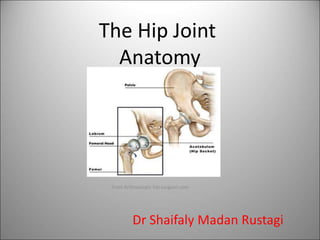
- 1. The Hip Joint Anatomy From Arthroscopic hip surgeon.com Dr Shaifaly Madan Rustagi
- 2. The Hip Joint • Ball-and-Socket variety of synovial joint • Articulation of the head of the femur with the acetabulum of the hip bone • A fibrocartilaginous ring called the acetabular labrum deepens the acetabulum . From Arthroscopic hip surgeon.com
- 3. The Acetabulum The acetabulum is formed by the pubis, ischium and ilium bones
- 5. The Joint Capsule • Anteriorly – proximally to the bone surrounding the acetabulum. – Distally to the trochanteric line • Posteriorly -to the margins of the acetabulum and surrounding bone -neck of the femur- not to the trochanteric crest Capsule has longitudinal and circular. • The circular fibers form a collar around the femoral neck called the zona orbicularis. • The longitudinal retinacular fibers travel along the neck and carry blood vessels. grays from wikipedia
- 6. Ischiofemoral ligament • It arises from the posteroinferior margin of the acetabular rim, passes laterally to the capsule and blends with the circular fibres of the capsule, the zona orbicularis. • Posterior joint capsule is reinforced by this ligament.
- 7. Iliofemoral ligament or ligament of Bigelow • It is the strongest ligament in the human body. • The apex is attached to the lower half of the anterior inferior iliac spine . • The base is attached to the intertrochanteric line. • It is inverted Y or V shaped. One limb goes to the base of the greater trochanter and the other to the base of the lesser trochanter. • It limits extension at the hip joint.
- 8. Pubofemoral ligament • It is attached to the superior ramus and obturator crest of the pubis superiorly and to the base of the lesser trochanter inferiorly. • It is inferior to the iliofemoral ligament and reinforces the inferior part of the hip joint capsule. • It also blends with the medial parts of the iliofemoral ligament Healthfavo.com
- 9. The round ligament or the ligamentum teres or the ligament of head of femur The round ligament of the head of the femur is attached to the transverse acetabular ligament and extends to the fovea centralis on the head of the femur Grays wikipedia
- 10. Synovial membrane • Lines fibrous capsule intracapsular portion of neck of femur Acetabular labrum Transverse ligament Round ligament of head of femur
- 11. Relations of hip joint www.ganfyd.org
- 12. Blood supply • Medial Circumflex • Lateral Circumflex • Obturator • Inferior gluteal
- 13. Nerves • Femoral (from nerve to rectus femoris) • Obturator (anterior division) • Sciatic (articular twigs) • Nerve to quadratus femoris Pain arising in hip joint may be referred to the knee.
- 14. Movements The hip joint is the most mobile joint in the lower limb. It is capable of flexion and extension, abduction and adduction, medial and lateral rotation and all of these in a circular motion- circumduction
- 15. Movements • Flexion-the head of femur rotates about a transverse axis that passes through the acetabula . • It is limited by the thigh touching the abdomen, the range is 120 degrees. • It is mainly due to contraction of the iliopsoas muscle, with help from the sartorius, rectus femoris, and pectineus. • Extension- it is limited by tension in the iliofemoral ligament ,range is 20 degrees. • It is brought about chiefly by the guteus maximus muscles with help by the hamstrings. In Adduction and abduction- the femoral head rotates in the acetabulum about an anteroposterior axis. Adduction is limited by contact with the other leg, range is 30 degrees. It is produced by the adductor longus, brevis, magnus and the gracilis and pectineus. Abduction- is limited by tension in the adductors and in the pubofemoral ligament.,range is 60 degrees. It is brought about by the gluteus medius and gluteus minimus
- 16. • Lateral rotation- by the gluteus maximus, quadratus femoris, piriformis, obturator internus and externus, gemelli • Medial rotation- by the anterior part of the glueteus minimus and medius and tensor fasciae latae muscles • Range is about 40 degrees for both the movements.
- 17. Applied anatomy • Head of femur shifts upwards. • Lurching gait • Tredlenburg test is positive
- 18. Perthes disease Destruction and flattening of head of femur
- 19. Coxa vara and Coxa Valga
- 20. Fracture neck of femur
- 22. Displacement of greater trochanter
- 23. Nelatons line and Bryants triangle - View of the outer surface of the bones of the hip. showing Roser-Nelaton line (a d); Bryant's triangle (a b c - b c being its base) chestofbooks.com