Cardiovasular system
•
14 gefällt mir•1,466 views

This system has three main components: the heart, the blood vessel and the blood itself. The heart is the system's pump and the blood vessels are like the delivery routes. Blood can be thought of as a fluid which contains the oxygen and nutrients the body needs and carries the wastes which need to be removed.

Empfohlen
Weitere ähnliche Inhalte
Was ist angesagt?
Was ist angesagt? (20)
Ähnlich wie Cardiovasular system
Ähnlich wie Cardiovasular system (20)
Mehr von DR .PALLAVI PATHANIA
Mehr von DR .PALLAVI PATHANIA (20)
Kürzlich hochgeladen
Kürzlich hochgeladen (20)
Cardiovasular system
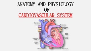
- 1. ANATOMY AND PHYSIOLOGY OF CARDIOVASCULAR SYSTEM
- 2. CIRCULATORY SYSTEM Submitted to: Submitted by: Dr. Pallavi Pathania Manisha Kumari Associate professor MSc nursing 1st year
- 3. INDEX S.NO. CONTENT 1 2 3 4 5 6 7 8 9 Cardiovascular system Blood vessels Vascular system of heart Blood Summarization Recapitalization Assignment Conclusion References
- 4. INTRODUCTION The circulatory system, also called the cardiovascular system or the vascular system, is an organ system that permits blood to circulate and transport nutrients (such as amino acid and electrolytes), . Oxygen, carbon dioxide, hormones and blood cells to from cells in the body to provide nourishment and help in fighting disease ,stablishing temperature and PH and maintain homeostasis
- 5. CIRCULATORY SYSTEM ORGAN/COMPONENTS PRIMARY FUNCTION HEART Propels blood, maintains blood pressure BLOOD VESSELS Distribute blood around the body ARTERIES Carry blood heart to capillaries CAPILLARIES Permit diffusion between blood and interstitial fluids VEINS Return blood from capillaries to the heart BLOOD Transport oxygen, carbon dioxide and blood cells, delivers nutrients and hormones, remove waste products, assists in temperature regulation and defence against disease
- 6. PROCESS OF BLOOD FLOW Heart Arteries Arterioles Capillaries Venules Veins
- 7. DEFINTION • The circulatory system, sometimes called the cardiovascular system, consists of the heart, blood vessels, and blood. • It transports oxygen, hormones and nutrients to all the cells in the body • The circulatory system is composed of the heart, arteries, capillaries, and veins. This remarkable system transports oxygenated blood from the lungs and heart throughout the body via the arteries
- 10. CIRCULATION BEFORE BIRTH • Oxygen and nutrients from the mother's blood are transferred across the placenta to the fetus through the umbilical cord.
- 12. CONT… At birth, major changes take place. The umbilical cord is clamped and the baby no longer receives oxygen and nutrients from the mother. With the first breaths of air, the lungs start to expand, and the ductus arteriosus and the foramen ovale both close. The baby's circulation and blood flow through the heart now function like an adult's.
- 14. HEART
- 15. SIZE OF HEART
- 16. LOCATION AND SURFACE PROJECTION • Apex- lower, cone shaped • Base- border, superior portion • The heart is the hollow, cone shaped about the size of closed fist • It lies in the mediastinum between the lungs and rests upon the diaphragm • Two- third of its mass lies to left of the midline
- 17. POSTION OF HEART • Normally located in the middle and slightly to the left side of the thoracic • The apex is about 9 cm to the left of the midline at the level of the 5th intercostal space and the base extends to the levels of the 2nd rib . • Weighs about 325 gm is males and about 275 gm in females.
- 18. LAYERS OF HEART WALLS
- 19. CONT… • PERICARDUIM – membrane (sac)that surrounds and protects the heart by the help of two layers. a. Fibrous pericardium- superficial layer, tough, inelastic, prevents overstretching, provide protection and anchors the heart in place. b. Serous pericardium- 1. partial layer-fused to the fibrous pericardium 2. visceral layer- or epicardium adheres to the heart itself c. Pericardial cavity –present between two layer is filled with pericardial fluids which reduce friction
- 20. CONT…
- 21. CONT… • EPICARDUIM is the layer immediately outside of the heart muscle proper (the myocardium). The epicardium is largely made of connective tissue and functions as a protective layer
- 22. CONT… MYOCARDUIM The myocardium of the left ventricle is the thickest, as this ventricle is responsible for generating the power needed to pump oxygenated blood from the heart to the rest of the body
- 23. CONT… ENDOCARDUIM • Endocardium (endo-cardium) is the thin inner layer of the heart wall. This layer lines the inner heart chambers, covers heart valves, and is continuous with the endothelium of large blood vessels. • The endocardium of heart atria consists of smooth muscle, as well as elastic fibers.
- 24. CHAMBERS OF HEART Four chambers • Right Atrium • Right Ventricle • Left atrium • Left ventricle
- 25. CONT… RIGHT ATRIUM • Receives venous blood from whole of the body via the superior vena cava(SVC) at its upper end and inferior vena cava (IVC) at its lower end. • It pumps into Right ventricle (RV)through the tricuspid valve during the ventricular diastole. • RIGHT VENRTICLE • Triangular shaped or crescent shaped • Opens into pulmonary artery through pulmonary valve • Most anterior chamber
- 26. CONT… LEFT ATRIUM • Posterior most chamber • Receives oxygenated blood from pulmonary veins • Pulmonary veins open into LA from the posterior wall • RIGHT VENRTICLE • chambers and is responsible for pumping oxygenated blood to tissues all over the body
- 27. VALVES OF HEART • There are also 4 one-way valves that direct flow of blood through the heart in one direction
- 28. CONT… 2 Atrioventricular (AV) valves • Bicuspid (Mitral) valve - separates left atrium and ventricle, consists of two flaps of tissues • Tricuspid valve – separates right atrium and ventricle - consists of three flaps of tissue ( prevent backflow (eversion) keeps valves pointed in direction of flow )
- 29. CONT… 2 Semilunar valves • at beginning of arteries leaving the ventricles Aortic SL valve- at beginning of aorta Pulmonary SL valve-at beginning of pulmonary trunk
- 31. PULMONARY CIRCULATION • Transports oxygen-poor blood from the right ventricle to the lungs where blood picks up a new oxygen supply.
- 32. SYSTEMATIC CIRCULATION • It returns oxygen rich blood and nutrients to the left atrium and is pumped out all over the body It also picks up carbon dioxide and other waste products.
- 33. CORONARY CIRCUALTION • Coronary arteries supply blood to the heart muscle. Like all other tissues in the body, the heart muscle needs oxygen-rich blood to function. Also, oxygen-depleted blood must be carried away. The coronary arteries wrap around the outside of the heart. Small branches divide into the heart muscle to bring it blood.
- 34. CONT… • Left main coronary artery (LMCA)-supplies blood to the left side of the heart muscle (the left ventricle and left atrium). The left main coronary divides into branches: • The left anterior descending artery branches off the left coronary artery and supplies blood to the front of the left side of the heart. • The circumflex artery branches off the left coronary artery and encircles the heart muscle. This artery supplies blood to the outer side and back of the heart.
- 35. CONT… • Right coronary artery (RCA)-supplies blood to the right ventricle, the right atrium, and the SA (sinoatrial) and AV (atrioventricular) nodes, which regulate the heart rhythm. • The right coronary artery divides into smaller branches, including the right posterior descending artery and the acute marginal artery. • Together with the left anterior descending artery, the right coronary artery helps supply blood to the middle or septum of the heart.
- 37. CONT… • To pump blood throughout the body, the muscles of the heart must be coordinated perfectly — squeezing the blood in the right direction, at the right time, at the right pressure. The heart’s activity is coordinated by electrical impulses.
- 38. CONT… • Electrical signals arising in the SA node (located in the right atrium) stimulate the atria to contract. Then the signals travel to the atrioventricular node (AV node), which is located in the interatrial septum.
- 39. CONT… • After a delay, the electrical signal diverges and is conducted through the left and right bundle of His to the respective Purkinje fibers for each side of the heart, as well as to the endocardium at the apex of the heart, then finally to the ventricular epicardium; causing its contraction. • These signals are generated rhythmically, which in turn results in the coordinated rhythmic contraction and relaxation of the heart.
- 40. CARDIC CYCLE • The cardiac cycle is the performance of the human heart from the ending of one heartbeat to the beginning of the next. It consists of two periods: one during which the heart muscle relaxes and refills with blood, called diastole following a period of robust contraction and pumping of blood, dubbed systole
- 42. CARDIC OUTPUT • The cardiac output is the amount of the blood ejected from each ventricle every minutes. • The amount of expelled by each contraction of each ventricles is the stroke volume cardiac output = stroke volume x heart rate • For a resting adult CO = 70mL/beat x75beats/min = 5250 mL/min = 5.25 L/min
- 43. DISEASE OF CARDIOVASCULAR DISEASE • Heart failure • Stenosis • Ischemic heart disease • Myocardial infraction • Rheumatic heart disease • Infective endocarditis
- 44. Assessment of cardiovascular system • Elicit a description of present illness and chief complaint Include: • Onset • Duration • Location
- 45. CONT… Common symptoms of cardiovascular disease • Chest discomfort or pains • Palpations • Fatigue • Dyspnoea • Nocturia
- 46. CONT… Past medical history • Rheumatic fever, kidney disease, diabetes , lung disorders • Evaluation use of the OTC drugs, herbs, recreational drugs Family health history • Any diabetes, kidney disease, hypertension, heart disease etc Psychosocial history • Education, occupation, martial status , children and relationships • Health habits –diet, exercise, smoking , alcohol use
- 47. CONT… • General appearance ( build appearance of the client, skin colour, distress level ,LOC) • Client with chronic heart failure may appear malnourished, thin. Latest skin signs going to appear ascites, jaundice etc • Inspection of finger and nails
- 48. CONT… assess vitals signs Measure BP in both arms initially Assess pulse -rate -Rhythm -Quality
- 49. Cardiac auscultation • Produced from blood turbulence caused by closing of the heart valves • S1 – atrioventricular valve closure LUB • S2 – semilunar valve closure DUB • S3 – rapid ventricular filling • S4 – atrial systole
- 50. BLOOD VESSELS
- 51. INTRODUCTION • The blood vessels are the components of the circulatory system that transport blood throughout the human body. These vessels transport blood cells, nutrients, and oxygen to the tissues of the body. They also take waste and carbon dioxide away from the tissues. Blood vessels are needed to sustain life, because all of the body's tissues rely on their functionality
- 52. DEFINITION • Blood vessel, a vessel in the human or animal body in which blood circulates. The vessels that carry blood away from the heart are called arteries, and their very small branches are arterioles. Very small branches that collect the blood from the various organs and parts are called venules, and they unite to form veins, which return the blood to the heart.
- 53. TYPES OF BLOOD VESSELS 1. Arteries & Arterioles 2. Veins & Venules 3. Capillaries
- 54. CONT… 1.Arteries & Arterioles • built to withstand the greatest pressure of the system a. strong resilient walls, b. thick layers of connective tissues c. more muscular than veins
- 55. CONT… • Arteries and arterioles typically contain ~25% of all blood in circulation (15% in arteries; 10% in arterioles ) • Pressure is variable MAP ~ 93 varies from 100 – 40 mmHg • Most organs receive blood from >1 arterial branch provides alternate pathways
- 56. CONT… Veins & Venules • Generally have a greater diameter than arteries but thinner walls, flaccid • Three layer are all thinner than in arteries tunica adventitia is thickest of three • But not as elastic as arteries • Little smooth muscle
- 57. CONT… • 70% of all blood is in veins & venules (~60% in veins, ~10% in venules) • low pressure: • 12 – 8 mmHg venules • 6 – 1 mmHg veins larger veins near 0
- 58. CONT… 3. Capillaries: • Actual site of exchange of materials the rest is just pumps and plumbing • Consist of only a single layer of squamous epithelium= endothelial layer (=tunica intima)
- 59. CONT… • Arranged into capillary beds = functional units of circulatory system • Capillaries are extremely abundant in almost every tissue of the body • Only 5% of blood at any one time is in capillaries
- 60. MEMBRANE OF VESSELS walls of arteries and veins consist of three layers: a. Tunica Externa b. Tunica Media c. Tunica Interna
- 61. CONT… a. Tunica Externa (= T. adventitia) • outer loose connective tissue • anchors the vessel and provides passage for small nerves, lymphatic vessels and smaller blood vessels
- 62. CONT… b. Tunica Media • middle, made mainly of smooth muscle with some elastic tissue and collagen fibers strengthens vessel walls • prevent high pressure from rupturing them allows vasodilation and vasoconstriction • usually the thickest layer, especially in arteries
- 63. CONT… c. Tunica Interna (=T. Intima) • inner endothelium • exposed to blood when damaged or inflamed induce platelets or • WBC’s to adhere • may lead to plaque build up and atherosclerosis
- 64. AORTA • The aorta is the main and largest artery in the human body originating from the left ventricle of the heart and extending down to the abdomen, where it splits into two smaller arteries (the common iliac arteries). The aorta distributes oxygenated blood to all parts of the body through the systemic circulation
- 65. CONT… • part of the aorta is by anatomical compartment, where the • 1. Thoracic aorta (or thoracic portion of the aorta) runs from the heart to the diaphragm. • 2. Abdominal aorta (or abdominal portion of the aorta) from the diaphragm to the aortic bifurcation
- 66. THORACIC AORTA • This part of the aorta lies above the diaphragm ana is described into 3 parts : • Ascending aorta • Arch of the aorta • Descending aorta in the thorax
- 67. CONT.. • ASCENDING AROTA • It is a portion of the aorta commencing at the upper part of the base of the left ventricle, on a level with the lower border of the third costal cartilage behind the left half of the sternum • Right and left coronary arties are branches of ascending aorta.
- 68. CONT.. • ARCH OF AORTA • The aortic arch is the connection between the ascending and descending aorta, and its central part is formed by the left 4th aortic arch during early development • Three branches arises from upper aspect : • Brachiocephalic artery or trunk • Left common carotid artery • Left subclavian artery
- 69. CONT…
- 70. CONT.. • DESENDING AROTA • The descending aorta is the part of the aorta, the largest artery in the body, that runs down through the chest and the abdomen. The descending aorta starts after the arch of the aorta and ends by splitting into two great arteries (the common iliac arteries) that go to the legs.
- 71. ABDOMINALAROTA • The abdominal aorta, is the final section of the aorta. It begins at the diaphragm as a continuation of the thoracic aorta and runs down to where the aorta ends (by splitting into the two leg arteries)or right/left common iliac artery. The abdominal aorta supplies oxygenated blood to all of the abdominal and pelvic organs and the legs.
- 72. VENAE CAVAE • The venae cavae from the Latin for "hollow veins", singular "vena cava" are two large veins (venous trunks) that return deoxygenated blood from the body into the heart. In humans there are the superior vena cava and the inferior vena cava and both empty into the right atrium. They are located slightly off- center, toward the right side of the body.
- 73. CONT… • The superior vena cava (SVC) is the superior of the two venae cave, the great venous trunks that return deoxygenated blood from the systemic circulation to the right atrium of the heart. • It is a large-diameter (24 mm) short length vein that receives venous return from the upper half of the body, above the diaphragm
- 74. CONT… • The inferior vena cava (or IVC) is a large vein that carries the deoxygenated blood from the lower and middle body into the right atrium of the heart. • Its walls are rigid and it has valves so the blood does not flow down via gravity. It is formed by the joining of the right and the left common iliac veins, usually at the level of the fifth lumbar vertebra.
- 75. PULMONARYARTERIES AND VEINS • Key facts • Pulmonary arteries • Right ventricle -> pulmonary trunk -> right and left pulmonary arteries Purpose: deliver deoxygenated blood to the respective lung • Pulmonary veins • Lung capillaries -> 4 pulmonary veins (2 from each lung) -> left atrium Purpose: deliver oxygenated blood to the left ventricle which will distribute it to the body
- 76. VASCULAR SYSTEM • Provide conduits for blood to travel from the heart to nourish the body. • Carry cellular wastes to the excretory organs. • Return blood to the heart for recirculation.
- 77. ARTIAL SYSTEM • Delivers blood to various tissues for nourishment • Transport of cellular wastes • Contribute to thermal regulation
- 78. CONT… Arteries of the Thoracic Region Vessel Description Visceral branches A group of arterial branches of the thoracic aorta; supplies blood to the viscera (i.e., organs) of the thorax Bronchial artery Systemic branch from the aorta that provides oxygenated blood to the lungs; this blood supply is in addition to the pulmonary circuit that brings blood for oxygenation Pericardial artery Branch of the thoracic aorta; supplies blood to the pericardium Esophageal artery Branch of the thoracic aorta; supplies blood to the esophagus Mediastinal artery Branch of the thoracic aorta; supplies blood to the mediastinum Parietal branches Also called somatic branches, a group of arterial branches of the thoracic aorta; include those that supply blood to the thoracic wall, vertebral column, and the superior surface of the diaphragm Intercostal artery Branch of the thoracic aorta; supplies blood to the muscles of the thoracic cavity and vertebral column Superior phrenic artery Branch of the thoracic aorta; supplies blood to the superior surface of the diaphragm
- 79. CONT… Aortic Arch Branches and Brain Circulation Vessel Description Brachiocephalic artery Single vessel located on the right side of the body; the first vessel branching from the aortic arch; gives rise to the right subclavian artery and the right common carotid artery; supplies blood to the head, neck, upper limb, and wall of the thoracic region Subclavian artery The right subclavian artery arises from the brachiocephalic artery while the left subclavian artery arises from the aortic arch; gives rise to the internal thoracic, vertebral, and thyrocervical arteries; supplies blood to the arms, chest, shoulders, back, and central nervous system Internal thoracic artery Also called the mammary artery; arises from the subclavian artery; supplies blood to the thymus, pericardium of the heart, and anterior chest wall Vertebral artery Arises from the subclavian artery and passes through the vertebral foramen through the foramen magnum to the brain; joins with the internal carotid artery to form the arterial circle; supplies blood to the brain and spinal cord Thyrocervical artery Arises from the subclavian artery; supplies blood to the thyroid, the cervical region, the upper back, and shoulder Common carotid artery The right common carotid artery arises from the brachiocephalic artery and the left common carotid artery arises from the aortic arch; each gives rise to the external and internal carotid arteries; supplies the respective sides of the head and neck External carotid artery Arises from the common carotid artery; supplies blood to numerous structures within the face, lower jaw, neck, esophagus, and larynx Internal carotid artery Arises from the common carotid artery and begins with the carotid sinus; goes through the carotid canal of the temporal bone to the base of the brain; combines with the branches of the vertebral artery, forming the arterial circle; supplies blood to the brain
- 80. CONT…
- 81. VENOUS SYSTEM • Series of veins located adjacent to arterial system • Veins collect blood from the capillaries & terminal arterioles • Acts as reservoir for blood
- 82. CONT… Major Veins of the Head and Neck Vessel Description Internal jugular vein Parallel to the common carotid artery, which is more or less its counterpart, and passes through the jugular foramen and canal; primarily drains blood from the brain, receives the superficial facial vein, and empties into the subclavian vein Temporal vein Drains blood from the temporal region and flows into the external jugular vein Maxillary vein Drains blood from the maxillary region and flows into the external jugular vein External jugular vein Drains blood from the more superficial portions of the head, scalp, and cranial regions, and leads to the subclavian vein
- 83. CONT… Veins of the Thoracic Region Vessel Description Superior vena cava Large systemic vein; drains blood from most areas superior to the diaphragm; empties into the right atrium Subclavian vein Located deep in the thoracic cavity; formed by the axillary vein as it enters the thoracic cavity from the axillary region; drains the axillary and smaller local veins near the scapular region and leads to the brachiocephalic vein Brachiocephalic veins Pair of veins that form from a fusion of the external and internal jugular veins and the subclavian vein; subclavian, external and internal jugulars, vertebral, and internal thoracic veins flow into it; drain the upper thoracic region and lead to the superior vena cava Vertebral vein Arises from the base of the brain and the cervical region of the spinal cord; passes through the intervertebral foramina in the cervical vertebrae; drains smaller veins from the cranium, spinal cord, and vertebrae, and leads to the brachiocephalic vein; counterpart of the vertebral artery Internal thoracic veins Also called internal mammary veins; drain the anterior surface of the chest wall and lead to the brachiocephalic vein Intercostal vein Drains the muscles of the thoracic wall and leads to the azygos vein Esophageal vein Drains the inferior portions of the esophagus and leads to the azygos vein Bronchial vein Drains the systemic circulation from the lungs and leads to the azygos vein
- 84. CONT…
- 85. BLOOD
- 86. INTRODUCTION • Blood, fluid that transports oxygen and nutrients to the cells and carries away carbon dioxide and other waste products. Technically, blood is a transport liquid pumped by the heart (or an equivalent structure) to all parts of the body, after which it is returned to the heart to repeat the process.
- 87. DEFINITION • Blood is a body fluid in humans and other animals that delivers necessary substances such as nutrients and oxygen to the cells and transports metabolic waste products away from those same cells. • Blood is a special type of fluid connective tissue derived from mesoderm. The branch of science concerned with the study of blood, blood-forming tissues, and the disorders associated with them is called haematology. (Gk: haeme – blood and logos - study)
- 88. CONT.. • The average human has 5 liters of blood(Average Blood Volume is 4 to 6 liters). • It carries vital substances to all parts of the body • Blood is the only fluid tissue. • Blood is a complex connective tissue in which living cells, the formed elements, are suspended in the nonliving fluid called plasma.
- 89. PROPERTIES OF BLOOD Content Properties Colour Bright red in arteries & dark red in veins Mass 8 % of the body mass PH Slightly alkaline (pH = 7.35 – 7.45) Taste Salty Temperature 38° C (100.4° F) Volume 5 – 6 litre
- 90. COMPOSTION OF BLOOD 55% Plasma (fluid matrix of water, salts, proteins, etc.) 45% Cellular elements: • Red Blood Cells (RBCs): 5-6 million RBCs/ml of blood. Contain hemoglobin which transport oxygen and CO2. • White Blood Cells (WBCs): 5,000-10,000 WBCs/ml of blood.
- 91. CONT.. Play an essential role in immunity and defense. Include: 1. Lymphocytes: T cells and B cells 2. Macrophages: (phagocytes) 3. Granulocytes: Neutrophils, basophils, and eosinophils. • Platelets: Cellular fragments, 250,000- 400,000/ml of blood. Important in blood clotting
- 92. PLASMA • Plasma is a pale yellow colored liquid component of a blood that holds the cellular elements of blood in suspension
- 93. COMPONENTS OF PLASMA • 90% of plasma is water: • other substances in plasma: salts (electrolytes), nutrients , gases, hormones, plasma proteins, various wastes and products of cell metabolism. • Plasma proteins: • Constitute 7-9% of plasma. • Provide the colloid osmotic pressure needed to draw H2O from interstitial fluid to capillaries.
- 94. CONT..
- 95. PLASMA PROTEINS • Constitute 7-9% of plasma • Three types of plasma proteins: albumins, globulins, & fibrinogen • Albumin accounts for 60-80, plasma protein made by the liver, Creates colloid osmotic pressure that draws H20 from interstitial fluid into capillaries to maintain blood volume & pressure
- 96. CONT… • Globulins carry lipids • alpha globulin: Transport lipids and fat soluble vitamins. • beta globulin: Transport lipids and fat soluble vitamins. • gamma globulin: Antibodies that function in immunity. • Gamma globulins are antibodies • Fibrinogen Constitutes 4% of plasma proteins. Important clotting factor. Converted into fibrin during the clotting process.
- 97. HEMATOPOSIS
- 98. RBCs/ ERYTHROCYTES • Red blood cell, also called erythrocyte, cellular component of blood, millions of which in the circulation of vertebrates give the blood its characteristic colour and carry oxygen from the lungs to the tissues. • The mature human red blood cell is small, round, and biconcave; it appears dumbbell-shaped in profile.
- 99. COMPONENTS OF RBCs • Anucleate - they lack a nucleus • Filled with hemoglobin which carries oxygen • Biconcave discs = greater surface area for gas exchange. • Half-life ~ 120 days. • Contain 280 million hemoglobin with 4 heme chains (contain iron).
- 101. RBC (RED BLOOD CELLS) • Shape -Circular biconcave non-nucleated • Diameter= 7-8um • Thickness=2.5 um • Colour =Red (haemoglobin pigment) • Count = Adult male = 5.4 million RBCs/μL Adult female = 4.8 million RBCs/μL • Life span=120 days
- 102. ERYTHROPOIESIS The production of RBCs is known as erythropoiesis • Adult- Red bone marrow of long bones (hip bone, breast bone & ribs) • Child(up to 5 year)-Bone marrow of all the bones • Foetus-Liver & spleen • Increase in number of RBCs is known as polycythaemia • Decrease in number of RBCs is known as erythropenia
- 103. CONT..
- 104. FUNCTIONS OF RBCs • Transport of oxygen from lungs to tissues • Transport of carbon dioxide from tissues to lungs • Regulate acid base balance
- 105. DISODERS RELATED TO RBCs • Aplastic anemia • Iron-deficiency anemia • Sickle cell anemia • Hemolytic anemia
- 106. WHITE BLOOD CELLS(WBCs) / LEUKOCYTES • White blood cells (WBCs), also called leukocytes or leucocytes, are the cells of the immune system that are involved in protecting the body against both infectious disease and foreign invaders. • All white blood cells are produced and derived from multipotent cells in the bone marrow known as hematopoietic stem cells. Leukocytes are found throughout the body, including the blood and lymphatic system.
- 107. COMPONENTS • Complete cells (nuclei, mitochondria and organelles) • Almost invisible, so named after stains. • Neutrophils are the most abundant WBC, accounts for 50 – 70% of WBCs.
- 108. CONT… • Involved in immune function. Crucial for defense. • Positive chemotaxis: they respond to chemical signals and move toward damage or threats. • Body increases amount in response to infection
- 109. WHITE BLOOD CELLS • Shape-Amoeboid nucleated • Size-12 – 15 μm • Colour-Colourless & translucent • Count-5000 – 10000 WBCs/μL • Life span-10-13 days
- 110. LEUCOPOIESIS • The production of WBCs is known as leucopoiesis Adult Liver, spleen, tonsils, bone marrow Foetus Liver, spleen
- 111. TYPES Of WBCs
- 112. GRANULAR WBCs
- 113. AGRANULAR WBCs
- 114. DISODERS RELATED TO WBCS • Neutropenia • Leukaemia • Leukopenia • Leucocytosis
- 115. PLATELETS • Platelets, also called thrombocytes , are a component of blood whose function (along with the coagulation factors) is to react to bleeding from blood vessel injury by clumping, thereby initiating a blood clot. • Platelets have no cell nucleus: they are fragments of cytoplasm that are derived from the megakaryocytes of the bone marrow which then enter the circulation
- 116. COMPONENTS • Derived from ruptured multinucleate cells (megakaryocytes) • Smallest of formed elements. • Are fragments of megakaryocytes. • Lack nuclei. • Normal platelet count = 300,000/mm3 • Survive 5-9 days
- 117. CONT…. • Have amoeboid movement. • Important in blood clotting: a. Constitute most of the mass of the clot. b. Release serotonin to reduce blood flow to area. c. Secrete growth factors d. Maintain the integrity of blood vessel wall.
- 118. PLATELETS
- 119. THROMBOPOIESIS
- 120. FUNCTIONS • The functions of platelets are primarily related to haemostasis. • Serotonin released by platelets contributes to the vasoconstriction observed immediately after vascular injury • Platelets aggregates to plug the vascular integrity • Contractile proteins of the platelets brings about clot retraction • It has growth factors which stimulates mitosis in vascular wall thus repair the damage of vessels walls
- 122. BLOOD CLOTTING
- 123. PROCESS OF BLOOD CLOTTING
- 124. WOUND HEALING • Cutaneous wound healing is the process by which the skin repairs itself after damage. It is important in restoring normal function to the tissue. • There are two main types of healing, primary intention and secondary intention. In both types, there are four stages which occur; hemostasis, inflammation, proliferation, and remodeling.
- 127. ANTICOAGULANT
- 128. BLOOD GROUPING • There are 4 main blood groups (types of blood) – A, B, AB and O. Your blood group is determined by the genes you inherit from your parents.
- 129. CONT… • Antibodies and antigens • Blood is made up of red blood cells, white blood cells and platelets in a liquid called plasma. Your blood group is identified by antibodies and antigens in the blood. • Antibodies are proteins found in plasma. They're part of your body's natural defenses. They recognize foreign substances, such as germs, and alert your immune system, which destroys them. • Antigens are protein molecules found on the surface of red blood cells.
- 130. CONT… The ABO system There are 4 main blood groups defined by the ABO system: • blood group A – has A antigens on the red blood cells with anti-B antibodies in the plasma • blood group B – has B antigens with anti-A antibodies in the plasma • blood group O – has no antigens, but both anti-A and anti-B antibodies in the plasma • blood group AB – has both A and B antigens, but no antibodies • Blood group O is the most common blood group. Almost half of the UK population (48%) has blood group O.
- 131. CONT…
- 132. CONT… • The universal red cell donor has Type O negative blood. • The universal plasma donor has Type AB blood.
- 133. SUMMERIZARTION
- 134. CONCULSION The heart and blood vessels form an intricate network throughout the human body. The anatomy of the four-chambered heart is central to the network and functions to maintain blood moving throughout the body. Arteries, capillaries, and veins work in conjunction to keep all tissues healthy by providing oxygen and other nutrients while removing carbon dioxide and other waste through continuous blood flow. As the body encounters parasites or infections, the immune response is triggered and the appropriate blood components initiate a response to remove the undesirable items. In cases of mutations or injuries to the circulatory system, other systems in the body can be affected, thus demonstrating the importance of how the body requires the interaction of all the systems to maintain proper function.
- 136. ANSWER THE FOLLOWING QUESTIONS • What is the weight of heart • Enlist layer of heart • Enlist valves of heart • What are the types come under blood vessels • What do you understand by T. adventitia • What are the 3 branches arises from arc of aorta • What do you understand by leucopoiesis • What is life span for RBCs • What is PH of blood
- 137. ASSISGNMENT • Draw structure of heart • Draw diagram on pulmonary and systemic circulation • Draw structure of aorta • Draw diagram on atrial system • Draw diagram on WBCs, RBCs, Platelets • Draw flow chart on haematopoiesis
- 138. REFERANCES • https://www.medicinenet.com/script/main/art.asp?articlekey=273 viewed on 27/5/2020 • https://en.wikipedia.org/wiki/Heart viewed on 26/5/2020 • https://www.healthline.com/human-body-maps/heart#1 viewed on 26/5/2020 • https://courses.lumenlearning.com/suny-ap2/chapter/circulatory-pathways/ viewed on 28/5/2020 • https://www.britannica.com/science/blood-vessel viewed on 28/5/2020 • https://en.wikipedia.org/wiki/Blood_vessel viewed on 28/5/2020 • https://www.britannica.com/science/blood-biochemistry viewed on 31/5/2020 • https://www.oneblood.org/about-donating/blood-donor-basics/what-is-blood/ viewed on 1/6/2020 • https://en.wikipedia.org/wiki/Blood viewed on 1/6/2020