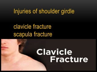
Clavicle fracture
•Als PPTX, PDF herunterladen•
167 gefällt mir•54,389 views

1. Fractures of the clavicle and scapula are uncommon but can result from high-energy trauma. 2. Clavicle fractures most commonly occur in the middle third and are usually treated conservatively with sling immobilization. 3. Scapula fractures involve the body, neck, glenoid, coracoid, or acromion and are often associated with life-threatening injuries requiring assessment by ATLS protocols. Most are also treated initially with sling immobilization.

Empfohlen
Weitere ähnliche Inhalte
Was ist angesagt?
Was ist angesagt? (20)
Ähnlich wie Clavicle fracture
Ähnlich wie Clavicle fracture (20)
Mehr von MONTHER ALKHAWLANY
Mehr von MONTHER ALKHAWLANY (20)
Kürzlich hochgeladen
Kürzlich hochgeladen (20)
Clavicle fracture
- 1. Injuries of shoulder girdle clavicle fracture scapula fracture
- 2. CLAVICLE : Is an S-shape long, curved ,tubular bone , lies horizontally a cross the root of neck . It articulate with sternum medially to form sternoclavicular joint. Also articulate with acromion process of scapula at acromioclavicular joint and acromioclavicular ligament . the muscles inserting on clavicle are : sternocleidomastoid, And subclavius muscles . The subclavian vessels and brachial plexus lie posterior to clavicle .
- 6. Fractures of the clavicle: common fracture in all ages especially in children . It is 2 – 10% of all fractures .
- 8. Mechanism of injury : Direct traumatic impact or fall on the shoulder 87% . Direct impact to clavicle 07% . Fall on outstretched hand 06% . From fall on the side . Vigorous muscle contraction , seizures [rare] . Pathological fracture [rare] .
- 10. Most common causes are : Road traffic accident [RTA] Sporting injuries
- 11. Allman classification : according to site of fracture : group 1: Fracture mostly occur in the middle one third of clavicle 80% . group 2: The fractures of outer third is 15% . Fractures involving the acromioclavicular joint 28% . group 3: fracture of inner [medial] third 5% .
- 12. Why does the fracture occur in middle third more ? It is the thinnest part of the bone . It is the junction of the tow main curves of shaft . Site of entrance of nutrient artery .
- 14. common pattern of fractures of clavicle are : 1 - Green stick fracture : Common at the junction between middle and outer third . Common in children .
- 15. 2 - Un displaced fracture in :
- 16. 3 - Separation of bone end :
- 17. 4 - With greater displacement : •There is over lapping and shortening .
- 18. Clinical presentation : pain and tenderness at site of injury . Obvious deformity and swelling sometimes occur . Patient come support his injured limb with other hand and head tilted toward injured side . Local bruising .
- 19. vascular compilication are rare , but we must look for it by : check pulse , gently palpate root of neck . Outer third # are easily missed for acromioclavicular joint .
- 20. Diagnosis : - Clinical picture examination . investigation : x-ray [AP view ] : # is usually in middle third, outer fragment below the inner . #of outer third may be missed . CT scan : useful for non union assessment . arteriography : if vascular injury suspected .
- 22. Treatment : The aim is to provide support for the weight of the arm . Fracture of clavicle unite with or without treatment . Healing occurs usually in 3-6 weeks . It may be : conservative or surgical .
- 23. Conservative treatment : Support the arm in a sling until the pain subsides , usually 1-3 weeks . Figure of 8- bandage . Clavicle ring . Analgesics .
- 24. Rehabilitation : The patient should be instructed regarding hand wrist and elbow exercises during immobilization . And regarding shoulder exercises once fracture healed .
- 28. Surgical treatment : Rarely indicated , except in : - lateral one third fracture . - presence of neurovascular injury . - non union cases . Internal fixation plate .
- 31. Complication: late : Malunion . Ununion : treated by internal fixation and bone grafting . Neurovascular injury [rare] . . Stiffness of shoulder in elderly . Ulnar neuropathy . Refracture . Early : [subclavian or carotid artery injury ,pneumothorax and hemothorax ,brachial injury ]
- 32. Scapula
- 33. Fractures of scapula …
- 35. Scapula : Is a flat triangular bone that lies on the posterior thorax wall between 2-7 rib. It envelope by : supraspinatus muscle infraspinatus muscle subscapularis muscle Attached to clavicle at acromioclavicular joint ,secured by acromioclavicular ligament . Articulate with humerus at glenohumeral joint . Attached to thorax in scapulothoraxic joint .
- 39. Fracture of scapula : Fractures of scapula are uncommon because of scapula location and surrounding muscles whitch protect it . -Fractures of scapula are result of high energy trauma with high incidence Of associated injuries by 60-98 % .
- 40. Associated life threatening injuries with scapula # : pneumothorax pulmonary contusion arterial injury abdominal injury head injury splenic or liver laceration brachial plexus injury
- 41. Fractures of scapula are classified according to location : body fracture 50 % . neck fracture 5-30 % . glenoid fracture 10 % . Coracoid fracture 8 % . Acromion fracture 7 % .
- 42. Mechanism of injury : # of body : from sever direct trauma - fall from height with direct landing on posterior aspect of trunk . - motor vehicle crush . # of neck : direct blow to shoulder - fall on shoulder . - fall on outstretched hand . # of glenoid : direct blow to lateral aspect of shoulder . or impaction of humeral head in to glenoid fossa .
- 43. # of coracoid process : direct blow or shoulder dislocation . # of acromion : direct down ward blow to shoulder .
- 44. Clinical picture : Sight > swelling deformity ecchymosis erosion . Touch > pain tenderness crepitation . Pain exacerbated by movment .
- 45. Clinical picture : -Brusing over scapula or chest area . -Pain in movement . -Swelling around back of shoulder . -Tenderness at site of # . Arm is held immobile .
- 46. Diagnosis : After initial assessment , according to advanced trauma life support [ATLS] principles , radiograghic evaluation is indicated as soon as possible as patient stable . X – ray : Anteroposterior view lateral axillary view . C T scan :is useful in glenoid or body # . .
- 50. Treatment : Reduction is usually unnecessary . Patient wears a sling for comfort and from start movement. Check repeatedly for dislocation of the shoulder .
- 51. # of body by : conservatively by analgesics and simple sling to rest shoulder for 2-3 weeks . # of acromion process : Un displaced : sling for 3-4 weeks for rest shoulder. displaced : acromion should be reduced and fixed .
- 52. # of coracoid : conservatively in major , using a sling for 2-3 weeks. Vigorous exercises should be prohibited for 2 m . If there is marked displacement > open reduction . # of neck and glenoid : - sling for 2-3 weeks - if there is displacement > shoulder spica after reduction . - open reduction > indicated if there is isolated glenoid rim fractures associated with dislocation or subluxation of shoulder .
- 58. Complication : Malunion non union > rare Glenohumeral arthritis . Limitation in range of motion . After surgery : local dyscomfort infection nerve injuries post traumatic arthritis rotator cuff dysfunction
- 59. Notes : Scapular fracture should alert the surgeon to presence of other injuries . Sever chest injury should also raise suspicion of possible scapular injury .