SlideShare ist ein Scribd-Unternehmen logo
1 von 64
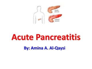
Acute Pancreatitis
By: Amina A. Al-Qaysi
Objectives
• Introduction
• Definition
• Epidemiology
• Aetiology & Pathogenesis
• Signs & Symptoms
• Investigations
• Management
• Complications
• Mortality
Pancreatitis
• Inflammation of the pancreatic parenchyma.
• Types:
1. Acute: Emergency condition.
2. Chronic: Prolonged & frequently lifelong
disorder resulting from the development of
fibrosis within the pancreas.
Acute Pancreatitis
• Definition:
Acute condition of diffuse pancreatic
inflammation & autodigestion, presents with
abdominal pain, and is usually associated with
raised pancreatic enzyme levels in the blood &
urine.
• Reversible inflammation of
the pancreas
• Ranges from mild to severe.
Epidemiology
• Acute pancreatitis accounts for 3% of all cases
of abdominal pain among patients admitted
to hospital in the UK.
• Affect 2 – 28 per 100 000 of population.
• It may occur at any age, peak incidence is
between 50 and 60 years.
• Women are affected more the men, but men
are more likely to suffer recurrent attacks.
Etiology
• 80% of the cases are due to gallstones & alcohol.
• The remaining 20 % of cases are due to:
1. Congenital: Pancreatic divisum
2. Metabolic: Hyperlipidemia, Hypercalcemia.
3. Toxic: Scorpion venom
4. Infective: Mumps, Coxsackie B, EBV, CMV.
5. Drugs: Azathioprine,
Sulfonamides, Steroids,
Thiazides, Estrogens.
6. Vascular: Ischemia, Vasculitis
(SLE, PAN).
7. Autoimmune: Hereditary
pancreatitis.
8. Traumatic.
9. Miscellaneous: CF,
Hypothermia, Periampullary
Tumors.
10. Idiopathic.
• Mnemonic for the causes of Acute Pancreatitis:
‘I get smashed‘
Idiopathic
Gallstones
Ethanol
Trauma
Steroids
Mumps
Autoimmune
Scorpion / Snakes
Hyperlipidaemia / Hypercalcaemia
ERCP
Drugs
• Biliary Pancreatitis:
1. Common channel
theory
2. Incompetent
sphincter of Oddi
3. Obstruction of the
pancreatic duct
Alcoholic Pancreatitis:
- Direct toxic effect on the pancreatic acinar
cells
- Stimulation of the pancreatic secretion
- Constriction of the sphincter of Oddi
Symptoms
• Upper Abdominal pain,
sudden onset, sharp, severe,
continuous, radiates to the
back, reduced by leaning
forward.
Generalized abdominal pain,
radiates to the shoulder tips.
Patient lies very still.
• Nausea, non-projactile
vomiting, retching
• Anorexia
• Fever, weakness
Signs
• Distressed, moving continuously, or sitting still
• Pale, diaphoretic. Confusion
• Low grade fever
• Tachycardia, Tachypnea
• Shallow breathing
• Hypotension
• Mild icterus
• Abdominal distension (Ileus, Ascites)
• Grey Turner’s sign, Cullen’s sign, Fox’s sign
• Rebound tenderness, Rigidity
• Shifting dullness, reduced bowel sounds
Cullen’s Sign Grey Turner’s Sign
Fox’s Sign
Panniculitis
• Subcutaneous nodular fat necrosis
• Tender red nodules
• Usually measures 0.5 – 2 cm
• Usually over the extremities
Differential Diagnosis
 Perforated viscus (DU)
 Acute cholecystits, Biliary colic
 Acute intestinal obstruction
 Esophageal rupture
 Mesenteric vascular obstruction
 Renal colic
 Dissecting aortic aneurysm
 Myocardial infarction
 Basal pneumonia
 Diabetic ketoacidosis
Investigations
Should be aimed at answering three
questions:
1. Is a diagnosis of acute pancreatitis correct ?
2. How severe is the attack ?
3. What is the aetiology ?
Investigations
Blood tests:
• Complete Blood Count
• Serum amylase & lipase
• C-reactive Protein
• Serum electrolytes
• Blood glucose
• Renal Function Tests
• Liver Function Tests
• LDH
• Coagulation profile
• Arterial Blood Gas Analysis
Serum Amylase:
• Sensitivity: 72% Specificity: 99%
• Released within 6-12 hours of the
onset, & Remains elevated for 3-5
days.
• Elevation ˃ 3X normal is significant.
• Undergoes renal clearance. After its
serum levels decline, its urinary
level remains elevated.
• Its level doesn't correlate with the
disease activity.
Serum Lipase:
• More pancreatic-specific than s. Amylase.
• Sensitivity: about 100% Specificity: 96%
• Remains elevated longer than amylase (up to
week).
• Useful in patients presenting late to the
physician.
• S. Amylase tends to be higher in gallstone
pancreatitis
• S. Lipase tend to be higher in alcoholic
pancreatitis
Imaging Investigations:
• Plain erect chest X-ray: not diagnostic on
pancreatitis, but to rule out other D/D
• Pleural effusion, diffuse alveolar infiltrate (ARDS)
Sentinel Loop Sign
Colon cut-off sign
• CT Scan: not indicated in every patient, only
in:
1. Diagnostic uncertainty.
2. Severe acute pancreatitis.
3. Clinical deterioration, with multi-organ failure,
sepsis, progressive deterioration.
4. Local complications occurs (fluid collection,
pseuodocyst, pseudo-aneurysm).
Axial CT Scan: Peripancreatic stranding
(arrow). Multiple gallstones in the
gallbladder
Contrast-enhanced CT: acute necrotising
pancreatitis. Pancreatic area of reduced
enhancement, peripancreatic edema and
stranding of the fatty tissue
Pancreatic pseudocyst occupying the
head of the pancreas. The pancreatic
duct (arrow) is dilated
CT Severity Index = Balthazar
Grade + Necrosis Score
MRCP
• Endoscopic Ultrasound, MRCP: CBD
stones detection, assessment of pancreatic
parenchyma. Not widely available.
• ERCP: CBD stones identification & removal.
Urgent ERCP in severe acute gallstone
pancreatitis & signs of ongoing biliary
obstruction & cholangitis.
ERCP
ERCP in Acute Pancreatitis
Biliary
sphincterotomy with
stone removal
Goals of Treatment
• Aggressive supportive care
• Decrease inflammation
• Limit superinfection
• Identify and treat complications
(of pancreatitis & its treatment)
• Treat cause if possible
Conservative Management
• Gain IV access, obtain blood sample, rapid fluid
resuscitation & electrolytes replacement.
• Give analgesics (IM pethidine).
• Give Anti-emetics.
• Keep the patient NPO (until pain free/2-3 days).
• NGT insertion to relieve vomiting.
• Urinary catheterization is done.
• Monitor the vital signs.
• Injection Ranitidine 50 mg IV 8 hourly, or
Omeprazole 40 mg IV BD.
• Somatostatin or octreotide (pancreatic
secretions inhibitors).
• Respiratory support: oxygen supplementation,
or Venti mask
• ICU admission if severe acute pancreatitis.
Role of Antibiotics
• Prophylactic antibiotics have shown No decrease in
mortality in severe acute pancreatitis.
• Antibiotics are justified if:
1. Gas in retroperitoneal space
2. Needle aspiration of necrotic material confirms
infection
3. Sepsis
4. CRP of ˃ 120 mg/L
5. Peri-pancreatic fluid collection
6. Organ dysfunction
7. APACHE II Score of ˃ 6
Operative Management
• Surgery has no immediate role in acute
pancreatitis.
• Aggressive surgical pancreatic debridement
(Necrosectomy) should be undertaken soon
after confirmation of the presence of infected
necrosis.
• Pseudocyst: Cystogastrostomy,
Cystodudenostomy, Roux-en-Y
cystojejunostomy.
Complications
Complications
Systemic Complications:
• Cardiovascular: Shock, Arrhythmias, Pericardial
effusion
• Pulmonary: Basal atelactasis, pleural effusion, ARDS
• Renal: ATN, Renal failure
• Haematological: DIC
• Metabolic: Hypocalcemia, Hyperglycemia,
Hyperlipidemia
• GIT: Ileus
• Neurological: Confusion, Irritability, Encephalopathy
• Miscellaneous: Subcutaneous fat necrosis, Arthralgia
Local Complications
Acute fluid collection:
• Occurs early in the course of acute pancreatitis
• Located in or near the pancreas, the wall encompassing
the collection is ill defined, the fluid is sterile.
• Most of such collections resolve, & no intervention is
necessary unless a large collection causes symptoms or
pressure effects, in which case it can be percutaneously
aspirated under ultrasound or CT guidance.
• Transgastric drainage under EUS guidance is another
option.
• An acute fluid collection that does not resolve can
evolve into a pseudocyst or an abscess if it becomes
infected.
Pancreatic
Pseudocyst:
• Wall formed by granulation
tissue & fibrosis
• typically presents as
abdominal pain, abdominal
mass, & persistent
hyperamylasemia in a patient
with prior pancreatitis.
Transgastric Endoscopic Pseudocyst
Drainage
Sterile and infected pancreatic necrosis:
• Diffuse or focal area of non-viable parenchyma,
typically associated with peripancreatic fat
necrosis. These areas can be identified by an
absence of contrast enhancement on CT.
• They’re sterile to begin with, but can become
subsequently infected, due to the gut bacterial
translocation.
• Sterile necrotic material should not be drained
or interfered with.
• If the patient shows signs of sepsis, then one
should determine whether the necrosis is
infected.
Mortality
• Mild acute pancreatitis: Mortality rate of 1%
• Severe pancreatitis: Mortality rate of 75-90%
• Overall mortality rate of 15-20%
• First week of illness -> MODS
• Subsequent weeks -> infection
References
• Bailey’s & Love’s Short Practice of Surgery, 25th
Edition, Page 1138 – 1146.
• Essential Surgery, Burkitt, 4th Edition, Page 380 –
388.
• Robbins & Cotran Pathologic Basis of Disease, 8th
Edition.
• http://www.aafp.org/afp/2007/0515/p1513.html
• http://www.aafp.org/afp/2000/0701/p164.html
Pancreatitis

Weitere ähnliche Inhalte

Was ist angesagt? (20)

Hernia
Hernia Hernia
Hernia
 
Hiatal hernia
Hiatal hernia Hiatal hernia
Hiatal hernia
 
Anal Fissure
Anal FissureAnal Fissure
Anal Fissure
 
Peritonitis
PeritonitisPeritonitis
Peritonitis
 
Intestinal obstruction
Intestinal obstructionIntestinal obstruction
Intestinal obstruction
 
Hydrocele
HydroceleHydrocele
Hydrocele
 
Achalasia cardia
Achalasia cardiaAchalasia cardia
Achalasia cardia
 
gastrointestinal bleeding ( GI Bleed)
gastrointestinal bleeding ( GI Bleed)gastrointestinal bleeding ( GI Bleed)
gastrointestinal bleeding ( GI Bleed)
 
Chronic pancreatitis
Chronic pancreatitisChronic pancreatitis
Chronic pancreatitis
 
Gastritis
GastritisGastritis
Gastritis
 
Cholecystitis and cholelithiasis
Cholecystitis and cholelithiasis Cholecystitis and cholelithiasis
Cholecystitis and cholelithiasis
 
Cushings syndrome
Cushings syndromeCushings syndrome
Cushings syndrome
 
Presentation cholelithiasis
Presentation cholelithiasisPresentation cholelithiasis
Presentation cholelithiasis
 
Liver cirrhosis
Liver cirrhosisLiver cirrhosis
Liver cirrhosis
 
Urine Retention
Urine RetentionUrine Retention
Urine Retention
 
Inguinal hernia
Inguinal herniaInguinal hernia
Inguinal hernia
 
Varicose vein
Varicose veinVaricose vein
Varicose vein
 
Pancreatitis
PancreatitisPancreatitis
Pancreatitis
 
chronic liver disease (CLD)
chronic liver disease (CLD)chronic liver disease (CLD)
chronic liver disease (CLD)
 
Intestinal obstruction
Intestinal obstructionIntestinal obstruction
Intestinal obstruction
 

Andere mochten auch

Acute Pancreatitis (According to American College of Gastroenterology 2013 gu...
Acute Pancreatitis (According to American College of Gastroenterology 2013 gu...Acute Pancreatitis (According to American College of Gastroenterology 2013 gu...
Acute Pancreatitis (According to American College of Gastroenterology 2013 gu...Jibran Mohsin
 
Acute pancreatitis 2015
Acute pancreatitis   2015Acute pancreatitis   2015
Acute pancreatitis 2015samirelansary
 
Acute Pancreatitis
Acute PancreatitisAcute Pancreatitis
Acute PancreatitisSimmedic UKM
 
Acute pancreatitis
Acute  pancreatitisAcute  pancreatitis
Acute pancreatitisbarun kumar
 
Acute Pancreatitis
Acute PancreatitisAcute Pancreatitis
Acute PancreatitisMa Wady
 
Acute Pancreatitis Managment
Acute Pancreatitis ManagmentAcute Pancreatitis Managment
Acute Pancreatitis ManagmentNouman Memon
 
Acute pancreatitis atlanta classification & management
Acute pancreatitis   atlanta classification & managementAcute pancreatitis   atlanta classification & management
Acute pancreatitis atlanta classification & managementSeneeth Peramuna
 
Acute pancreatitis
Acute pancreatitisAcute pancreatitis
Acute pancreatitisRahul Garg
 
Acute and Chronic Pancreatitis
Acute and Chronic PancreatitisAcute and Chronic Pancreatitis
Acute and Chronic Pancreatitismarcosmachado
 
Acute pancreatitis.ppt
Acute pancreatitis.pptAcute pancreatitis.ppt
Acute pancreatitis.pptIbrahim Odeh
 
Acute pancreatitis
Acute pancreatitisAcute pancreatitis
Acute pancreatitisAtit Ghoda
 
Acute Pancreatitis Management Conference
Acute Pancreatitis Management ConferenceAcute Pancreatitis Management Conference
Acute Pancreatitis Management Conferencejcm MD
 
Acute pancreatitis
Acute pancreatitisAcute pancreatitis
Acute pancreatitisSam George
 
22 acute pancreatitis
22 acute pancreatitis22 acute pancreatitis
22 acute pancreatitisinternalmed
 
ACUTE PANCREATITIS
ACUTE PANCREATITISACUTE PANCREATITIS
ACUTE PANCREATITISRaj Kumar
 
Acute pancreatitis
Acute pancreatitisAcute pancreatitis
Acute pancreatitisIbrahim Odeh
 

Andere mochten auch (20)

Acute Pancreatitis (According to American College of Gastroenterology 2013 gu...
Acute Pancreatitis (According to American College of Gastroenterology 2013 gu...Acute Pancreatitis (According to American College of Gastroenterology 2013 gu...
Acute Pancreatitis (According to American College of Gastroenterology 2013 gu...
 
Acute pancreatitis 2015
Acute pancreatitis   2015Acute pancreatitis   2015
Acute pancreatitis 2015
 
Acute Pancreatitis
Acute PancreatitisAcute Pancreatitis
Acute Pancreatitis
 
Pancreatitis
PancreatitisPancreatitis
Pancreatitis
 
Acute pancreatitis
Acute pancreatitisAcute pancreatitis
Acute pancreatitis
 
Acute pancreatitis
Acute  pancreatitisAcute  pancreatitis
Acute pancreatitis
 
Acute Pancreatitis
Acute PancreatitisAcute Pancreatitis
Acute Pancreatitis
 
Acute Pancreatitis Managment
Acute Pancreatitis ManagmentAcute Pancreatitis Managment
Acute Pancreatitis Managment
 
Acute pancreatitis atlanta classification & management
Acute pancreatitis   atlanta classification & managementAcute pancreatitis   atlanta classification & management
Acute pancreatitis atlanta classification & management
 
Acute pancreatitis
Acute pancreatitisAcute pancreatitis
Acute pancreatitis
 
Acute and Chronic Pancreatitis
Acute and Chronic PancreatitisAcute and Chronic Pancreatitis
Acute and Chronic Pancreatitis
 
Acute pancreatitis.ppt
Acute pancreatitis.pptAcute pancreatitis.ppt
Acute pancreatitis.ppt
 
Acute pancreatitis
Acute pancreatitisAcute pancreatitis
Acute pancreatitis
 
Acute Pancreatitis
Acute PancreatitisAcute Pancreatitis
Acute Pancreatitis
 
Biliary pancreatitis
Biliary pancreatitisBiliary pancreatitis
Biliary pancreatitis
 
Acute Pancreatitis Management Conference
Acute Pancreatitis Management ConferenceAcute Pancreatitis Management Conference
Acute Pancreatitis Management Conference
 
Acute pancreatitis
Acute pancreatitisAcute pancreatitis
Acute pancreatitis
 
22 acute pancreatitis
22 acute pancreatitis22 acute pancreatitis
22 acute pancreatitis
 
ACUTE PANCREATITIS
ACUTE PANCREATITISACUTE PANCREATITIS
ACUTE PANCREATITIS
 
Acute pancreatitis
Acute pancreatitisAcute pancreatitis
Acute pancreatitis
 

Ähnlich wie Pancreatitis

pancreatitis Gi disorder diagnosis management
pancreatitis Gi disorder diagnosis managementpancreatitis Gi disorder diagnosis management
pancreatitis Gi disorder diagnosis managementTHaripriya1
 
pancreatitis1970-160120092229 (1).pptx
pancreatitis1970-160120092229 (1).pptxpancreatitis1970-160120092229 (1).pptx
pancreatitis1970-160120092229 (1).pptxabeerarajput
 
Pancreatitis -a detailed study ( medical information )
Pancreatitis -a detailed study ( medical information )Pancreatitis -a detailed study ( medical information )
Pancreatitis -a detailed study ( medical information )martinshaji
 
Pancreatitis DEFINITION, COMPLICATIONS, TREATMENT .pptx
Pancreatitis DEFINITION, COMPLICATIONS, TREATMENT .pptxPancreatitis DEFINITION, COMPLICATIONS, TREATMENT .pptx
Pancreatitis DEFINITION, COMPLICATIONS, TREATMENT .pptxrohanbijarnia2
 
Acute pancreatitis
Acute pancreatitis Acute pancreatitis
Acute pancreatitis Mohsin Khan
 
Pancreatitis & pancreatic pseudocyst
Pancreatitis & pancreatic pseudocystPancreatitis & pancreatic pseudocyst
Pancreatitis & pancreatic pseudocystShweta Kutty
 
Appendicitis, diverticulitis, peptic ulcer disease, chron's disease
Appendicitis, diverticulitis, peptic ulcer disease, chron's diseaseAppendicitis, diverticulitis, peptic ulcer disease, chron's disease
Appendicitis, diverticulitis, peptic ulcer disease, chron's diseaseharon taufiq
 
acutepancreatitis-190207120812 (1).pdf (1).pptx
acutepancreatitis-190207120812 (1).pdf (1).pptxacutepancreatitis-190207120812 (1).pdf (1).pptx
acutepancreatitis-190207120812 (1).pdf (1).pptxmaleehazainab01
 
Liver Disease Important Question And Answers.pdf
Liver Disease Important Question And Answers.pdfLiver Disease Important Question And Answers.pdf
Liver Disease Important Question And Answers.pdfsainavlefusion
 
Chronic Pancreatitis
Chronic PancreatitisChronic Pancreatitis
Chronic PancreatitisAbdul Basit
 
chronic pancreatitis anoop k r
chronic pancreatitis anoop k rchronic pancreatitis anoop k r
chronic pancreatitis anoop k ranoop k r
 
Diverticular disease of the colon hegazy
Diverticular disease of the colon hegazyDiverticular disease of the colon hegazy
Diverticular disease of the colon hegazymostafa hegazy
 
Billiary tract
Billiary tractBilliary tract
Billiary tractRMLIMS
 

Ähnlich wie Pancreatitis (20)

pancreatitis Gi disorder diagnosis management
pancreatitis Gi disorder diagnosis managementpancreatitis Gi disorder diagnosis management
pancreatitis Gi disorder diagnosis management
 
pancreatitis1970-160120092229 (1).pptx
pancreatitis1970-160120092229 (1).pptxpancreatitis1970-160120092229 (1).pptx
pancreatitis1970-160120092229 (1).pptx
 
Pancreatitis -a detailed study ( medical information )
Pancreatitis -a detailed study ( medical information )Pancreatitis -a detailed study ( medical information )
Pancreatitis -a detailed study ( medical information )
 
Pancreatitis DEFINITION, COMPLICATIONS, TREATMENT .pptx
Pancreatitis DEFINITION, COMPLICATIONS, TREATMENT .pptxPancreatitis DEFINITION, COMPLICATIONS, TREATMENT .pptx
Pancreatitis DEFINITION, COMPLICATIONS, TREATMENT .pptx
 
Acute pancreatitis
Acute pancreatitisAcute pancreatitis
Acute pancreatitis
 
Acute pancreatitis
Acute pancreatitis Acute pancreatitis
Acute pancreatitis
 
PANCREAS.pptx
PANCREAS.pptxPANCREAS.pptx
PANCREAS.pptx
 
Surgical jaundice
Surgical jaundiceSurgical jaundice
Surgical jaundice
 
Acute pancreatitis
Acute pancreatitisAcute pancreatitis
Acute pancreatitis
 
Pancreatitis & pancreatic pseudocyst
Pancreatitis & pancreatic pseudocystPancreatitis & pancreatic pseudocyst
Pancreatitis & pancreatic pseudocyst
 
Diseases of the liver
Diseases of the liverDiseases of the liver
Diseases of the liver
 
Appendicitis, diverticulitis, peptic ulcer disease, chron's disease
Appendicitis, diverticulitis, peptic ulcer disease, chron's diseaseAppendicitis, diverticulitis, peptic ulcer disease, chron's disease
Appendicitis, diverticulitis, peptic ulcer disease, chron's disease
 
acutepancreatitis-190207120812 (1).pdf (1).pptx
acutepancreatitis-190207120812 (1).pdf (1).pptxacutepancreatitis-190207120812 (1).pdf (1).pptx
acutepancreatitis-190207120812 (1).pdf (1).pptx
 
Liver Disease Important Question And Answers.pdf
Liver Disease Important Question And Answers.pdfLiver Disease Important Question And Answers.pdf
Liver Disease Important Question And Answers.pdf
 
Pancreatic disorders
Pancreatic disordersPancreatic disorders
Pancreatic disorders
 
Chronic Pancreatitis
Chronic PancreatitisChronic Pancreatitis
Chronic Pancreatitis
 
chronic pancreatitis anoop k r
chronic pancreatitis anoop k rchronic pancreatitis anoop k r
chronic pancreatitis anoop k r
 
Diverticular disease of the colon hegazy
Diverticular disease of the colon hegazyDiverticular disease of the colon hegazy
Diverticular disease of the colon hegazy
 
Pancreatic diseases
Pancreatic diseasesPancreatic diseases
Pancreatic diseases
 
Billiary tract
Billiary tractBilliary tract
Billiary tract
 

Mehr von meducationdotnet

Mehr von meducationdotnet (20)

No Title
No TitleNo Title
No Title
 
Spondylarthropathy
SpondylarthropathySpondylarthropathy
Spondylarthropathy
 
Diagnosing Lung cancer
Diagnosing Lung cancerDiagnosing Lung cancer
Diagnosing Lung cancer
 
Eczema Herpeticum
Eczema HerpeticumEczema Herpeticum
Eczema Herpeticum
 
The Vagus Nerve
The Vagus NerveThe Vagus Nerve
The Vagus Nerve
 
Water and sanitation and their impact on health
Water and sanitation and their impact on healthWater and sanitation and their impact on health
Water and sanitation and their impact on health
 
The ethics of electives
The ethics of electivesThe ethics of electives
The ethics of electives
 
Intro to Global Health
Intro to Global HealthIntro to Global Health
Intro to Global Health
 
WTO and Health
WTO and HealthWTO and Health
WTO and Health
 
Globalisation and Health
Globalisation and HealthGlobalisation and Health
Globalisation and Health
 
Health Care Worker Migration
Health Care Worker MigrationHealth Care Worker Migration
Health Care Worker Migration
 
International Institutions
International InstitutionsInternational Institutions
International Institutions
 
Haemochromotosis brief overview
Haemochromotosis brief overviewHaemochromotosis brief overview
Haemochromotosis brief overview
 
Ascities overview
Ascities overviewAscities overview
Ascities overview
 
Overview of the Liver
Overview of the LiverOverview of the Liver
Overview of the Liver
 
Overview of Antidepressants
Overview of AntidepressantsOverview of Antidepressants
Overview of Antidepressants
 
Gout Presentation
Gout PresentationGout Presentation
Gout Presentation
 
Review of orthopaedic services: Prepared for the Auditor General for Scotland...
Review of orthopaedic services: Prepared for the Auditor General for Scotland...Review of orthopaedic services: Prepared for the Auditor General for Scotland...
Review of orthopaedic services: Prepared for the Auditor General for Scotland...
 
Sugammadex - a revolution in anaesthesia?
Sugammadex - a revolution in anaesthesia?Sugammadex - a revolution in anaesthesia?
Sugammadex - a revolution in anaesthesia?
 
Ophthamology Revision
Ophthamology RevisionOphthamology Revision
Ophthamology Revision
 

Pancreatitis

  • 2. Objectives • Introduction • Definition • Epidemiology • Aetiology & Pathogenesis • Signs & Symptoms • Investigations • Management • Complications • Mortality
  • 3. Pancreatitis • Inflammation of the pancreatic parenchyma. • Types: 1. Acute: Emergency condition. 2. Chronic: Prolonged & frequently lifelong disorder resulting from the development of fibrosis within the pancreas.
  • 4. Acute Pancreatitis • Definition: Acute condition of diffuse pancreatic inflammation & autodigestion, presents with abdominal pain, and is usually associated with raised pancreatic enzyme levels in the blood & urine. • Reversible inflammation of the pancreas • Ranges from mild to severe.
  • 5. Epidemiology • Acute pancreatitis accounts for 3% of all cases of abdominal pain among patients admitted to hospital in the UK. • Affect 2 – 28 per 100 000 of population. • It may occur at any age, peak incidence is between 50 and 60 years. • Women are affected more the men, but men are more likely to suffer recurrent attacks.
  • 6. Etiology • 80% of the cases are due to gallstones & alcohol. • The remaining 20 % of cases are due to: 1. Congenital: Pancreatic divisum 2. Metabolic: Hyperlipidemia, Hypercalcemia. 3. Toxic: Scorpion venom 4. Infective: Mumps, Coxsackie B, EBV, CMV.
  • 7. 5. Drugs: Azathioprine, Sulfonamides, Steroids, Thiazides, Estrogens. 6. Vascular: Ischemia, Vasculitis (SLE, PAN). 7. Autoimmune: Hereditary pancreatitis. 8. Traumatic. 9. Miscellaneous: CF, Hypothermia, Periampullary Tumors. 10. Idiopathic.
  • 8. • Mnemonic for the causes of Acute Pancreatitis: ‘I get smashed‘ Idiopathic Gallstones Ethanol Trauma Steroids Mumps Autoimmune Scorpion / Snakes Hyperlipidaemia / Hypercalcaemia ERCP Drugs
  • 9. • Biliary Pancreatitis: 1. Common channel theory 2. Incompetent sphincter of Oddi 3. Obstruction of the pancreatic duct
  • 10. Alcoholic Pancreatitis: - Direct toxic effect on the pancreatic acinar cells - Stimulation of the pancreatic secretion - Constriction of the sphincter of Oddi
  • 11.
  • 12.
  • 13. Symptoms • Upper Abdominal pain, sudden onset, sharp, severe, continuous, radiates to the back, reduced by leaning forward. Generalized abdominal pain, radiates to the shoulder tips. Patient lies very still. • Nausea, non-projactile vomiting, retching • Anorexia • Fever, weakness
  • 14. Signs • Distressed, moving continuously, or sitting still • Pale, diaphoretic. Confusion • Low grade fever • Tachycardia, Tachypnea • Shallow breathing • Hypotension • Mild icterus • Abdominal distension (Ileus, Ascites) • Grey Turner’s sign, Cullen’s sign, Fox’s sign • Rebound tenderness, Rigidity • Shifting dullness, reduced bowel sounds
  • 15. Cullen’s Sign Grey Turner’s Sign Fox’s Sign
  • 16. Panniculitis • Subcutaneous nodular fat necrosis • Tender red nodules • Usually measures 0.5 – 2 cm • Usually over the extremities
  • 17. Differential Diagnosis  Perforated viscus (DU)  Acute cholecystits, Biliary colic  Acute intestinal obstruction  Esophageal rupture  Mesenteric vascular obstruction  Renal colic  Dissecting aortic aneurysm  Myocardial infarction  Basal pneumonia  Diabetic ketoacidosis
  • 18. Investigations Should be aimed at answering three questions: 1. Is a diagnosis of acute pancreatitis correct ? 2. How severe is the attack ? 3. What is the aetiology ?
  • 19. Investigations Blood tests: • Complete Blood Count • Serum amylase & lipase • C-reactive Protein • Serum electrolytes • Blood glucose • Renal Function Tests • Liver Function Tests • LDH • Coagulation profile • Arterial Blood Gas Analysis
  • 20. Serum Amylase: • Sensitivity: 72% Specificity: 99% • Released within 6-12 hours of the onset, & Remains elevated for 3-5 days. • Elevation ˃ 3X normal is significant. • Undergoes renal clearance. After its serum levels decline, its urinary level remains elevated. • Its level doesn't correlate with the disease activity.
  • 21. Serum Lipase: • More pancreatic-specific than s. Amylase. • Sensitivity: about 100% Specificity: 96% • Remains elevated longer than amylase (up to week). • Useful in patients presenting late to the physician. • S. Amylase tends to be higher in gallstone pancreatitis • S. Lipase tend to be higher in alcoholic pancreatitis
  • 22.
  • 23. Imaging Investigations: • Plain erect chest X-ray: not diagnostic on pancreatitis, but to rule out other D/D • Pleural effusion, diffuse alveolar infiltrate (ARDS)
  • 24.
  • 27.
  • 28. • CT Scan: not indicated in every patient, only in: 1. Diagnostic uncertainty. 2. Severe acute pancreatitis. 3. Clinical deterioration, with multi-organ failure, sepsis, progressive deterioration. 4. Local complications occurs (fluid collection, pseuodocyst, pseudo-aneurysm).
  • 29. Axial CT Scan: Peripancreatic stranding (arrow). Multiple gallstones in the gallbladder
  • 30. Contrast-enhanced CT: acute necrotising pancreatitis. Pancreatic area of reduced enhancement, peripancreatic edema and stranding of the fatty tissue
  • 31. Pancreatic pseudocyst occupying the head of the pancreas. The pancreatic duct (arrow) is dilated
  • 32. CT Severity Index = Balthazar Grade + Necrosis Score
  • 33.
  • 34.
  • 35. MRCP
  • 36. • Endoscopic Ultrasound, MRCP: CBD stones detection, assessment of pancreatic parenchyma. Not widely available. • ERCP: CBD stones identification & removal. Urgent ERCP in severe acute gallstone pancreatitis & signs of ongoing biliary obstruction & cholangitis.
  • 37. ERCP
  • 38. ERCP in Acute Pancreatitis
  • 39.
  • 41.
  • 42. Goals of Treatment • Aggressive supportive care • Decrease inflammation • Limit superinfection • Identify and treat complications (of pancreatitis & its treatment) • Treat cause if possible
  • 43. Conservative Management • Gain IV access, obtain blood sample, rapid fluid resuscitation & electrolytes replacement. • Give analgesics (IM pethidine). • Give Anti-emetics. • Keep the patient NPO (until pain free/2-3 days). • NGT insertion to relieve vomiting.
  • 44. • Urinary catheterization is done. • Monitor the vital signs.
  • 45. • Injection Ranitidine 50 mg IV 8 hourly, or Omeprazole 40 mg IV BD. • Somatostatin or octreotide (pancreatic secretions inhibitors). • Respiratory support: oxygen supplementation, or Venti mask • ICU admission if severe acute pancreatitis.
  • 46.
  • 47. Role of Antibiotics • Prophylactic antibiotics have shown No decrease in mortality in severe acute pancreatitis. • Antibiotics are justified if: 1. Gas in retroperitoneal space 2. Needle aspiration of necrotic material confirms infection 3. Sepsis 4. CRP of ˃ 120 mg/L 5. Peri-pancreatic fluid collection 6. Organ dysfunction 7. APACHE II Score of ˃ 6
  • 48. Operative Management • Surgery has no immediate role in acute pancreatitis. • Aggressive surgical pancreatic debridement (Necrosectomy) should be undertaken soon after confirmation of the presence of infected necrosis. • Pseudocyst: Cystogastrostomy, Cystodudenostomy, Roux-en-Y cystojejunostomy.
  • 50. Complications Systemic Complications: • Cardiovascular: Shock, Arrhythmias, Pericardial effusion • Pulmonary: Basal atelactasis, pleural effusion, ARDS • Renal: ATN, Renal failure • Haematological: DIC • Metabolic: Hypocalcemia, Hyperglycemia, Hyperlipidemia • GIT: Ileus • Neurological: Confusion, Irritability, Encephalopathy • Miscellaneous: Subcutaneous fat necrosis, Arthralgia
  • 51.
  • 52. Local Complications Acute fluid collection: • Occurs early in the course of acute pancreatitis • Located in or near the pancreas, the wall encompassing the collection is ill defined, the fluid is sterile. • Most of such collections resolve, & no intervention is necessary unless a large collection causes symptoms or pressure effects, in which case it can be percutaneously aspirated under ultrasound or CT guidance. • Transgastric drainage under EUS guidance is another option. • An acute fluid collection that does not resolve can evolve into a pseudocyst or an abscess if it becomes infected.
  • 53. Pancreatic Pseudocyst: • Wall formed by granulation tissue & fibrosis • typically presents as abdominal pain, abdominal mass, & persistent hyperamylasemia in a patient with prior pancreatitis.
  • 54.
  • 56.
  • 57.
  • 58.
  • 59. Sterile and infected pancreatic necrosis: • Diffuse or focal area of non-viable parenchyma, typically associated with peripancreatic fat necrosis. These areas can be identified by an absence of contrast enhancement on CT. • They’re sterile to begin with, but can become subsequently infected, due to the gut bacterial translocation. • Sterile necrotic material should not be drained or interfered with. • If the patient shows signs of sepsis, then one should determine whether the necrosis is infected.
  • 60.
  • 61.
  • 62. Mortality • Mild acute pancreatitis: Mortality rate of 1% • Severe pancreatitis: Mortality rate of 75-90% • Overall mortality rate of 15-20% • First week of illness -> MODS • Subsequent weeks -> infection
  • 63. References • Bailey’s & Love’s Short Practice of Surgery, 25th Edition, Page 1138 – 1146. • Essential Surgery, Burkitt, 4th Edition, Page 380 – 388. • Robbins & Cotran Pathologic Basis of Disease, 8th Edition. • http://www.aafp.org/afp/2007/0515/p1513.html • http://www.aafp.org/afp/2000/0701/p164.html

Hinweis der Redaktion

  1. Disorder of the exocrine pancreas Predominantly d/t activation of intracelluler trypsinogen to trypsin
  2. peak in young men and older women.
  3. Gallstones: females Alcohol: males
  4. Widespread fat necrosis of the omentum. A test tube has been filled with blood-stained peritoneal fluid. This specimen was rich in amylase. Fat necroses are dull, opaque, yellow-white areas suggestive of drops of wax. They are most abundant in the vicinity of the pancreas, but are widespread in the greater omentum and the mesentery. At necropsy, they can sometimes be demonstrated beneath the pleura and pericardium, and even in the subsynovial fat of the knee joint. Fat necroses consist of small islands of saponification caused by the liberation of lipase, which splits into glycerol and fatty acids. Free fatty acids combine with calcium to form soaps (fatty necrosis) Acute pancreatitis. The microscopic field shows a region of fat necrosis on the right and focal pancreatic parenchymal necrosis (center).
  5. pancreatic position
  6. Jaundice: obstruction of CBD d/t pancreatic head edema or cholidocholithiasis
  7. d/t fat necrosis & retroperitoneal bleeding Severe necrotizing pancreatitis Fox’s sign: bruising over the inguinal ligament
  8. Esophageal rupture: HX of emesis
  9. Leukocytosis, hemoconcentration (elevated hgb & hct) Hyperglycemia: reduced insulin release, increase glucagon release, increase adrenal corticosteroids & catecholamines Hyperbilirubinemia Ionized Ca ECG: abnormalities simulating MI Dx: 1. typical abdominal pain, with 2. increased amylase or lipase (3X normal), or 3. imaging finding compatible with pancreatitis
  10. Half life in the blood is about 10 hrs Renal failure: reduced renal clearance Normal serum amylase level doesn't rule out Acute pancreatitis, and the level poorly correlates with the severity. Prolonged amylase elevation (>1 week) may be a clue to the presence of pancreatic abscess, pseudocyst, or ascites.
  11. Slightly greater sensitivity, specially when measured after 24 hrs of the presentation
  12. d/d esophageal rupture
  13. Calcified gallstones
  14. Doesn’t establish the Dx. Edematous pancreas, extra-pancreatic fluid collection, gallstones, dilated CBD. To be done in the first 24 hrs. Limited value d/t presence of intestinal gas.
  15. CT is the most important imaging investigation in A.P. It assess severity, complications Areas of necrosis: non enhancing regions more than 3 cm, on IV contrast if renal function permits CT or US guided aspiration to differentiate sterile from infected pancreatic necrosis or pseudocyst CRP ˃110 mg/L Ranson’s score ˃ 3, APACHE II score ˃8
  16. Early intervention (˂72 hrs) is favoured in severe biliary pancreatitis In acute biliary pancreatitis but without obstructive jaundice, early ERCP & papillotomy are not beneficial
  17. If 3 or more factors are present in the patient, it indicates severe pancreatitis.
  18. Gain IV access, rapid fluid resuscitation & electrolytes replacement (key factor in the tt of acute pancreatitis). Pancreatic rest
  19. Hourly urine output
  20. Somatostatin or octreotide: reduce mortality rate but no change in the complications
  21. Cefuroxime : 2nd generation cephalosporin Enteral nutrition is preferred to parental nutrition for improving patient outcomes
  22. Purtscher retinopathy (funduscopy ) ischemic injury to the retina causing temporary/permanent blindness
  23. CT of pancreatic pseudocyst. A large pseudocyst is seen in the head of the pancreas. A nasojejunal feeding tube is seen anterolaterally.
  24. MC cause of early death in A. P. Patients is hypovolemic shock