SlideShare ist ein Scribd-Unternehmen logo
1 von 22
Downloaden Sie, um offline zu lesen
THE NERVOUS SYSTEM
The nervous system, along with the endocrine system, responds to changes in the external or internal
environment. The nervous system is the co-ordination system for rapid actions.
INITIAL EVOLUTION OF THE NERVOUS SYSTEM
To coordinate the initial rod shaped organisms some form of communication system would have been
required. Chemical messengers that spread by diffusion might suffice initially but, for rapid responses, a
nervous system would be better and, probably at much the same time as the rod organisms were being
evolved in a segmental fashion, a midline coordinating structure (the spinal cord) developed. Vertebrates
eventually evolved from such segmented ancestors (possibly flatworms). Segmentation was useful to
allow sequential contraction of the muscle groups (myotomes), for defensive reflexes and for propulsion
in water or on land. The development of similar segments also minimized the need for genetic
information - all that was required was a genetic instruction “repeat this basic pattern n times.”
Presumably vertebrates living on the seafloor were governed by gravitational considerations, and
because muscles which conferred mobility work best if attached to a rigid structure like the backbone,
and because muscle groups became ventrally suspended from the backbone, it made sense for the motor
output to skeletal muscles to be ventrally situated in the spinal cord. The sensory nerves would mostly
serve the outside of the rod-like animal and would have been dorsal to avoid pushing through the muscle
groups (Fig. 1).
Initially there was a neural tube, the front of which then divided into three functional areas (Fig.2).
Three important early features in mammalian evolution were:
• bilateral symmetry (humans are mostly symmetrical)
• segmentation (to allow sequential contraction for swimming purposes)
• cephalization (a major focalisation of organisation in a head)
Nearly all nervous pathways, except cerebellar and vestibular pathways,cross the midline either in the
spinal cord, the brainstem or in the brain. There seem to be three possible explanations.
Firstly the primitive coiling reflex to noxious stimuli (Fig. 3) required that nerve fibres crossed the
midline. Impulses from the sensory nerve fibres had to be conducted to the motor neurones on the
immediate opposite side of the spinal cord to allow the primitive rod-like animal to bend away from the
noxious stimulus.
Secondly stereoscopic sight using two eyes (which allows accurate distance appreciation) would have
offered great evolutionary advantages. Integration of the information from the two eyes demands that
information crosses the midline (Figs. 4a, 4b and 4c). A threat to the right of the head is appreciated by
the left hand side of each retina and information from one eye has to cross the midline (in the optic
chiasma) and the visual input from both retinas is coordinated in the left occipital cortex (Fig. 4c). Thus
if there is to be a coordinated response, then a quicker and more efficient response to the threat demands
that the right side (specifically the right arm) be moved. To move the right arm the left motor cortex has
to initiate action and to do this information from the left occipital cortex has to cross the midline.
Thirdly swimming or walking requires coordination of the two sides and this requires that nervous
information crosses the midline.
The basic structure of the human nervous system is illustrated in Fig. 2. In anatomical terms the central
nervous system comprises the brain and spinal cord (Fig. 5 ) whilst the peripheral nervous system
comprises 12 pairs of cranial nerves and 31 pairs of nerves arising from the spinal cord.
The brain and spinal cord are surrounded by three membranes, called the meninges. The outer layer is
the dura mater, the middle the arachnoid mater (interior to which is the cerebrospinal fluid) and the inner
pia mater. Blood or infection in the cerebrospinal fluid causes meningeal irritation which results in neck
stiffness.
The capillary blood vessels supplying the central nervous system are anatomically and functionally
different from capillaries elsewhere in the body. They constitute the blood-brain barrier which prevents
certain large molecules and certain drugs from crossing into the central nervous system. On electron
microscopy the CNS endothelial cells are joined by tight junctions which are impermeable to large
molecules. The are fenestrations (holes) between endothelial cells outside the CNS.
NERVES IN GENERAL
Nerve cells (Fig.6)
The 1011
nerve cells and their processes are the building blocks of the nervous system. Nerve cells, once
dead, cannot be replaced. Each nerve cell body has one or more dendrites which collect impulses from
other nerve cell bodies or from sensory receptors. Axons are long outgrowths from nerve cells which
transmit impulses away from the nerve cell. Axons may branch at their terminations and impact upon
other nerve cells or their dendrites. In the central nervous system such impulses can be excitatory or
inhibitory. In some nerve cells a single processemerges which splits into two in a T-shaped manner -
(pseudo) unipolar nerve cells. For example the cells of the sensory spinal ganglia lateral to the dorsal
surface of the spinal cord have neurones which have sensory gathering nervous fibres extending
peripherally and a nerve fibre extending centrally (Fig. 7). Nerve cells do not divide except in the
embryo.
There are four types of nerve fibres:
• Myelinated with neurolemma sheaths (mostly in peripheral nerves)
• Myelinated with no neurolemma sheaths (in brain and spinal cord)
Axon (=hillock): an outgrowth, usually a single outgrowth, from a nerve cell which conducts
impulses along the nerve cell to the other nerve cells or their dendrites. Synonymous with nerve fibre
Dendrite (=tree): the branching process of a nerve cell which conducts impulses to synapses on other
nerve cells
Decussation: nerve fibres which cross the midline in bundles of fibres (commisures pass to the same
side)
Ganglia (=knotlike) collections of nerve cell bodies outwith the central nervous system (except the
basal ganglia which are within the central nervous system). They are the seat of visceral motor
reflexes which affect some internal organs
Neurone: nerve cell, axon, and dendrites
Nerve fibre: axon and its myelin sheath.
• No myelination but a neurolemma sheath (autonomic nerves and fine efferents of corticospinal
nerves)
• No myelination and no neurolemma sheath (the gray matter of brain and spinal cord)
There are three types of glial cells within the CNS, astrocytes, oligodendrocytes and ependymal lining
cells. These support neurones structurally and metabolically. In the peripheral nervous system neurones
are supported by Schwann cells which ensheath axons. In the CNS the oligodendrocytes produce myelin
which ensheaths CNS axons. Whilst all axons have Schwann cells surrounding them the speed of
propagation along axons is enhanced if the axons are wrapped up with a myelin sheath by Schwann cells
(Fig. 6). This “plastic covering of the electric wire” is interrupted at intervals by Nodes of Ranvier.
Myelinated axons appear white (constituting the white matter of the brain and spinal cord) whereas
unmyelinated axons appear gray (constituting the gray matter (neuronal cell bodies) of the brain and
spinalcord). White matter conducts impulses but gray matter processes and interprets. The cortex of the
cerebral hemispheres and cerebellum are mostly infolded sheets of nerve cells (gray matter). Areas of
the brain called nuclei are (more or less) spherically shaped collections of functionally related gray
matter in the central nervous system. Multiple sclerosis produces sensory and motor symptoms and
signs caused by patchy demyelination “plaques” in medullated axons “white matter” of the central but
not the peripheral nervous system.
There are 31 pairs of spinal nerves - eight cervical, 12 thoracic, five lumbar, five sacral and one
rudimentary coccygeal. Peripheral nerves are formed by the unification of the dorsal (sensory) and
ventral (motor) roots. In the cervical and lumbar regions these roots join to form the cords of the brachial
and lumbrosacral plexuses and from these arise mixed sensory and motor peripheral nerves. The motor
fibres supply voluntary (striated muscle). Autonomic nerves arise from sympathetic plexuses to supply
cardiac muscle, smooth muscle of the gut and blood vessels, and the salivary and some other glands.
Depending on which roots are predominantly affected there may be:
• Segmental muscle wasting
• Reflex loss
• Dermatomal pattern sensory loss (Fig. 8a, 8b for peripheral nerve losses and Fig. 8c for the likely
explanation of the actual dermatome distributions)
• Radiating root pain (localization often poor)
• Tingling
• Oversensitive skin (hyperaethesiae)
If nerve root problems are caused by pathologies that tether the root then anything which moves the root
(movements, coughing, straining) will exacerbate symptoms, especially pain. Depending on the
pathology there may also be signs of spinal cord compression. If the nerve roots leaving the end of the
spinal cord (conus and cauda equina) are involved then bladder sensation may be reduced and there may
be difficulty voiding with a poor stream (motor) and rectal function may be similarly affected leading to
incontinence and faecal soiling.
Synapses (=junction)
The junction between a “sending” axon and a “receiving dendrite or nerve cell is a synapse (Fig.6).
Synapses are only found in the central nervous system or in autonomic ganglia and only conducts
information in one direction. Some axons terminate with a single synapse whilst others may have
thousand of terminations. A typical motor neurone in the spinal cord has about 10,000 synaptic
contacts. Whether the receiving nerve cell responds usually depends on the number of inputs from other
axons that it is receives. The more synapses that impinge on a nerve cell or its dendrites the more
modifying influences there are on the final output of the receiving nerve cell. This is the basis of the
integrated neural networking that forms the brain.
Frequent use of particular synapses in certain situations may result in the establishment of a regular
response pattern. In the central nervous system this constitutes a basis for learning (consciously or
unconsciously). Synapse damage leads to forgetting.
Nerves pass on information by putting out chemicals - neurotransmitters - at their synaptic endings.
There are four main groups of neurotransmitters (Fig. 6):
• Acetylcholine (in cholinergic neurones)
• Amines Noradrenaline (in noradrenergic neurones)
Serotonin (an important transmitter in sensory channels relevant to emotions)
Dopamine (in the motor systems, limbic system and in the hypothalamus)
• Amino acids Glutamic acid (always excitatory)
Gamma aminobutyric acid (always inhibitory)
• Peptides Enkephalins
Endorphins
These transmitters can be reabsorbed, broken down, or competitively inhibited or totally blocked before
they can act. Nitric oxide (a gas) has recently been shown to be a neurotransmitter.
Neuromuscular junctions
When a motor nerve arrives at its muscle it loses its myelinated sheath and the axon forms a pre-synaptic
terminal (motor end plate). Acetylcholine is released to bridge the gap (Fig. 9). When sufficient
acetylcholine accumulates the muscle fibre depolarizes and an action potential spreads across the muscle
to cause contraction. Acetycholine release from the pre-synaptic membrane is enhanced by calcium and
its action is limited by destruction by an enzyme, cholinesterase, within the neuromuscular junction.
Problems may arise if acetycholine release is modified, its action blocked, or if acetyl choline is broken
down by cholinesterase. The final result is muscle weakness.
Antibodies may damage the acetylcholine receptor and prevent acetylcholine binding causing poor
depolarisation of nerve impulses to muscle which leads to muscle fatigue and weakness on repeated use
(myasthenia gravis). This may be focal (often causing drooping of the eyelids), or may affect other
brainstem-derived cranial nerves or may be more widespread. In the Eaton-Lambert syndrome there are
antibodies to calcium channels in the pre-synaptic terminals leading to failure of acetylcholine release
(and perhaps also in the autonomic nerves to cause parasympathetic failure). Often the weakness
improves on repeated use.
With botulism the toxin binds to the terminals of nerves such that acetylcholine cannot be released and
there is widespread muscle paralysis and autonomic dysfunction.
Nerve impulses are electric waves of depolarisationwhich propagate along axons. Normally sodium is
pumped out of the axon whereas potassium is kept at a high concentration inside the axon. This causes a
60 mVolts positive charge outside the axon. If the axon is excited sodium briefly enters the axon, the
inside briefly becoming 40 mvolts positive, and a current flows ahead and initiates further sodium
penetration “down the line” and thus a wave of depolarisation travels along the axon. An axon either
transmits an impulse or not, “all or nothing,” and information conveyed depends on alterations in the
frequency of impulses plus the nature and situation of the sending nerve cell body or receptor. On
arrival centrally the sensory inputs are allowed to progress depending on their density and timing from
the periphery. This wave of depolarisation is followed by a refractory period in which the nerve is
absolutely or relatively resistant to the passage of other impulses. After conducting an impulse the
larger nerve fibres recover in over one millisecond (up to 1,000 vibrations per second can be
appreciated). In practice most sensory fibres can conduct 300-400 impulses per second and motor fibres
can conduct at 100-150 impulses a second. If the axon is unmyelinated then the wave flows
continuously to its destination but if there are nodes of Ranvier the wave jumps from node to node,
increasing the velocity of conduction. Unmyelinated axons can conduct as slowly as 50 cms per second
whereas myelinated fibres can conduct at about 100 metres per second (Fig. 10).
In some situations there can be pain appreciation without touch (from the bladder for example) but in
other situations touch becomes pain if afferent nerves convey an excessive number of similar impulses.
Within limits, the larger the diameter of the axon the quicker the impulse conduction. Large nerve fibres
mostly transmit touch, pressure, two point discrimination, and joint position sensation but usually not
pain and temperature. Smaller fibres (which are more likely to be affected by diabetes) mostly transmit
pain and temperature but not other modalities (thus neuropathic foot ulcers caused by loss of pain
sensation may result). Axons normally only transmit away from the nerve cell. Nerves that carry
important messages tend to have myelinated axons and a minimum of synapses to allow rapid
conduction (for example in the corticospinal tract).
Nerve cell bodies in the central nervous system, usually in the cortex, can give rise to unusual,
paroxysmal, electrical activity which may spread to cause epilepsy, the manifestations of which depend
upon the initial area of gray matter involved and the extent of spread. Localized seizure discharges in
the frontal lobe motor cortex produce a localized jerking of skeletal muscle on the contralateral side but
generalized epilepsy produces tonic (continuous contraction) and then clonic (jerking) contraction of all
skeletal muscle along with loss of consciousness. Epileptic foci in areas of the brain that do not
normally cause skeletal muscle action may present in other ways (temporal lobe epilepsy for example
may present with autonomic sensations of taste, smell or déjà vu with loss of awareness without loss of
consciousness.
Peripheral neuropathy
Peripheral nerves are surrounded by neurilemma (Fig. 11), groups of axons by perineurium, and groups
of groups (the nerves) are surrounded by epineurium which also contains the nourishing blood vessels.
The nerve fibres of brain and spinal cord have no neurolemma and are incapable of regeneration
whereas peripheral nerve axons can regrow along the “empty” neurolemmal tubes after nerve damage.
The nutrient-providing blood vessels may be damaged by metabolic disorders (such as diabetes mellitus)
and cause axonal degeneration or demyelination. The longer the nerve then the more cumulative low
grade damage, explaining why symptoms and signs of neuropathy (nerve damage without inflammation)
often begin in the feet. Nerve compression often causes both axonal degeneration and demyelination.
Neuropathy may affect individual nerves or nerves (mononeuropathy) or all nerves (generalized
neuropathy). In either motor symptoms may include:
• Weakness
• Wasting
• Fasciculation (twitching of small areas of muscle caused by spontaneous electrical activity in the motor
unit so that individual muscle fasciculi contract)
In either sensory symptoms may include:
• Loss of sensation (but only in the area exclusively supplied by an individual nerves in a
mononeuropathy)
• Pain
• Tingling
With a generalized sensory neuropathy there will be glove and stocking sensory loss (Fig. 12). Reflexes
may be reduced by damage to either sensory or motor nerve fibres or both.
Peripheral nerves usually contain a mixture of sensory (afferent= to carry to) and motor (efferent= to
carry away) fibres. If a perip heral nerve is cut then the result usually include:
• Flaccid paralysis of the muscles receiving motor innervation, followed by muscle wasting and
contractures
• Loss of cutaneous sensation in the areas exclusively supplied by the nerve (most cutaneous areas
exhibit overlap of cutaneous sensation)
• Loss of reflexes (if there are reflexes associated with the nerve concerned)
• Loss of sympathetic and parasympathetic responses (causing initial vasodilatation, lack of
sweating and piloerection)
The cell bodies of the motor nerves (motor horn cells) may be affected in isolation within the spinal cord
(polio specifically damages the anterior horn cells of the lower motor neurone) to produce lower motor
neurone signs.
If a peripheral nerve is damaged the neurone fibres degenerate proximally, usually up to the proximal
node of Ranvier, and distally. The Schwann cells (Fig. 6) then remain, surrounding an empty tube. The
intact proximal neurone then grows distally, hopefully along its previous tube, at a rate of about one mm
a day. If a nerve is cut but the two ends remain approximately opposed an axon may regrow along the
wrong tube and this can cause partial or altered sensation or false motor innervation leading to persisting
dysfunction.
Malignant tumours of nerves are rare but benign tumours are not uncommon, either in isolation (as
neuromas, notably acoustic neuromas) or as part of a more generalized problem (e.g. neurofibromatosis).
Generalized peripheral neuropathy can be post-infective, metabolic, or related to malignancy.
• diabetes mellitus
• vitamin deficiency (notably vitamin B12 or B1)
• infections (including leprosy and HIV)
• toxic (including alcohol)
• drug induced
• hereditary
• vasculitis
• compression at certain points: wrist (median nerve), elbow (ulnar nerve), supper arm (radial nerve), head
of fibula(lateral popliteal nerve), groin (lateral cutaneous nerve of thigh) and buttock (sciatic nerve).
Groups of functionally similar axons are called tracts, fascicles, peduncles, or lemnisci.
The first part specifies the site of origin, the second the destination e.g. corticospinal =
from the cortex to the spinal cord

Weitere ähnliche Inhalte

Was ist angesagt?

Anatomy and physiology of Central Nervous System
Anatomy and physiology of Central Nervous SystemAnatomy and physiology of Central Nervous System
Anatomy and physiology of Central Nervous Systemiffat aisha
 
Nervous System Presentation
Nervous System PresentationNervous System Presentation
Nervous System Presentationjrfisher78
 
Parts of nervous system
Parts of nervous systemParts of nervous system
Parts of nervous systemHeena Parveen
 
Central Nervous System
Central Nervous SystemCentral Nervous System
Central Nervous SystemKevin Young
 
Central Nervous System 1
Central Nervous System 1Central Nervous System 1
Central Nervous System 1MBBS IMS MSU
 
Review Of Anatomy And Physiology Of The Nervous
Review Of Anatomy And Physiology Of The NervousReview Of Anatomy And Physiology Of The Nervous
Review Of Anatomy And Physiology Of The Nervousmycomic
 
Lauren reny laurens nervous system show
Lauren reny laurens nervous system showLauren reny laurens nervous system show
Lauren reny laurens nervous system showMrs Seo
 
F:\Biology Form 5\Chp 3 Coordination And Response\3 2 The Role Of The Human N...
F:\Biology Form 5\Chp 3 Coordination And Response\3 2 The Role Of The Human N...F:\Biology Form 5\Chp 3 Coordination And Response\3 2 The Role Of The Human N...
F:\Biology Form 5\Chp 3 Coordination And Response\3 2 The Role Of The Human N...racheleasaw
 
Nervous system ( anatomy and physiology)
Nervous system ( anatomy and physiology)Nervous system ( anatomy and physiology)
Nervous system ( anatomy and physiology)Ravish Yadav
 
Ch11 Ppt Lect
Ch11 Ppt LectCh11 Ppt Lect
Ch11 Ppt Lectbholmes
 
The nervous system presentation dawn part 1
The nervous system presentation dawn part 1The nervous system presentation dawn part 1
The nervous system presentation dawn part 1DAWN V TOMY
 
Nervous System.ppt
Nervous System.pptNervous System.ppt
Nervous System.pptShama
 
Anatomy and physiology of nervous system
Anatomy and physiology of nervous systemAnatomy and physiology of nervous system
Anatomy and physiology of nervous systemShweta Sharma
 
Central nervous-system-09-10
Central nervous-system-09-10Central nervous-system-09-10
Central nervous-system-09-10Isyafiq qamaal
 
CENTRAL NERVOUS SYSTEM : EXERCISE PHYSIOLOGY
CENTRAL NERVOUS SYSTEM : EXERCISE PHYSIOLOGY CENTRAL NERVOUS SYSTEM : EXERCISE PHYSIOLOGY
CENTRAL NERVOUS SYSTEM : EXERCISE PHYSIOLOGY J. Priyanka
 

Was ist angesagt? (19)

Anatomy and physiology of Central Nervous System
Anatomy and physiology of Central Nervous SystemAnatomy and physiology of Central Nervous System
Anatomy and physiology of Central Nervous System
 
Nervous System Presentation
Nervous System PresentationNervous System Presentation
Nervous System Presentation
 
Nervous system
Nervous systemNervous system
Nervous system
 
Parts of nervous system
Parts of nervous systemParts of nervous system
Parts of nervous system
 
Nervous system
Nervous systemNervous system
Nervous system
 
Central Nervous System
Central Nervous SystemCentral Nervous System
Central Nervous System
 
Central Nervous System 1
Central Nervous System 1Central Nervous System 1
Central Nervous System 1
 
Review Of Anatomy And Physiology Of The Nervous
Review Of Anatomy And Physiology Of The NervousReview Of Anatomy And Physiology Of The Nervous
Review Of Anatomy And Physiology Of The Nervous
 
Lauren reny laurens nervous system show
Lauren reny laurens nervous system showLauren reny laurens nervous system show
Lauren reny laurens nervous system show
 
F:\Biology Form 5\Chp 3 Coordination And Response\3 2 The Role Of The Human N...
F:\Biology Form 5\Chp 3 Coordination And Response\3 2 The Role Of The Human N...F:\Biology Form 5\Chp 3 Coordination And Response\3 2 The Role Of The Human N...
F:\Biology Form 5\Chp 3 Coordination And Response\3 2 The Role Of The Human N...
 
Nervous system ( anatomy and physiology)
Nervous system ( anatomy and physiology)Nervous system ( anatomy and physiology)
Nervous system ( anatomy and physiology)
 
The Nervous System
The Nervous SystemThe Nervous System
The Nervous System
 
Ch11 Ppt Lect
Ch11 Ppt LectCh11 Ppt Lect
Ch11 Ppt Lect
 
The nervous system presentation dawn part 1
The nervous system presentation dawn part 1The nervous system presentation dawn part 1
The nervous system presentation dawn part 1
 
Nervous System.ppt
Nervous System.pptNervous System.ppt
Nervous System.ppt
 
Anatomy and physiology of nervous system
Anatomy and physiology of nervous systemAnatomy and physiology of nervous system
Anatomy and physiology of nervous system
 
Central nervous-system-09-10
Central nervous-system-09-10Central nervous-system-09-10
Central nervous-system-09-10
 
CENTRAL NERVOUS SYSTEM : EXERCISE PHYSIOLOGY
CENTRAL NERVOUS SYSTEM : EXERCISE PHYSIOLOGY CENTRAL NERVOUS SYSTEM : EXERCISE PHYSIOLOGY
CENTRAL NERVOUS SYSTEM : EXERCISE PHYSIOLOGY
 
NERVOUS SYSTEM
NERVOUS SYSTEMNERVOUS SYSTEM
NERVOUS SYSTEM
 

Andere mochten auch

General Physiology - The nervous system, basic functions of synapses
General Physiology - The nervous system, basic functions of synapsesGeneral Physiology - The nervous system, basic functions of synapses
General Physiology - The nervous system, basic functions of synapsesHamzeh AlBattikhi
 
The nervous system
The nervous systemThe nervous system
The nervous systemSteve Bishop
 
Organization of the nervous system
Organization of the nervous systemOrganization of the nervous system
Organization of the nervous systemCsilla Egri
 
autonomic nervous system Ppt
autonomic nervous system Pptautonomic nervous system Ppt
autonomic nervous system Pptdrbskamble
 
SCIENCE GRADE 10 LEARNER'S MODULE
SCIENCE GRADE 10 LEARNER'S MODULESCIENCE GRADE 10 LEARNER'S MODULE
SCIENCE GRADE 10 LEARNER'S MODULEPRINTDESK by Dan
 
The Nervous System (Slide Show)
The Nervous System (Slide Show)The Nervous System (Slide Show)
The Nervous System (Slide Show)William Banaag
 
Unit 3 - Science 10 Learner’s Material
Unit 3 - Science 10 Learner’s MaterialUnit 3 - Science 10 Learner’s Material
Unit 3 - Science 10 Learner’s MaterialPRINTDESK by Dan
 

Andere mochten auch (9)

Cns2007
Cns2007Cns2007
Cns2007
 
General Physiology - The nervous system, basic functions of synapses
General Physiology - The nervous system, basic functions of synapsesGeneral Physiology - The nervous system, basic functions of synapses
General Physiology - The nervous system, basic functions of synapses
 
The nervous system
The nervous systemThe nervous system
The nervous system
 
Organization of the nervous system
Organization of the nervous systemOrganization of the nervous system
Organization of the nervous system
 
autonomic nervous system Ppt
autonomic nervous system Pptautonomic nervous system Ppt
autonomic nervous system Ppt
 
SCIENCE GRADE 10 LEARNER'S MODULE
SCIENCE GRADE 10 LEARNER'S MODULESCIENCE GRADE 10 LEARNER'S MODULE
SCIENCE GRADE 10 LEARNER'S MODULE
 
The Nervous System (Slide Show)
The Nervous System (Slide Show)The Nervous System (Slide Show)
The Nervous System (Slide Show)
 
Nervous system notes
Nervous system notesNervous system notes
Nervous system notes
 
Unit 3 - Science 10 Learner’s Material
Unit 3 - Science 10 Learner’s MaterialUnit 3 - Science 10 Learner’s Material
Unit 3 - Science 10 Learner’s Material
 

Ähnlich wie Nervous system summary

MÔ HỌC HỆ THẦN KINH
MÔ HỌC HỆ THẦN KINHMÔ HỌC HỆ THẦN KINH
MÔ HỌC HỆ THẦN KINHSoM
 
13 Nervous Tissue
13 Nervous Tissue13 Nervous Tissue
13 Nervous Tissueguest334add
 
Nerve tissue . The Nervous System
Nerve tissue . The Nervous SystemNerve tissue . The Nervous System
Nerve tissue . The Nervous SystemEneutron
 
The Nervous System ppt.pdf
The Nervous System ppt.pdfThe Nervous System ppt.pdf
The Nervous System ppt.pdfAyeGob
 
General histology || Nervous tissue
General histology || Nervous tissueGeneral histology || Nervous tissue
General histology || Nervous tissueAli Nasser
 
NERVOUS SYSSS
NERVOUS SYSSSNERVOUS SYSSS
NERVOUS SYSSSYanRecto2
 
Histology of nerve system
Histology of nerve systemHistology of nerve system
Histology of nerve systemMBBS IMS MSU
 
Nerve1
Nerve1Nerve1
Nerve1HQAED
 
The Nervous System
The Nervous SystemThe Nervous System
The Nervous SystemVijay A Raj
 
Anatomy-Nervous-System Anatomy and Physiology updated.pptx
Anatomy-Nervous-System Anatomy and Physiology updated.pptxAnatomy-Nervous-System Anatomy and Physiology updated.pptx
Anatomy-Nervous-System Anatomy and Physiology updated.pptxJRRolfNeuqelet
 
8 nervous system
8  nervous system8  nervous system
8 nervous systemHome
 
Human Anatomy and Physiology 1 - Chapter 7 and 8.pptx
Human Anatomy and Physiology 1 - Chapter 7 and 8.pptxHuman Anatomy and Physiology 1 - Chapter 7 and 8.pptx
Human Anatomy and Physiology 1 - Chapter 7 and 8.pptxRuchithChandeepa
 
The nervous system
The nervous systemThe nervous system
The nervous systemMukul Kumar
 

Ähnlich wie Nervous system summary (20)

MÔ HỌC HỆ THẦN KINH
MÔ HỌC HỆ THẦN KINHMÔ HỌC HỆ THẦN KINH
MÔ HỌC HỆ THẦN KINH
 
13 Nervous Tissue
13 Nervous Tissue13 Nervous Tissue
13 Nervous Tissue
 
Nerve tissue . The Nervous System
Nerve tissue . The Nervous SystemNerve tissue . The Nervous System
Nerve tissue . The Nervous System
 
Neurophysiology complete note (hphy 305) 2
Neurophysiology complete note (hphy 305) 2Neurophysiology complete note (hphy 305) 2
Neurophysiology complete note (hphy 305) 2
 
The Nervous System ppt.pdf
The Nervous System ppt.pdfThe Nervous System ppt.pdf
The Nervous System ppt.pdf
 
General histology || Nervous tissue
General histology || Nervous tissueGeneral histology || Nervous tissue
General histology || Nervous tissue
 
NERVOUS SYSSS
NERVOUS SYSSSNERVOUS SYSSS
NERVOUS SYSSS
 
Histology of nerve system
Histology of nerve systemHistology of nerve system
Histology of nerve system
 
Cerebellum.pptx
Cerebellum.pptxCerebellum.pptx
Cerebellum.pptx
 
Nervous System Anatomy
Nervous System Anatomy Nervous System Anatomy
Nervous System Anatomy
 
49 nervous systems
49   nervous systems49   nervous systems
49 nervous systems
 
Nerve1
Nerve1Nerve1
Nerve1
 
The Nervous System
The Nervous SystemThe Nervous System
The Nervous System
 
Anatomy-Nervous-System Anatomy and Physiology updated.pptx
Anatomy-Nervous-System Anatomy and Physiology updated.pptxAnatomy-Nervous-System Anatomy and Physiology updated.pptx
Anatomy-Nervous-System Anatomy and Physiology updated.pptx
 
The nervous system
The nervous systemThe nervous system
The nervous system
 
8 nervous system
8  nervous system8  nervous system
8 nervous system
 
Human Anatomy and Physiology 1 - Chapter 7 and 8.pptx
Human Anatomy and Physiology 1 - Chapter 7 and 8.pptxHuman Anatomy and Physiology 1 - Chapter 7 and 8.pptx
Human Anatomy and Physiology 1 - Chapter 7 and 8.pptx
 
Nervous System Overview.pptx
Nervous System Overview.pptxNervous System Overview.pptx
Nervous System Overview.pptx
 
The nervous system
The nervous systemThe nervous system
The nervous system
 
The Nervous system
The Nervous systemThe Nervous system
The Nervous system
 

Mehr von meducationdotnet

Mehr von meducationdotnet (20)

No Title
No TitleNo Title
No Title
 
Spondylarthropathy
SpondylarthropathySpondylarthropathy
Spondylarthropathy
 
Diagnosing Lung cancer
Diagnosing Lung cancerDiagnosing Lung cancer
Diagnosing Lung cancer
 
Eczema Herpeticum
Eczema HerpeticumEczema Herpeticum
Eczema Herpeticum
 
The Vagus Nerve
The Vagus NerveThe Vagus Nerve
The Vagus Nerve
 
Water and sanitation and their impact on health
Water and sanitation and their impact on healthWater and sanitation and their impact on health
Water and sanitation and their impact on health
 
The ethics of electives
The ethics of electivesThe ethics of electives
The ethics of electives
 
Intro to Global Health
Intro to Global HealthIntro to Global Health
Intro to Global Health
 
WTO and Health
WTO and HealthWTO and Health
WTO and Health
 
Globalisation and Health
Globalisation and HealthGlobalisation and Health
Globalisation and Health
 
Health Care Worker Migration
Health Care Worker MigrationHealth Care Worker Migration
Health Care Worker Migration
 
International Institutions
International InstitutionsInternational Institutions
International Institutions
 
Haemochromotosis brief overview
Haemochromotosis brief overviewHaemochromotosis brief overview
Haemochromotosis brief overview
 
Ascities overview
Ascities overviewAscities overview
Ascities overview
 
Overview of the Liver
Overview of the LiverOverview of the Liver
Overview of the Liver
 
Overview of Antidepressants
Overview of AntidepressantsOverview of Antidepressants
Overview of Antidepressants
 
Gout Presentation
Gout PresentationGout Presentation
Gout Presentation
 
Review of orthopaedic services: Prepared for the Auditor General for Scotland...
Review of orthopaedic services: Prepared for the Auditor General for Scotland...Review of orthopaedic services: Prepared for the Auditor General for Scotland...
Review of orthopaedic services: Prepared for the Auditor General for Scotland...
 
Sugammadex - a revolution in anaesthesia?
Sugammadex - a revolution in anaesthesia?Sugammadex - a revolution in anaesthesia?
Sugammadex - a revolution in anaesthesia?
 
Ophthamology Revision
Ophthamology RevisionOphthamology Revision
Ophthamology Revision
 

Nervous system summary

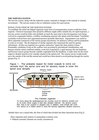
  • 1. THE NERVOUS SYSTEM The nervous system, along with the endocrine system, responds to changes in the external or internal environment. The nervous system is the co-ordination system for rapid actions. INITIAL EVOLUTION OF THE NERVOUS SYSTEM To coordinate the initial rod shaped organisms some form of communication system would have been required. Chemical messengers that spread by diffusion might suffice initially but, for rapid responses, a nervous system would be better and, probably at much the same time as the rod organisms were being evolved in a segmental fashion, a midline coordinating structure (the spinal cord) developed. Vertebrates eventually evolved from such segmented ancestors (possibly flatworms). Segmentation was useful to allow sequential contraction of the muscle groups (myotomes), for defensive reflexes and for propulsion in water or on land. The development of similar segments also minimized the need for genetic information - all that was required was a genetic instruction “repeat this basic pattern n times.” Presumably vertebrates living on the seafloor were governed by gravitational considerations, and because muscles which conferred mobility work best if attached to a rigid structure like the backbone, and because muscle groups became ventrally suspended from the backbone, it made sense for the motor output to skeletal muscles to be ventrally situated in the spinal cord. The sensory nerves would mostly serve the outside of the rod-like animal and would have been dorsal to avoid pushing through the muscle groups (Fig. 1). Initially there was a neural tube, the front of which then divided into three functional areas (Fig.2). Three important early features in mammalian evolution were: • bilateral symmetry (humans are mostly symmetrical)
  • 2. • segmentation (to allow sequential contraction for swimming purposes) • cephalization (a major focalisation of organisation in a head)
  • 3.
  • 4. Nearly all nervous pathways, except cerebellar and vestibular pathways,cross the midline either in the spinal cord, the brainstem or in the brain. There seem to be three possible explanations. Firstly the primitive coiling reflex to noxious stimuli (Fig. 3) required that nerve fibres crossed the midline. Impulses from the sensory nerve fibres had to be conducted to the motor neurones on the immediate opposite side of the spinal cord to allow the primitive rod-like animal to bend away from the noxious stimulus.
  • 5. Secondly stereoscopic sight using two eyes (which allows accurate distance appreciation) would have offered great evolutionary advantages. Integration of the information from the two eyes demands that information crosses the midline (Figs. 4a, 4b and 4c). A threat to the right of the head is appreciated by the left hand side of each retina and information from one eye has to cross the midline (in the optic chiasma) and the visual input from both retinas is coordinated in the left occipital cortex (Fig. 4c). Thus if there is to be a coordinated response, then a quicker and more efficient response to the threat demands that the right side (specifically the right arm) be moved. To move the right arm the left motor cortex has to initiate action and to do this information from the left occipital cortex has to cross the midline.
  • 6.
  • 7. Thirdly swimming or walking requires coordination of the two sides and this requires that nervous information crosses the midline. The basic structure of the human nervous system is illustrated in Fig. 2. In anatomical terms the central nervous system comprises the brain and spinal cord (Fig. 5 ) whilst the peripheral nervous system comprises 12 pairs of cranial nerves and 31 pairs of nerves arising from the spinal cord.
  • 8.
  • 9. The brain and spinal cord are surrounded by three membranes, called the meninges. The outer layer is the dura mater, the middle the arachnoid mater (interior to which is the cerebrospinal fluid) and the inner pia mater. Blood or infection in the cerebrospinal fluid causes meningeal irritation which results in neck stiffness. The capillary blood vessels supplying the central nervous system are anatomically and functionally different from capillaries elsewhere in the body. They constitute the blood-brain barrier which prevents certain large molecules and certain drugs from crossing into the central nervous system. On electron microscopy the CNS endothelial cells are joined by tight junctions which are impermeable to large molecules. The are fenestrations (holes) between endothelial cells outside the CNS. NERVES IN GENERAL
  • 11. The 1011 nerve cells and their processes are the building blocks of the nervous system. Nerve cells, once dead, cannot be replaced. Each nerve cell body has one or more dendrites which collect impulses from other nerve cell bodies or from sensory receptors. Axons are long outgrowths from nerve cells which transmit impulses away from the nerve cell. Axons may branch at their terminations and impact upon other nerve cells or their dendrites. In the central nervous system such impulses can be excitatory or inhibitory. In some nerve cells a single processemerges which splits into two in a T-shaped manner - (pseudo) unipolar nerve cells. For example the cells of the sensory spinal ganglia lateral to the dorsal surface of the spinal cord have neurones which have sensory gathering nervous fibres extending peripherally and a nerve fibre extending centrally (Fig. 7). Nerve cells do not divide except in the embryo. There are four types of nerve fibres: • Myelinated with neurolemma sheaths (mostly in peripheral nerves) • Myelinated with no neurolemma sheaths (in brain and spinal cord) Axon (=hillock): an outgrowth, usually a single outgrowth, from a nerve cell which conducts impulses along the nerve cell to the other nerve cells or their dendrites. Synonymous with nerve fibre Dendrite (=tree): the branching process of a nerve cell which conducts impulses to synapses on other nerve cells Decussation: nerve fibres which cross the midline in bundles of fibres (commisures pass to the same side) Ganglia (=knotlike) collections of nerve cell bodies outwith the central nervous system (except the basal ganglia which are within the central nervous system). They are the seat of visceral motor reflexes which affect some internal organs Neurone: nerve cell, axon, and dendrites Nerve fibre: axon and its myelin sheath.
  • 12. • No myelination but a neurolemma sheath (autonomic nerves and fine efferents of corticospinal nerves) • No myelination and no neurolemma sheath (the gray matter of brain and spinal cord) There are three types of glial cells within the CNS, astrocytes, oligodendrocytes and ependymal lining cells. These support neurones structurally and metabolically. In the peripheral nervous system neurones are supported by Schwann cells which ensheath axons. In the CNS the oligodendrocytes produce myelin which ensheaths CNS axons. Whilst all axons have Schwann cells surrounding them the speed of propagation along axons is enhanced if the axons are wrapped up with a myelin sheath by Schwann cells (Fig. 6). This “plastic covering of the electric wire” is interrupted at intervals by Nodes of Ranvier. Myelinated axons appear white (constituting the white matter of the brain and spinal cord) whereas unmyelinated axons appear gray (constituting the gray matter (neuronal cell bodies) of the brain and spinalcord). White matter conducts impulses but gray matter processes and interprets. The cortex of the cerebral hemispheres and cerebellum are mostly infolded sheets of nerve cells (gray matter). Areas of the brain called nuclei are (more or less) spherically shaped collections of functionally related gray matter in the central nervous system. Multiple sclerosis produces sensory and motor symptoms and signs caused by patchy demyelination “plaques” in medullated axons “white matter” of the central but not the peripheral nervous system. There are 31 pairs of spinal nerves - eight cervical, 12 thoracic, five lumbar, five sacral and one rudimentary coccygeal. Peripheral nerves are formed by the unification of the dorsal (sensory) and ventral (motor) roots. In the cervical and lumbar regions these roots join to form the cords of the brachial and lumbrosacral plexuses and from these arise mixed sensory and motor peripheral nerves. The motor fibres supply voluntary (striated muscle). Autonomic nerves arise from sympathetic plexuses to supply cardiac muscle, smooth muscle of the gut and blood vessels, and the salivary and some other glands. Depending on which roots are predominantly affected there may be: • Segmental muscle wasting • Reflex loss • Dermatomal pattern sensory loss (Fig. 8a, 8b for peripheral nerve losses and Fig. 8c for the likely explanation of the actual dermatome distributions) • Radiating root pain (localization often poor) • Tingling • Oversensitive skin (hyperaethesiae)
  • 13.
  • 14.
  • 15. If nerve root problems are caused by pathologies that tether the root then anything which moves the root (movements, coughing, straining) will exacerbate symptoms, especially pain. Depending on the
  • 16. pathology there may also be signs of spinal cord compression. If the nerve roots leaving the end of the spinal cord (conus and cauda equina) are involved then bladder sensation may be reduced and there may be difficulty voiding with a poor stream (motor) and rectal function may be similarly affected leading to incontinence and faecal soiling.
  • 17. Synapses (=junction) The junction between a “sending” axon and a “receiving dendrite or nerve cell is a synapse (Fig.6). Synapses are only found in the central nervous system or in autonomic ganglia and only conducts information in one direction. Some axons terminate with a single synapse whilst others may have thousand of terminations. A typical motor neurone in the spinal cord has about 10,000 synaptic contacts. Whether the receiving nerve cell responds usually depends on the number of inputs from other axons that it is receives. The more synapses that impinge on a nerve cell or its dendrites the more modifying influences there are on the final output of the receiving nerve cell. This is the basis of the integrated neural networking that forms the brain. Frequent use of particular synapses in certain situations may result in the establishment of a regular response pattern. In the central nervous system this constitutes a basis for learning (consciously or unconsciously). Synapse damage leads to forgetting. Nerves pass on information by putting out chemicals - neurotransmitters - at their synaptic endings. There are four main groups of neurotransmitters (Fig. 6): • Acetylcholine (in cholinergic neurones) • Amines Noradrenaline (in noradrenergic neurones) Serotonin (an important transmitter in sensory channels relevant to emotions) Dopamine (in the motor systems, limbic system and in the hypothalamus) • Amino acids Glutamic acid (always excitatory) Gamma aminobutyric acid (always inhibitory) • Peptides Enkephalins Endorphins These transmitters can be reabsorbed, broken down, or competitively inhibited or totally blocked before they can act. Nitric oxide (a gas) has recently been shown to be a neurotransmitter. Neuromuscular junctions When a motor nerve arrives at its muscle it loses its myelinated sheath and the axon forms a pre-synaptic terminal (motor end plate). Acetylcholine is released to bridge the gap (Fig. 9). When sufficient acetylcholine accumulates the muscle fibre depolarizes and an action potential spreads across the muscle to cause contraction. Acetycholine release from the pre-synaptic membrane is enhanced by calcium and its action is limited by destruction by an enzyme, cholinesterase, within the neuromuscular junction. Problems may arise if acetycholine release is modified, its action blocked, or if acetyl choline is broken down by cholinesterase. The final result is muscle weakness.
  • 18. Antibodies may damage the acetylcholine receptor and prevent acetylcholine binding causing poor depolarisation of nerve impulses to muscle which leads to muscle fatigue and weakness on repeated use (myasthenia gravis). This may be focal (often causing drooping of the eyelids), or may affect other brainstem-derived cranial nerves or may be more widespread. In the Eaton-Lambert syndrome there are antibodies to calcium channels in the pre-synaptic terminals leading to failure of acetylcholine release (and perhaps also in the autonomic nerves to cause parasympathetic failure). Often the weakness improves on repeated use. With botulism the toxin binds to the terminals of nerves such that acetylcholine cannot be released and there is widespread muscle paralysis and autonomic dysfunction. Nerve impulses are electric waves of depolarisationwhich propagate along axons. Normally sodium is pumped out of the axon whereas potassium is kept at a high concentration inside the axon. This causes a 60 mVolts positive charge outside the axon. If the axon is excited sodium briefly enters the axon, the inside briefly becoming 40 mvolts positive, and a current flows ahead and initiates further sodium penetration “down the line” and thus a wave of depolarisation travels along the axon. An axon either transmits an impulse or not, “all or nothing,” and information conveyed depends on alterations in the frequency of impulses plus the nature and situation of the sending nerve cell body or receptor. On arrival centrally the sensory inputs are allowed to progress depending on their density and timing from the periphery. This wave of depolarisation is followed by a refractory period in which the nerve is absolutely or relatively resistant to the passage of other impulses. After conducting an impulse the larger nerve fibres recover in over one millisecond (up to 1,000 vibrations per second can be appreciated). In practice most sensory fibres can conduct 300-400 impulses per second and motor fibres can conduct at 100-150 impulses a second. If the axon is unmyelinated then the wave flows continuously to its destination but if there are nodes of Ranvier the wave jumps from node to node, increasing the velocity of conduction. Unmyelinated axons can conduct as slowly as 50 cms per second whereas myelinated fibres can conduct at about 100 metres per second (Fig. 10).
  • 19. In some situations there can be pain appreciation without touch (from the bladder for example) but in other situations touch becomes pain if afferent nerves convey an excessive number of similar impulses. Within limits, the larger the diameter of the axon the quicker the impulse conduction. Large nerve fibres mostly transmit touch, pressure, two point discrimination, and joint position sensation but usually not pain and temperature. Smaller fibres (which are more likely to be affected by diabetes) mostly transmit pain and temperature but not other modalities (thus neuropathic foot ulcers caused by loss of pain sensation may result). Axons normally only transmit away from the nerve cell. Nerves that carry important messages tend to have myelinated axons and a minimum of synapses to allow rapid conduction (for example in the corticospinal tract). Nerve cell bodies in the central nervous system, usually in the cortex, can give rise to unusual, paroxysmal, electrical activity which may spread to cause epilepsy, the manifestations of which depend upon the initial area of gray matter involved and the extent of spread. Localized seizure discharges in the frontal lobe motor cortex produce a localized jerking of skeletal muscle on the contralateral side but generalized epilepsy produces tonic (continuous contraction) and then clonic (jerking) contraction of all skeletal muscle along with loss of consciousness. Epileptic foci in areas of the brain that do not normally cause skeletal muscle action may present in other ways (temporal lobe epilepsy for example may present with autonomic sensations of taste, smell or déjà vu with loss of awareness without loss of consciousness. Peripheral neuropathy Peripheral nerves are surrounded by neurilemma (Fig. 11), groups of axons by perineurium, and groups of groups (the nerves) are surrounded by epineurium which also contains the nourishing blood vessels. The nerve fibres of brain and spinal cord have no neurolemma and are incapable of regeneration whereas peripheral nerve axons can regrow along the “empty” neurolemmal tubes after nerve damage.
  • 20. The nutrient-providing blood vessels may be damaged by metabolic disorders (such as diabetes mellitus) and cause axonal degeneration or demyelination. The longer the nerve then the more cumulative low grade damage, explaining why symptoms and signs of neuropathy (nerve damage without inflammation) often begin in the feet. Nerve compression often causes both axonal degeneration and demyelination. Neuropathy may affect individual nerves or nerves (mononeuropathy) or all nerves (generalized neuropathy). In either motor symptoms may include: • Weakness • Wasting • Fasciculation (twitching of small areas of muscle caused by spontaneous electrical activity in the motor unit so that individual muscle fasciculi contract) In either sensory symptoms may include: • Loss of sensation (but only in the area exclusively supplied by an individual nerves in a mononeuropathy) • Pain • Tingling With a generalized sensory neuropathy there will be glove and stocking sensory loss (Fig. 12). Reflexes may be reduced by damage to either sensory or motor nerve fibres or both.
  • 21. Peripheral nerves usually contain a mixture of sensory (afferent= to carry to) and motor (efferent= to carry away) fibres. If a perip heral nerve is cut then the result usually include: • Flaccid paralysis of the muscles receiving motor innervation, followed by muscle wasting and contractures • Loss of cutaneous sensation in the areas exclusively supplied by the nerve (most cutaneous areas exhibit overlap of cutaneous sensation) • Loss of reflexes (if there are reflexes associated with the nerve concerned) • Loss of sympathetic and parasympathetic responses (causing initial vasodilatation, lack of sweating and piloerection) The cell bodies of the motor nerves (motor horn cells) may be affected in isolation within the spinal cord (polio specifically damages the anterior horn cells of the lower motor neurone) to produce lower motor neurone signs. If a peripheral nerve is damaged the neurone fibres degenerate proximally, usually up to the proximal node of Ranvier, and distally. The Schwann cells (Fig. 6) then remain, surrounding an empty tube. The intact proximal neurone then grows distally, hopefully along its previous tube, at a rate of about one mm a day. If a nerve is cut but the two ends remain approximately opposed an axon may regrow along the wrong tube and this can cause partial or altered sensation or false motor innervation leading to persisting dysfunction.
  • 22. Malignant tumours of nerves are rare but benign tumours are not uncommon, either in isolation (as neuromas, notably acoustic neuromas) or as part of a more generalized problem (e.g. neurofibromatosis). Generalized peripheral neuropathy can be post-infective, metabolic, or related to malignancy. • diabetes mellitus • vitamin deficiency (notably vitamin B12 or B1) • infections (including leprosy and HIV) • toxic (including alcohol) • drug induced • hereditary • vasculitis • compression at certain points: wrist (median nerve), elbow (ulnar nerve), supper arm (radial nerve), head of fibula(lateral popliteal nerve), groin (lateral cutaneous nerve of thigh) and buttock (sciatic nerve). Groups of functionally similar axons are called tracts, fascicles, peduncles, or lemnisci. The first part specifies the site of origin, the second the destination e.g. corticospinal = from the cortex to the spinal cord