PULSE OXIMETRY
•Download as PPTX, PDF•
19 likes•8,254 views

Pulse oximetry is a noninvasive test that uses light to measure the oxygen saturation level in a person's blood. A clip-like probe is placed on the finger or earlobe and uses red and infrared light wavelengths absorbed differently by oxygenated and deoxygenated hemoglobin to calculate the oxygen saturation percentage. This information helps healthcare providers determine if a patient needs supplemental oxygen or how well treatment is working. However, pulse oximetry has limitations as it does not measure other blood gas levels, ventilation, or oxygen metabolism and can be affected by factors like carbon monoxide, anemia, blood flow, and skin pigmentation.

Recommended
More Related Content
What's hot
What's hot (20)
Similar to PULSE OXIMETRY
Similar to PULSE OXIMETRY (20)
More from MAHESWARI JAIKUMAR
More from MAHESWARI JAIKUMAR (20)
Recently uploaded
Recently uploaded (20)
PULSE OXIMETRY
- 2. • Pulse oximetry is a test used to measure the oxygen level (oxygen saturation) of the blood. It is an easy, painless procedure to assess the oxygen saturation
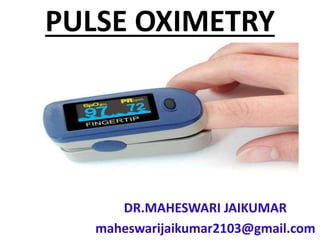
- 3. • Pulse oximetry is clip-like device called a probe is placed on a body part, such as a finger or ear lobe. The probe uses light to measure how much oxygen is in the blood. • This information helps the healthcare provider decide if a person needs extra oxygen.
- 4. PARTS OF PULSE OXIMETER
- 6. INDICATIONS FOR PULSE OXIMETRY • During or after surgery or procedures that use sedation • To see how well lung medicines are working
- 7. • To check a person’s ability to handle increased activity levels • To see if a ventilator is needed to help with breathing, or to see how well it’s working
- 8. • To check a person has moments when breathing stops during sleep (sleep apnea)
- 9. • Pulse oximetry is also used to check the health of a person with any condition that affects blood oxygen levels, such as: • Heart attack • Heart failure • Chronic obstructive pulmonary disease (COPD) • Anemia • Lung cancer • Asthma • Pneumonia
- 10. TYPES OF PUSE OXIMETRY • TRANSMISSIVE APPLICATION MODE • REFLECTANCE PULSE OXIMETRY
- 11. MODE OF ACTION • In its most common (transmissive) application mode, a sensor device is placed on a thin part of the patient's body, usually a fingertip or earlobe, or in the case of an infant, across a foot.
- 12. • The device passes two wavelengths of light through the body part to a photo detector. It measures the changing absorbance at each of the wavelengths, allowing it to determine the absorbances due to the pulsing arterial blood alone, excluding venous blood, skin, bone, muscle, fat, and (in most cases) nail polish.
- 13. FINGER SENSOR
- 14. • Reflectance pulse oximetry is a less common alternative to transmissive pulse oximetry. This method does not require a thin section of the person's body and is therefore well suited to a universal application such as the feet, forehead, and chest, but it also has some limitations.
- 15. • Vasodilation and pooling of venous blood in the head due to compromised venous return to the heart can cause a combination of arterial and venous pulsations in the forehead region and lead to spurious SpO2 results. • Such conditions occur while undergoing anesthesia with endotracheal intubation and mechanical ventilation or in patients in the Trendelenburg position
- 17. FUNCTION • A blood-oxygen monitor displays the percentage of blood that is loaded with oxygen. • More specifically, it measures what percentage of hemoglobin, the protein in blood that carries oxygen, is loaded.
- 19. • Acceptable normal ranges for patients without pulmonary pathology are from 95 to 99 percent. For a patient breathing room air at or near sea level, an estimate of arterial pO2 can be made from the blood-oxygen monitor "saturation of peripheral oxygen" (SpO2) reading.
- 20. • A typical pulse oximeter uses an electronic processor and a pair of small light-emitting diodes (LEDs) facing a photodiode through a translucent part of the patient's body, usually a fingertip or an earlobe.
- 22. • One LED is red, with wavelength of 660 nm, and the other is infrared with a wavelength of 940 nm. Absorption of light at these wavelengths differs significantly between blood loaded with oxygen and blood lacking oxygen.
- 24. • Oxygenated hemoglobin absorbs more infrared light and allows more red light to pass through • Deoxygenated hemoglobin allows more infrared light to pass through and absorbs more red light.
- 25. • The LEDs sequence through their cycle of one on, then the other, then both off about thirty times per second which allows the photodiode to respond to the red and infrared light separately and also adjust for the ambient light baseline.
- 26. • The amount of light that is transmitted (in other words, that is not absorbed) is measured, and separate normalized signals are produced for each wavelength. • These signals fluctuate in time because the amount of arterial blood that is present increases (literally pulses) with each heartbeat.
- 27. • By subtracting the minimum transmitted light from the transmitted light in each wavelength, the effects of other tissues are corrected for, generating a continuous signal for pulsatile arterial blood.
- 28. • The ratio of the red light measurement to the infrared light measurement is then calculated by the processor (which represents the ratio of oxygenated hemoglobin to deoxygenated hemoglobin), and this ratio is then converted to SpO2 by the processor via a lookup table based on the Beer–Lambert law
- 29. • The signal separation also serves other purposes: a plethysmograph waveform ("pleth wave") representing the pulsatile signal is usually displayed for a visual indication of the pulses as well as signal quality,and a numeric ratio between the pulsatile and baseline absorbance ("perfusion index") can be used to evaluate perfusion.
- 30. ADVANTAGES • Pulse oximetry is convenient for noninvasive continuous measurement of blood oxygen saturation. • Whereas blood gas levels must otherwise be determined in a laboratory on a drawn blood sample.
- 31. • Pulse oximetry is useful in any setting where a patient's oxygenation is unstable, including intensive care, operating, recovery, emergency and hospital ward settings, pilots in unpressurized aircraft, for assessment of any patient's oxygenation, and determining the effectiveness of or need for supplemental oxygen.
- 32. • Although a pulse oximeter is used to monitor oxygenation, it cannot determine the metabolism of oxygen, or the amount of oxygen being used by a patient.
- 33. • Therefore, it is necessary to also measure carbon dioxide (CO2) levels. • It can also be used to detect abnormalities in ventilation.
- 34. • However, the use of a pulse oximeter to detect hypoventilation is impaired with the use of supplemental oxygen. • Therefore, the routine administration of supplemental oxygen may be unwarranted if the patient is able to maintain adequate oxygenation in room air
- 35. • Because of their simplicity of use and the ability to provide continuous and immediate oxygen saturation values, pulse oximeters are of critical importance in emergency medicine and are also very useful for patients with respiratory or cardiac problems, especially COPD, or for diagnosis of some sleep disorders such as apnea and hypopnea.
- 36. LIMITATIONS • Pulse oximetry solely measures hemoglobin saturation, not ventilation and is not a complete measure of respiratory sufficiency. • It is not a substitute for blood gases checked in a laboratory, because it gives no indication of base deficit, carbon dioxide levels, blood pH, or bicarbonate (HCO3 −) concentration
- 37. • The metabolism of oxygen can be readily measured by monitoring expired CO2, but saturation figures give no information about blood oxygen content. • Most of the oxygen in the blood is carried by hemoglobin; in severe anemia, the blood contains less hemoglobin, which despite being saturated cannot carry as much oxygen.
- 38. • Erroneously low readings may be caused by hypoperfusion of the extremity being used for monitoring (often due to a limb being cold, or from vasoconstriction secondary to the use of vasopressor agents); incorrect sensor application; highly calloused skin; or movement (such as shivering), especially during hypoperfusion.
- 39. • To ensure accuracy, the sensor should return a steady pulse and/or pulse waveform. • Pulse oximetry also is not a complete measure of circulatory oxygen sufficiency.
- 40. • If there is insufficient bloodflow or insufficient hemoglobin in the blood (anemia), tissues can suffer hypoxia despite high arterial oxygen saturation.
- 41. • Since pulse oximetry measures only the percentage of bound hemoglobin, a falsely high or falsely low reading will occur when hemoglobin binds to something other than oxygen:
- 42. • Hemoglobin has a higher affinity to carbon monoxide than it does to oxygen, and a high reading may occur despite the patient's actually being hypoxemic. • In cases of carbon monoxide poisoning, this inaccuracy may delay the recognition of hypoxia (low cellular oxygen level).
- 43. SUMMARY • Limitations in Using a Pulse Oximeter • Carbon Monoxide. Carbon monoxide molecules, even in a small amount, can attach to the patient's hemoglobin replacing oxygen molecules. • Hemoglobin Deficiency (Anemia) • Blood Volume Deficiency. • Irregular Signals. • External Interference. • Fingernail Polish and Pressed on Nails. • Skin Pigmentation. • Intravenous Dyes.
- 44. NORMAL RANGE FOR PULSE OXIMETRY • Normal pulse oximeter readings usually range from 95 to 100 percent. Values under 90 percent are considered low.
- 45. FACTORS AFFECTING PULSE OXIMETRY READING • Blood pressure generally needs to be >80 SBP. • Vascular impingement from any cause. • AV fistula can decrease distal flow. • Elevation with respect to the heart. • Compression by the probe. • Cardiac arrest (don't use during arrest) • Heart Rate extremes <30 or >200. Cold. Fear (Endogenous catecholamines) Medications.
- 46. REFERENCES • Jørgensen JS, Schmid ER, König V, Faisst K, Huch A, Huch R (July 1995). "Limitations of forehead pulse oximetry". Journal of Clinical Monitoring. 11 (4): 253–6. doi:10.1007/bf01617520. PMID 7561999. • ^ Matthes K (1935). "Untersuchungen über die Sauerstoffsättigung des menschlichen Arterienblutes" [Studies on the Oxygen Saturation of Arterial Human Blood]. Naunyn-Schmiedeberg's Archives of Pharmacology (in German). 179 (6): 698–711. doi:10.1007/BF01862691. • ^ Millikan GA (1942). "The oximeter: an instrument for measuring continuously oxygen saturation of arterial blood in man". Review of Scientific Instruments. 13(10): 434– 444. Bibcode:1942RScI...13..434M. doi:10.1063/1.1769941. • ^ Jump up to:a b Severinghaus JW, Honda Y (April 1987). "History of blood gas analysis. VII. Pulse oximetry". Journal of Clinical Monitoring. 3 (2): 135– 8. doi:10.1007/bf00858362. PMID 3295125. • Jopling MW, Mannheimer PD, Bebout DE (January 2002). "Issues in the laboratory evaluation of pulse oximeter performance". Anesthesia and Analgesia. 94 (1 Suppl): S62–8. PMID 11900041.
- 47. THANK YOU