
Recommended
More Related Content
What's hot
What's hot (20)
Similar to Excretory system:Kidneys,ureter,urethra and urinary bladder
Similar to Excretory system:Kidneys,ureter,urethra and urinary bladder (20)
More from Jasleen Kaur
More from Jasleen Kaur (20)
Recently uploaded
Recently uploaded (20)
Excretory system:Kidneys,ureter,urethra and urinary bladder
- 2. Excretion: It is removal of the waste products of metabolism,toxic material and substances from the body. Micturition: Micturition or urination is the process of expelling urine from the bladder.This act is also known as voiding of bladder. Nephrology:Nephrology is the scientific study of anatomy , physiology and pathology of the kidneys.
- 3. KIDNEYS-These are the primary excretory organs throwing out excretory products in the form of urine. LARGE INTESTINE-Undigested food is removed from large intestine and thrown out of body through anus. SKIN-It eliminate excess of water,urea,salts and other metabolic waste in the form of sweat.Sweat is secreted from sweat gland.
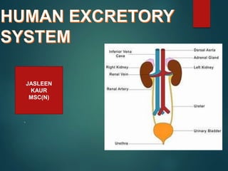
- 4. The urinary system is the main excretory system and consists of the following structures- 2 kidneys,which secrete urine 2 ureters, which conveys the urine from the kidneys to the urinary bladder. The urinary bladder, where urine collects and is temporarily stored The urethra through which the urine passes from urinary bladder to the exterior.
- 5. .
- 6. FUNCTIONS OF KIDNEYS- The main functions of kidneys are- • Formation and excretion of urine,which regulates total body water,electrolytes and acid base balance and enables excretion of waste products. • Production and secretion of ERYTHROPOIETIN, the harmone that stimulates formation of red blood cells. • Production and secretion of RENIN, an important enzyme in the control of blood pressure. • Activation of VITAMIN D. • Regulation of BLOOD GLUCOSE LEVEL.
- 7. - WHERE ARE THE KIDNEYS SITUATED IN THE HUMAN BODY? The kidneys lie on the posterior abdominal wall,one each side of the vertebral column,behind the peritoneum and below the diaphragm.They extend from the level of the 12th thoracic vertebra to the 3rd lumbar vertebra.
- 8. The right kidney is usually slightly lower than the left kidney, probably because of the space occupied by liver`
- 9. SHAPE- Kidneys are bean shaped organ. SIZE- About 11cm long,6cm wide and 3 cm thick and weigh 150gms each Kidneys are held in position by a mass of fat. A Sheath of fibrous connective tissue,also known as renal fascia,encloses the kidneys and renal Fat.
- 10. SURFACE ANATOMY- Each kidney is surrounded by three layers- Renal fascia- fibrous tissues which mainly provides support to the kidneys Adipose tissues- Protects kidney Renal capsule –it anchors kidney to body wall continuous with peritoneum.
- 11. 3 layers of connective tissue: Inner layer- Renal capsule Middle layer- Adipose capsule Outer layer-Renal fascia Renal cortex Retroperitoneal space
- 12. There are three areas of tissues that can be distinguished when distinguished when a longitudinal section of the kidney is viewed with a naked eye- An outer fibrous capsule, surrounding the kidney The cortex, a reddish brown layer of tissue immediately below the capsule The medulla, the innermost layer, consisting of pale conical shaped striations, the renal pyramids. The hilum is the concave medial border of the kidney where the renal blood and lymph vessels, the ureters and nerve enters.
- 13. .
- 14. A longitudinal section of right kidney
- 15. What volume of blood enters the renal arteries per minute? The renal arteries deliver 20-25% 0f the resting cardiac output to the kidneys. Hence in adult,renal blood flow through both kidneys,is about 1000-1200ml per minute. NOTE-The blood vessels of kidney are supplied by both sympathetic and parasympathetic nerves.
- 16. MICROSCOPIC STRUCTURE OF THE KIDNEY- The kidney is composed of about 1-2 million functional units, The Nephrons and a smaller number of collecting ducts. The collecting ducts transport urine through the pyramids to the calyces and renal pelvis,giving the pyramids their striped appearance. These collecting ducts are suported by a small amount of connective tissue,containing blood vessels,nerve and lymph vessels.
- 17. The Nephron:
- 18. The nephron consists of: 1)Renal corpuscles-The head of nephron. The renal corpuscles is composed of bowman’s capsule- Bowman’s capsule is the cover of corpuscles that surrounds the glomerulus The glomerulus is the network of cappillaries found inside the corpuscles Blood enters at glomurules by the way of affarent arteriole and leaves in an efferent arterioles.
- 19. 2)Renal Tubule-the tubular passageway of nephrons.It cosists of- *PROXIMAL CONVOLUTED TUBULE(PCT) *LOOP OF HENLE *DISTAL CONVOLUTED TUBULE
- 20. There are two types of Nephrons: 1)Cortical Nephrons:85% of the nephrons located in the cortex 2)Juxtamedullary Nephrons:closer to renal medulla and extends deep into renal pyramids
- 21. = Blood enters the kidney through the renal artery at the site of the hilum The renal artery divides in to ever smaller arteries and arterioles Afferent arterioles take blood to the glomerulus to be filtered Once blood is filtered efferent arterioles take blood away from the glomerulus The glomerulus is a network of capillaries which filters the blood Products which are filtered out: water, mineral salts, amino acids, glucose, hormones, urea, toxins Products which do not filter and remain in the blood: Leukocytes, erythrocytes, platelets, plasma proteins The filtered substances move into the proximal convoluted tubule The PCT is concerned with reabsorption- organic nutrients are reabsorbed and water follows because there is a concentration gradient The remaining filtrate moves into the descending loop of henle. This is lined with thin cells so water moves out Because water has been reabsorbed the concentration of the filtrate is not very high The walls of the ascending loop of henle are lined with thicker cells, so water can’t pass in or out. Instead sodium and chloride is pumped out actively The filtrate now enters the distal convoluted tubule- is it now only 20% of what it originally was. In the DCT the volume and composition of the filtrate can be adjusted but this is controlled by hormones From the DCT the filtrate now passes into the collecting duct. A number of other nephrons join up to the cleectig duct which travels through the medulla to the renal papilla wher the filtrate is emptied in the minor calyx 4-5 minor calyces join up to make a major calyx 2-3 major calyces join up to form the renal pelvis The renal pelvis joins the ureter at the hilum The ureter transport the filtrate/urine from the kidney to the bladder
- 22. THE FORMATION OF URINE:3 processes involved in the formation of urine.
- 23. It takes place through the semipermeable walls of glomerulus and glomerular capsule. Water and other small molecules pass through while blood cells,plasma proteins and other large molecules are too large to filter though and therefore remain in the capillaries.
- 24. FILTERATION TAKES PLACE BECAUSE THERE IS A DIFFERENCE BETWEEN THE BLOOD PRESSURE IN THE GLOMERULUS AND THE FILTERATE IN THE GLOMERULAR
- 25. NET FILTERATION PRESSURE=Glomerular hydrostatic pressure-(blood osmotic pressure+capsular hydrostatic pressure NFP=55-(30+15) =10mmHG
- 27. FACTORS AFFECTING GFR: Renal Blood flow Environmental conditions Constriction of afferent and efferent arteriole Disease condition e.g ureteral stricture,dehydration Biochemical changes under the influence of sympathetic nd parasympathetic stimulation
- 28. Selective reabsorption is the process by which certain molecules e.g. ions,glucose and amino acid after being filterd out of the capillaries along with nitrogeneous waste products i.e. urea and water in the glomerulus,are reabsorbed back into the blood circulation from the filtrate as they pass through the nephron. Only 60-70% filtrate reaches the loop of nephron.much of this is reabsorbed in the loop of henle. So only 15-20% of the original filtrate reaches the DCT. Now the composition of filtrate is very different from its starting values.
- 30. Hormones that influence Selective Reabsorption: PARATHYROID HORMONE ANTIDIURETIC HORMONE ALDOSTERONE HORMONE ATRIAL NATRIURETIC PEPTIDE
- 31. RAAS MECHANISM
- 32. Substances not required and foreign material .e.g. drugs including penicillin and aspirin, may not be cleared from the body by filtration because of short time it remain in the glomerulus. Such substances are cleared by secretion from the peritubular capillaries into the convoluted tubules and excreted from blood in the urine. Tubular secretion of hydrogen ion is important in maintaining normal blood pH.
- 33. COMPOSITION OF URINE: Urine is clear and amber in colour due to presence of urobilin,a bile pigment . Water-96% specific gravity of urine=1.020-1.030 Urea-2% Uric acid urine output/day=1000-1500ml/day Creatinine Ammonia pH=4.5-8 sodium Potassium 2% Chlorides Phosphates Sulphates Oxolates
- 34. ureters
- 35. DIMENSIONS:
- 37. BLOOD SUPPLY
- 40. The urinary blader is reservoir for urine..it lies in the pelvic cavity and its size and position vary,depending on the volume of urine it contains.when distended,the bladder rises into the abdominal cavity.
- 41. The appearance of bladder varies depending on the amount of urine stored.when full,it exhibits an , and when empty it is flattened and pear shaped. The external features of bladder are: 1.APEX-IS LOCATED SUPERIORLY,POINTING TOWARDS THE SYMPHYSIS PUBIS. 2.BODY-MAIN PART OF BLADDER,LOCATED BETWEEN THE APEX AND FUNDUS. 3.FUNDUS (BASE)-LOCATED POSTERIORLY.IT IS TRIANGULAR SHAPED,WITH THE TIP OF TRIANGLE POINTING BACKWARDS. 4.NECK-IT IS CONTINUOUS WITH THE URETHRA.
- 42. The bladder wall is composed of three main layers: * Outer loose connective tissues * The middle layer consisting of interlacing smooth muscle fibres and elastic tissues loosely arranged in three layers .this is called detrusor muscle and when it contracts , it empties the bladder. * The mucosa,composed that readily permits distension of bladder as it fills with urine.
- 43. When the bladder is empty the inner lining is arranged in folds or rugae which gradually disappear as it fills. Bladder capacity-250-300ml Filling upto 500ml may be tolerated ,but beyond this it becomes painful.
- 44. The urethra is a tube that connects the urinary bladder to the urinary meatus for the removal of urine from the body of both male and female.it is longer in male than in female. Length of urethra in male and female: male-16-18cm long Female- 3-4cm long Diameter-5-6mm respectively
- 45. The male urethra is generally associated with urinary and reproductive system The female urethra is associated with urinary system only.it runs downwards and forwards behind the symphysis pubis and opens at external uretheral sphincter just in front of the vagina. The internal uretheral sphincter , a thickening of uretheral smooth muscles and is under involuntary control The external urethral orifice is guarded by the external urethral sphincter,which is under voluntary control.
- 47. Micturition reflex is the neurological pathways involved in urination. When your bladder fills, the detrusor muscles expand and proprioceptors within the walls of the muscle activate. It sends signals (afferent nerves) that go to your medulla and also to the parasympathetic system in the sacral spinal cord.