Intro to ANS
•
5 gefällt mir•633 views

A short introduction to the Autonomic Nervous System (Pharmacology for BDS)

Empfohlen
Weitere ähnliche Inhalte
Was ist angesagt?
Was ist angesagt? (20)
Andere mochten auch
Andere mochten auch (11)
Ähnlich wie Intro to ANS
Ähnlich wie Intro to ANS (20)
Mehr von Hiba Hamid
Mehr von Hiba Hamid (16)
Kürzlich hochgeladen
Kürzlich hochgeladen (20)
Intro to ANS
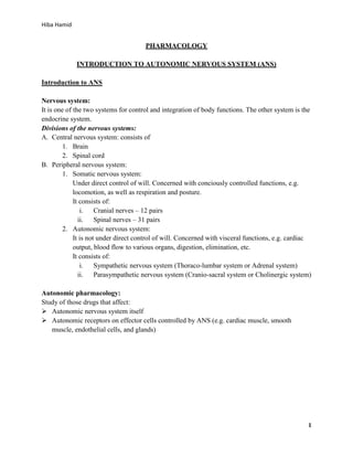
- 1. Hiba Hamid PHARMACOLOGY INTRODUCTION TO AUTONOMIC NERVOUS SYSTEM (ANS) Introduction to ANS Nervous system: It is one of the two systems for control and integration of body functions. The other system is the endocrine system. Divisions of the nervous systems: A. Central nervous system: consists of 1. Brain 2. Spinal cord B. Peripheral nervous system: 1. Somatic nervous system: Under direct control of will. Concerned with conciously controlled functions, e.g. locomotion, as well as respiration and posture. It consists of: i. Cranial nerves – 12 pairs ii. Spinal nerves – 31 pairs 2. Autonomic nervous system: It is not under direct control of will. Concerned with visceral functions, e.g. cardiac output, blood flow to various organs, digestion, elimination, etc. It consists of: i. Sympathetic nervous system (Thoraco-lumbar system or Adrenal system) ii. Parasympathetic nervous system (Cranio-sacral system or Cholinergic system) Autonomic pharmacology: Study of those drugs that affect: Autonomic nervous system itself Autonomic receptors on effector cells controlled by ANS (e.g. cardiac muscle, smooth muscle, endothelial cells, and glands) 1
- 2. Hiba Hamid Neurotransmitter aspects of the ANS Synthesis, storage, release, receptor interactions, and termination of action of the neurotransmitters all contribute to the action of autonomic drugs. Cholinergic transmission Acetycholine (ACh) is the primary transmitter in all autonomic ganglia and at the synapses between parasympathetic postganglionic neurons and their effector cells. Transmitter at postganglionic sympathetic neurons to the thermoregulatory sweat glands. Primary transmitter at the somatic (voluntary) skeletal muscle neuromuscular junction. 1) Synthesis and storage a) Synthesised in the nerve terminal by enzyme choline acetyltransferase (ChAT) from acetyl CoA (produced in mitochondria) and choline (transported across cell membrane). b) Transport of choline from outside the membrane to inside can be inhibited by the research drug hemicholinium. c) ACh is actively transported into vesicles for storage. This process can be inhibited by another research drug, vesamicol. 2) Release of acetylcholine a) Release of ACh from vesicles at the nerve ending requires entry of calcium through calcium channels. This process can be blocked off by botulinum toxins. 3) Termination of action of ACh a) Action of ACh in the synapse is normally terminated by metabolism to acetate and choline by the enzyme acetylcholinesterase in the synaptic cleft. b) Products are not excreted by recycled in the body. c) Inhibition of AChE is an important therapeutic (and potentially toxic) effect of several drugs. Adrenergic transmission Norepinephrine (NE) the primary transmitter at the sympathetic postganglionic neuroneffector cell synapses in most tissues. Important exceptions include sympathetic fibers to thermoregulatory (eccrine) sweat glands and probably vasodilator sympathetic fibers in skeletal muscle, which release ACh. Dopamine may be a vasodilator transmitter in renal blood vessels, but norepinephrine is a vasoconstrictor of these vessels. 1) Synthesis and storage a. After transport across cell membrane, tyrosine is hydroxylated by tyrosine hydroxylase (the rate-limiting step) to DOPA (dihydroxyphenylalanine), decarboxylated to dopamine, and (inside the vesicle) hydroxylated to NE. 2
- 3. Hiba Hamid b. Tyrosine hydroxylase can be inhibited by metyrosine. c. Storage of dopamine into vesicles can be inhibited by reserpine, resulting in depletion of transmitter stores. 2) Release and termination of action a. Dopamine and NE released from nerve endings by same calcium-dependent mechanism responsible for release of acetylcholine release. b. Metabolism is not responsible for termination of action of the catecholamine transmitters, NE and dopamine. Rather, diffusion and reuptake reduce their concentrations in the synaptic cleft and stop their actions. c. Outside the cleft, these transmitters are metabolised by enzymes and their products are excreted. d. Release of NE can be inhibited by guanethidine. e. Reuptake can be blocked by cocaine and tricyclic antidepressants. Actions of sympathetic and parasympathetic nervous systems on effector organs Red= sympathetic actions (red means alert, danger, flight fight fright mode) Blue= parasympathetic actions (blue means rest digest, calm and peaceful mode) EYE Contration of iris radial muscle (pupil dilates= mydriasis) (darr mein aapko zada dekhna hota hai bhaagne ke liye :P ) Contraction of iris sphincter muscle (pupil contracts= miosis) LACRIMAL GLANDS Stimulation of tears SALIVARY GLANDS Thick, viscous secretion (moun sookh jata hai darr mein) Copious, watery secretion HEART Increased rate; increased contractility (heart starts beating fast when stressed and afraid) Decreased rate; decreased contractility (sukun mein ho) TRACHEA AND BRONCHIOLES Dilation (darr mein saansen barh jaati hain :D ) Constriction, increased secretions 3
- 4. Hiba Hamid ADRENAL MEDULLA Secretion of epinephrine and norepinephrine GASTROINTESTINAL SYSTEM Decreased muscle motility and tone; contraction of spincters Increased muscle motility and tone KIDNEY Secretion of renin (β1 increases; α1 decreases) URETERS AND BLADDER Relaxation of detrusor; contraction of trigone and sphincter (darr mein bathroom bhaagne ke khyalat ghanta nai aate) Contraction of detrusor; relaxation of trigone and sphincter (happy bathroom ) GENITALIA (MALE) Stimulation of ejaculation Stimulation of erection GENITALIA (FEMALE) Relaxation of uterus BLOOD VESSELS (SKELETAL MUSCLE) Dilation BLOOD VESSELS (SKIN, MUCOUS MEMBRANES, AND SPLANCHNIC AREA) Constriction (you get pale when scared) 4
- 5. Hiba Hamid Characteristics of sympathetic and parasympathetic systems Sympathetic Sites of origin Thoracic and lumbar region of the spinal cord (thoracolumbar) Length of fibers Short preganglionic Long postganglionic Location of ganglia Close to spinal cord Preganglionic fiber Extensive branching Distribution Wide Type or response Diffuse Parasympathetic Brain and sacral area of spinal cord (cranio-sacral) Long preganglionic Short postganglionic Within or near effector organs Minimal Limited Discrete Receptor characteristics Major receptor systems in the ANS include: 1) Cholinoceptors 2) Adrenoceptors 3) Dopamine receptors Cholinoceptors 1) Muscarinic receptors: Agonist: muscarine, ACh Antagonist: Atropine Subtypes: a) M1: located in CNS, sympathetic postganglionic neuron (at sympathetic ganglia), gastric parietal cells, some presynaptic sites b) M2: located in CNS, end-organs (e.g. myocardium, smooth muscle, eccrine or thermoregulatory sweat glands, skeletal muscle blood vessels), some presynaptic sites 2) Nicotinic receptors: Agonists: ACh, nicotine, Antagonists: d-tubocurarine (blocks at autonomic ganglia and skeletal muscle), hexamethonium (blocks at autonomic ganglia) Location: a) Spinal cord b) Autonomic ganglia (sympathetic + parasympathetic) c) Skeletal muscle NMJ Types: a) NG: located in autonomic ganglia 5
- 6. Hiba Hamid b) NM: located in skeletal muscle Adrenoceptors 1) Alpha-adrenoceptors: Agonists: acc. to potency of action= epinephrine ≥ norepinephrine > isoproterenol Antagonists: phentolamine, phenoxybenzamine Subtypes: a) Alpha-1: blocked by prazosin. Located postsynaptically in radial muscle of eye, heart, most vascular smooth muscle, bronchial glands, GIT, rat liver, splenic capsule, urinary bladder, pilomotor smooth muscle b) Alpha-2: not blocked by prazosin and isoproterenol is ineffective on them. Located: i. Presynaptically on adrenergic and cholinergic nerve terminals ii. Postsynaptically on platelets, lipocytes, some vascular smooth muscles (noninnervated), brain 2) Beta-adrenoceptors Agonists: acc. to potency of action= isoproterenol > norepinephrine ≥ epinephrine Antagonist: propranolol Subtypes: a) Beta-1: have equal affinity for epinephrine and norepinephrine Located: i. Presynaptically on noradrenergic nerve terminals ii. Postsynaptically on heart, lipocytes, and kidneys. b) Beta-2: have higher affinity for epinephrine than norepinephrine. Located postsynaptically on following sites: bronchial smooth muscle, bronchial glands, GIT wall, human liver, urinary bladder, pregnant uterus, skeletal muscle blood vessels. Dopaminergic receptors Stimulated by dopamine. 1) D1: Agonist: phenothiazine 2) D2: Agonist: phenothiazine, butyrophenone Extra-cerebral locations of dopaminergic receptors: Presynaptically on terminals of nerves supplying heart, blood vessels, and GIT Postsynaptically on effector organs, especially vascular smooth muscles of splanchnic, coronary, and renal blood vessels. 6