SlideShare ist ein Scribd-Unternehmen logo
1 von 58
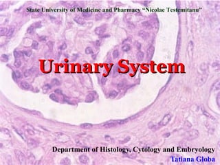
State University of Medicine and Pharmacy “Nicolae Testemitanu”


    Urinary System


         Department of Histology, Cytology and Embryology
                                             Tatiana Globa
Urinary System Functions
 Clear the blood of nitrogenous and other waste metabolic
  products (urea, uric acid, toxic stuff, drugs) by filtration and
  excretion
 Regulation of
   – blood volume
   – concentration of blood solutes
   – pH of extracellular fluid
 Endocrine   function: synthesis of erythropoietin, renin,
  prostaglandins
 Makes calcitrol (from Vit D3: stim Ca2+ absorption by
  intestinal epithelium)
 Recovers by reabsorbtion small molecules (amino acids,
  glucose, and peptides), ions (Na, Cl, Ca, PO), and water, in
  order to maintain blood homeostasis.
 Assists liver in detoxification of poisons
Urinary System

Consists:
 Kidneys
 Ureters
 Urinary bladder
  (storage)
 Urethra
Kidneys
 Kidney   are paired, bean-shaped organs, enveloped by
  a thin capsule of connective tissue
 Renal hilum is the concavity on the medial border of
  the kidney where there are:
   – Renal artery, vein; nerves, lymphatic vessels and
     ureter
 Sizes:10 cm X 5.5 cm X 3 cm
 Each kidney is divided into an outer cortex and an
  inner medulla
 Each kidney contains about 2 million nephrons –
  morpho-functional units
Kidney consists of
 Cortex,   which is divided into inner and outer regions.
   – Renal corpuscles and convoluted tubules
 Medulla, which is formed by conical masses, the
  medullary pyramids, with their bases located at the
  cortico-medullary border.
   – 10-18 renal pyramids
   – Each renal pyramid opens into the renal papilla
   – A medullary pyramids, together with the associated
     covering cortical region, constitutes a renal lobe
 Minor calyx
 Major calyx
 Renal pelvis (connected to ureter)
Medulla

          Medulla divided
          into pyramids


           Tip of pyramid
           like top of
           salt shaker
Medulla
pyramid


   ….
   ….
   ….
          Minor calyx

  urine
Parts of the Kidney
    Within the kidney, utilize the diagram on the right to identify the
  capsule, cortex, renal corpuscles, and medulla, which has no renal
corpuscles. The slide on the left is a representative section from this part
                             of the kidney.

cortex


                           medulla


Slide B93 Monkey Kidney H&E X20
Parts of the Kidney
On the left, locate an area in the cortex where tubules run parallel to one
another and are cut longitudinally. This is a pars radiata or medullary
ray. On either side is a pars convoluta, which contains renal corpuscles
                            and coiled tubules.

   cortex
          M
           ed
              ull
                 ar
                yR
                   ay


Pars Convoluta
                           medulla

        B93 Monkey Kidney H&E X20
Kidney: Cortex versus Medulla
   With the same image, note the medullary rays are composed of
collecting tubules. On either side is a pars convoluta, which contains
                 renal corpuscles and coiled tubules.


                                                 Pars Rad
            M              cortex
             ed
                  ul
                     la         Pars Convoluta
                          ry


                                                    ata  i
                               R
                                ay


      Pars Convoluta


                                           medulla
Kidney Blood Supply
   The main function of the kidney is to filter the blood
   The kidneys receive 20-25% of the total cardiac output per
    minute and filter about 1.25 L of blood per minute. All the
    blood of the body passes through the kidneys every 5 minutes
   About 90% of the cardiac output goes to the renal cortex; 10%
    of the blood goes to the medulla
   Approximately 125 ml of filtrate are produced per minute, but
    124 ml of this amount are reabsorbed
   About 180 L of fluid ultrafiltrate are produced in 24 hours and
    transported through the uriniferous tubules. Of this amount,
    178.5 L are recovered by the tubular cells and returned to the
    blood circulation, whereas only 1.5 L are excreted as URINE
Kidney Blood Supply


   Renal artery
    –   Branches until afferent arterioles to nephrons
    –   GLOMERULI capillaries
    –   Efferent arterioles
    –   Secondary capillary network, surrounding the cortical segments of the
        superficial uriniferous tubules
   Venules    renal vein
Nephron- morphofunctional unit
Consists of 2 components:
1. Renal corpuscle
     –   Bowman’s capsule and
         glomerulus
1.Renal tubule
 Proximal thick segment               Proximal straight tubule

     –   Proximal convoluted
         tubule and proximal
         straight tubule
    Thin segment
                                      Distal straight tubule
     –   descending and ascending
         limbs of loop of Henle
    Distal thick segment
     –   Distal convoluted tubule
         and distal straight tubule
Glomerulus
Types of Nephrons
Depending on the distribution
  nephrons can be:
 Cortical (85%)
   – Is located in the outer region
     of the cortex
   – Its loop of Henle is short and
     does not enter the medulla
   – Most of reabsorption and
     secretion
 Juxtamedullary (15%)
   – Is located in the cortex
     region adjacent to the
     medulla
   – Its loop of Henle is longer
     and extends deep into the
     medulla
   – Create conditions for making
     concentrated urine
Terms
 Blood   filtration and formation of the primary
  urine:
  Cause: high pressure in glomeruli, glomeruli caps
  more permeable than others in body

 Reabsorption            Formation
                          of the
 Secretion               secondary
                          urine

 Peritubular   caps/vasa recta
Renal Corpuscle - site of filtration
Consists of :
1) Glomerulus
2) Bowman’s capsule
 Glomerulus
     – tufts of fenestrated capillaries; fed by afferent arteriole and drains to
         efferent arteriole
   Mesangial cells
     –   Support capillaries
     –   Phagosytose
     –   Contraction and regulation of blood flow
     –   Secretion of amorphous extracellular matrix
   Bowman’s capsule – double-walled (visceral and parietal) epithelial capsule
     – Visceral layer: is attached to the capillary glomerulus, is lined by podocytes
         – modified simple squamous epithelium;
     –   Parietal layer: is lined by simple squamous epithelium
     –   Bowman space (containing primary urine)
     –   Vascular pole: site of afferent (incoming) and efferent (outgoing) arterioles
         supplying glomerulus
     –   Urinary pole: leads to proximal convoluted tubule; route of filtrate
Renal Corpuscle
     Bowman’s
     Capsule
Kidney: Renal Corpuscle
Note the schematic of the renal corpuscle (glomerulus) on the
right and how it is suspended in the urinary (Bowman’s) space.
The afferent and efferent arterioles enter and leave the
glomerulus at the vascular pole.

   DCT


Slide B92 Human Kidney PAS X200
Renal filtration barrier
 Capillaryfenestrated endothelium
 Basement Membrane
  – Much thicker than typical basement membrane
      Lamina rara externa – an electron-lucent zone
      Lamina densa – an electron dense intermediate zone

      Lamina rara interna – an electron-lucent zone


 Bowman’s      Capsule Visceral Epithelium
  – Podocytes – have long and branching cell processes that
    completely encicle the surface of the glomerular capillary. The
    endings of the cell processes, the pedicels, from the same
    podocyte or adjacent podocytes, interdigitate to cover the basal
    lamina and are separated by gaps, the filtration slits (are bridged
    by a membranous material, the filtration slit diaphragm).
Capillary Endothelium


             Basement Membrane


Podocytes    Podocyte
Composition of the primary urine
 Water
 Ions (K, Ca, Mg, bicarbonate, phosphate,
  sulfate ions)
 Glucose
 Small-weight proteins (less than 69,000
  Daltons
 Amino acids
 Urea
Renal tubule – site of selective re-absorption /
                   secretion of solutes
   Proximal convoluted tubule

   Functions:
       receives filtrate from urinary space
       site of selective re-absorption of most solutes
        – all glucose and amino acids
       60 - 80% of NaCl (active) and water (passive)
      proteins absorbed by pinocytosis followed by lysosomal
       degradation and release of amino acids
      re-absorbed materials released to peritubular capillary
       network
      site of pH balance
      site of creatinine secretion
Proximal Convoluted Tubule
STRUCTURE
 tubules formed by simple cuboidal / columnar epithelia
 apical surface covered with microvilli creating LM brush
  border
   – increase surface area for ion absorption
 cells
      tightly bound to one another to seal off intercellular
  space from lumen
   – tight junctions and zonula adherens apically; interdigitating plicae (folds)
     laterally
 interdigitating
                basal processes contain numerous
  mitochondria; creates LM basal striations; associated with
  ion transport
Histological appearance
 most abundant tubule in cortex
 eosinophilic cytoplasm with basal nucleus (polarized)
   – brush border rarely preserved producing occluded lumen
   – Indistinct cell margins due to basal and lateral border interdigitations
Proximal Straight Tubule
 locatedwithin or near medulla, depending upon type
  of nephron
 lower cuboidal epithelium
   – microvilli and basal and lateral interdigitations
     simplified
Loop of Henle: Thin Segment
Descending thin tubule
   located within medulla
   low cuboidal to squamous epithelium
   microvilli and basal and lateral interdigitations
    poorly developed creating leaky cell
   site of passive transport of ions (inward) and
    water (outward) between lumen and
    interstitium

Ascending thin tubules
   located within medulla
   similar in appearance to descending thin tubules
   water impermeable; passive transport of
    NaCl into interstitium
Distal Convoluted & Straight Tubules
Distal straight tubule
   located within medulla and cortex
   simple cuboidal epithelium with sparse microvilli and lacking
    lateral interdigitations
    – apical nucleus
    – basal interdigitations present with abundant mitochondria
   function: water impermeable; site of ion transport from lumen
    to interstitum which establishes ion gradient of medulla

Distal convoluted tubule
   located within cortex
   approximately 1/3 as long as proximal
   contacts renal corpuscle at macula densa to form
    juxtaglomerular apparatus (below)
   morphology similar to straight portion
   function: ion exchange
Kidney: Convoluted Tubules
  Within the pars convoluta, identify proximal convoluted tubules (PCT)
   and distal convoluted tubules (DCT). The PCT is more than twice as
           long as the DCT, so the majority of tubules are PCT.
  DISTINGUISHING
 CHARACTERISTICS            PCT
                                          DCT             DCT
    PCT
  star-shaped lumen               DCT
 glycocalyx      debris                             DCT
    in lumen highly                        PCT
                                                                DCT
   eosinophilic tall                              PCT
      cuboidal cell
                                          DCT
  DCT                                                           PCT
  more cells per lumen
                                    PCT
clear lumen (no debris)                           PCT
          no or minimal
                                           PCT                    PCT
brush border
        less eosinophilic
  cells normal cuboidal           PCT
                                                      Slide B92 Human
        epithelium                                    Kidney PAS X200
Kidney: PCT versus DCT
  The diameter of the distal convoluted tubules (DCT) is much smaller
   than the proximal convoluted tubules (PCT), although the luminal
       diameter of the two tubules are approximately the same.
 DISTINGUISHING
CHARACTERISTICS
  PCT                                                           renal
                             renal
star-shaped lumen is                                          corpuscle
                           corpuscle         PCT
 due to the autolysis
of the brush border.
Fewer nuclei appear
in cross-section and
 cell boundaries are
  indistinct.
Basal infoldings due                   DCT         PCT
   to mitochondria
DCT
no precipitate in lumen
    more nuclei with
distinct cell boundaries                                 Slide B90 Human
    paler cytoplasm           DCT                        Kidney H&E X400
Collecting Tubules & Ducts
 Start in cortex and descend
  through medulla
 Is lined by a cuboidal
  epithelium composed of
  two cell types:
   – Principal cells – resorb Na
     and water and secrete K in a
     Na, K ATPase pump-
     depending manner
   – Intercalated cells – have
     abundant mitochondria and
     secrete either H and HCO3.
     they are important regulators
     of acid-base balance
Kidney: Collecting Ducts
 Photo of renal papilla projecting into renal calyx. The apex of the papilla
  contains openings, the collecting ducts (of Bellini). These ducts deliver
             urine from the renal pyramid to the minor calyx.

Collecting tubules,                                   renal calyx
   widen to form
  collecting ducts       Collecting           collecting ducts
     (columnar            tubules               (of Bellini).
 epithelium). The
outer portion of the                           renal papilla
minor calyx is lined
  with transitional
    epithelium.

                                 renal (minor) calyx
Kidney: Renal Papilla
Higher magnification photo of renal papilla projecting into renal calyx. The
   openings seen within the papilla are the collecting ducts (of Bellini).


       Note the                                    renal
     transitional                                  calyx
  epithelium lining
 the outer surface
                         *
of the minor calyx.                      *
 The renal papilla
    has a simple
      columnar               *       *
     epithelium              renal papilla
                                                   renal
                                                   (minor)
                                         *         calyx
Juxtaglomerular Apparatus - site of blood
 pressure regulation via renin-angiotensin-aldosterone system
 Macula   densa: specialized cells in distal convoluted
  tubule adjacent to renal corpuscle
   – these cells have receptors for Na. If it is necessary
     they stimulate production of aldosterone
 Juxtaglomerular cells: modified smooth muscle cells of
  afferent and efferent arterioles
   – produce renin. Renin provides the transformation
     of angiotensinogen into angiotensin I, which
     transforms into angiotensin II (in lungs) that
     elevates the blood pressure
 Juxtavascular cells: extraglomerular mesangial cells
   – their function is not well known. Probably they are
     involved in the renin and erythropoietin secretion
     and blood pressure regulation
Juxtaglomerular Apparatus
Mechanism
   macula densa cells monitor NaCl levels in afferent arteriole
   renin secretion juxtaglomerular cells is stimulated by
    paracrine activity from the macula densa
   renin is a protease that cleaves plasma angiotensinogen into
    angiotensin I
   angiotensin I converted to angiotensin II in the lung (by
    enzyme in capillaries)
   angiotensin II promotes vascular smooth muscle contraction
    and release of aldosterone from the adrenal cortex
   aldosterone stimulates absorption of NaCl and water in the
    distal convoluted tubule thus increasing blood volume
   net result is to increase blood pressure
Kidney: Vascular Pole
Search for an area within the renal corpuscle where a distal
convoluted tubule makes contact with the vascular pole of the
renal corpuscle. Note the macula densa and juxtaglomerular cells


       DCT
                                    DCT


Slide B94 Rabbit Kidney PAS X200       Wheater’s Fig.16.18b
Kidney: Vascular Pole
The macula densa of the distal convoluted tubule and the
juxtaglomerular (JG) cells constitute a juxtaglomerular apparatus
(JGA). The JG cells secrete renin and erythropoietin.


                                               PCT


                                   PCT
Ureters and Urinary Bladder
Ureter
  Drains urine from kidney to
   urinary bladder
Structure
 mucosa – lined by transitional
   epithelium over connect tissue
   lamina propria
     – transitional epithelium –
       impermeable to water and salts;
       distendable
     – lamina propria - loose connective
       tissue
   muscularis externa – smooth
    muscle layer
     – bi-laminar: inner longitudinal and
       outer circular; produce peristalsis
   adventitia / serosa – connective
    tissue coat with or without
    mesothelial covering
Ureter
Urinary Bladder


   Hollow muscular organ: distensible reservoir
   Full: ~1 liter
   receives bilateral ureters and empties via midline urethra
   smooth muscle forms detrussor muscle; specialized distally as
    internal urethral sphincter
Urinary Bladder
The gross regions of the urinary bladder are
       Fundus
       Body

       Neck


The histology of the urinary bladder is as follows:
 Mucosa - transitional epithelium and lamina
  propria
 Submucosa - connective tissue with blood supply
 Muscularis externa - 3 layers of smooth muscle
         termed the detrusor muscle
 Serosa/Adventitia
MUCOSA

SUBMUCOSA


MUSCULARIS
EXTERNA
Urethra


          urethra
             urethra
Urethra
   Neck of bladder to exterior
   Female:
    – Short: 1-1.5 in
        UTI (bacteria or fungus)

    – External urethral orifice: very close to
        vaginal orifice
   Male:
    – Long: 7-8 in
    – terminal duct for both urinary and genital
      systems
    – Prosthatic, membranous, penile
   Urogenital diaphragm: external urethral
    sphincter (skeletal muscle)
    –   resting to urinate
Urinary System
Male Sphincters                       Female Sphincters


                  Internal urethral

                  External Urethral
                  sphincter
                  Sphincter
Urinary system

Weitere ähnliche Inhalte

Was ist angesagt?

Renal physiology-1
Renal physiology-1Renal physiology-1
Renal physiology-1FarragBahbah
 
Histo urinary p pt
Histo urinary p ptHisto urinary p pt
Histo urinary p ptsmblum2
 
Renal Histo-Pathology (I) - Normal Kidney Light Microscopy - Dr. Gawad
Renal Histo-Pathology (I) - Normal Kidney Light Microscopy - Dr. GawadRenal Histo-Pathology (I) - Normal Kidney Light Microscopy - Dr. Gawad
Renal Histo-Pathology (I) - Normal Kidney Light Microscopy - Dr. GawadNephroTube - Dr.Gawad
 
12.08.08: Histology of the Urinary Tract
12.08.08: Histology of the Urinary Tract 12.08.08: Histology of the Urinary Tract
12.08.08: Histology of the Urinary Tract Open.Michigan
 
Renal Physiology
Renal PhysiologyRenal Physiology
Renal PhysiologyKern Rocke
 
Histology of the Respiratory System
Histology of the Respiratory SystemHistology of the Respiratory System
Histology of the Respiratory SystemGarry D. Lasaga
 
Renal Physiology (I) - Kidney Function & Physiological Anatomy - Dr. Gawad
Renal Physiology (I) - Kidney Function & Physiological Anatomy - Dr. GawadRenal Physiology (I) - Kidney Function & Physiological Anatomy - Dr. Gawad
Renal Physiology (I) - Kidney Function & Physiological Anatomy - Dr. GawadNephroTube - Dr.Gawad
 
The urinary system
The urinary systemThe urinary system
The urinary systemSohel Daria
 
Anatomy & physiology of Urinary System ppt
Anatomy & physiology of Urinary System pptAnatomy & physiology of Urinary System ppt
Anatomy & physiology of Urinary System pptDr. Deepika Kenjale
 
Histological structure of pancreas
Histological structure of pancreasHistological structure of pancreas
Histological structure of pancreaschet08
 
Histology of the Lymphatic System
Histology of the Lymphatic System Histology of the Lymphatic System
Histology of the Lymphatic System Garry D. Lasaga
 
histology of male reproductive system
histology of male reproductive systemhistology of male reproductive system
histology of male reproductive systemSubhajit Hajra
 
Nephron (The Guyton and Hall physiology)
Nephron (The Guyton and Hall physiology)Nephron (The Guyton and Hall physiology)
Nephron (The Guyton and Hall physiology)Maryam Fida
 

Was ist angesagt? (20)

Renal physiology-1
Renal physiology-1Renal physiology-1
Renal physiology-1
 
Histo urinary p pt
Histo urinary p ptHisto urinary p pt
Histo urinary p pt
 
Renal Histo-Pathology (I) - Normal Kidney Light Microscopy - Dr. Gawad
Renal Histo-Pathology (I) - Normal Kidney Light Microscopy - Dr. GawadRenal Histo-Pathology (I) - Normal Kidney Light Microscopy - Dr. Gawad
Renal Histo-Pathology (I) - Normal Kidney Light Microscopy - Dr. Gawad
 
12.08.08: Histology of the Urinary Tract
12.08.08: Histology of the Urinary Tract 12.08.08: Histology of the Urinary Tract
12.08.08: Histology of the Urinary Tract
 
Renal Physiology
Renal PhysiologyRenal Physiology
Renal Physiology
 
Renal physiology
Renal physiology Renal physiology
Renal physiology
 
Histology of the Respiratory System
Histology of the Respiratory SystemHistology of the Respiratory System
Histology of the Respiratory System
 
Renal Physiology (I) - Kidney Function & Physiological Anatomy - Dr. Gawad
Renal Physiology (I) - Kidney Function & Physiological Anatomy - Dr. GawadRenal Physiology (I) - Kidney Function & Physiological Anatomy - Dr. Gawad
Renal Physiology (I) - Kidney Function & Physiological Anatomy - Dr. Gawad
 
Renal Histology
Renal HistologyRenal Histology
Renal Histology
 
The urinary system
The urinary systemThe urinary system
The urinary system
 
Anatomy & physiology of Urinary System ppt
Anatomy & physiology of Urinary System pptAnatomy & physiology of Urinary System ppt
Anatomy & physiology of Urinary System ppt
 
Renal physiology
Renal physiology Renal physiology
Renal physiology
 
Histological structure of pancreas
Histological structure of pancreasHistological structure of pancreas
Histological structure of pancreas
 
Histology of the Lymphatic System
Histology of the Lymphatic System Histology of the Lymphatic System
Histology of the Lymphatic System
 
Renal physiology
Renal physiologyRenal physiology
Renal physiology
 
histology of male reproductive system
histology of male reproductive systemhistology of male reproductive system
histology of male reproductive system
 
Histology of Kidney
Histology of KidneyHistology of Kidney
Histology of Kidney
 
Anatomy of kidneys
Anatomy of  kidneysAnatomy of  kidneys
Anatomy of kidneys
 
Urinary system
Urinary systemUrinary system
Urinary system
 
Nephron (The Guyton and Hall physiology)
Nephron (The Guyton and Hall physiology)Nephron (The Guyton and Hall physiology)
Nephron (The Guyton and Hall physiology)
 

Andere mochten auch (20)

Rh factor
Rh factorRh factor
Rh factor
 
Insulin
InsulinInsulin
Insulin
 
Cochlea & its functions
Cochlea & its functionsCochlea & its functions
Cochlea & its functions
 
MACULAR DISEASE
MACULAR DISEASEMACULAR DISEASE
MACULAR DISEASE
 
Cochlea
CochleaCochlea
Cochlea
 
nematodes morphology
nematodes morphologynematodes morphology
nematodes morphology
 
Insulin
InsulinInsulin
Insulin
 
Juxtaglomerular Apparatus
Juxtaglomerular Apparatus Juxtaglomerular Apparatus
Juxtaglomerular Apparatus
 
Chylomicrons
ChylomicronsChylomicrons
Chylomicrons
 
Blood group
Blood groupBlood group
Blood group
 
Thermoregulation
ThermoregulationThermoregulation
Thermoregulation
 
Lung volumes
Lung volumesLung volumes
Lung volumes
 
Lung volumes and capacities
Lung volumes and capacitiesLung volumes and capacities
Lung volumes and capacities
 
Anatomy of macula
Anatomy of maculaAnatomy of macula
Anatomy of macula
 
Blood groups
Blood groupsBlood groups
Blood groups
 
control and coordination
control and coordinationcontrol and coordination
control and coordination
 
Rh Blood Group System
Rh Blood Group SystemRh Blood Group System
Rh Blood Group System
 
Blood group
Blood groupBlood group
Blood group
 
Blood pressure
Blood pressure Blood pressure
Blood pressure
 
Insulin presentation
Insulin presentationInsulin presentation
Insulin presentation
 

Ähnlich wie Urinary system

823987 urinary-system
823987 urinary-system823987 urinary-system
823987 urinary-systemYoAmoNYC
 
823987 urinary-system
823987 urinary-system823987 urinary-system
823987 urinary-systemabctutor
 
Ali Abbas (model lesson.ppt).pdf
Ali Abbas (model lesson.ppt).pdfAli Abbas (model lesson.ppt).pdf
Ali Abbas (model lesson.ppt).pdfBMLchannel
 
Human kidney,structure and functions of kidney
Human kidney,structure and functions of kidneyHuman kidney,structure and functions of kidney
Human kidney,structure and functions of kidneyAnand P P
 
Anatomy of urinary system
Anatomy of urinary systemAnatomy of urinary system
Anatomy of urinary systemjafarqamar
 
URINARY SYSTEM.pptx
URINARY SYSTEM.pptxURINARY SYSTEM.pptx
URINARY SYSTEM.pptxRameeThj
 
Histology of the Kidney part 1
Histology of the Kidney part 1Histology of the Kidney part 1
Histology of the Kidney part 1Maha Hammady
 
Anatomy of Nephron
Anatomy of NephronAnatomy of Nephron
Anatomy of NephronShruti Richa
 
the urinary system
the urinary systemthe urinary system
the urinary systemDeb Smu
 
Urinary system introduction.pptx
Urinary system introduction.pptxUrinary system introduction.pptx
Urinary system introduction.pptxSunaynaChoudhary
 
1-Renal Physiology 1 (Renal Functions & Basic Concepts).ppt
1-Renal Physiology 1 (Renal Functions & Basic Concepts).ppt1-Renal Physiology 1 (Renal Functions & Basic Concepts).ppt
1-Renal Physiology 1 (Renal Functions & Basic Concepts).pptShinilLenin
 
nephron-.pptx
nephron-.pptxnephron-.pptx
nephron-.pptxbhavanibb
 
16. urinary system
16. urinary system16. urinary system
16. urinary systemReach Na
 

Ähnlich wie Urinary system (20)

Urinary system
Urinary systemUrinary system
Urinary system
 
823987 urinary-system
823987 urinary-system823987 urinary-system
823987 urinary-system
 
823987 urinary-system
823987 urinary-system823987 urinary-system
823987 urinary-system
 
Ali Abbas (model lesson.ppt).pdf
Ali Abbas (model lesson.ppt).pdfAli Abbas (model lesson.ppt).pdf
Ali Abbas (model lesson.ppt).pdf
 
Physiology
PhysiologyPhysiology
Physiology
 
Physiology
PhysiologyPhysiology
Physiology
 
Human kidney,structure and functions of kidney
Human kidney,structure and functions of kidneyHuman kidney,structure and functions of kidney
Human kidney,structure and functions of kidney
 
Histology of urinary system
Histology of urinary systemHistology of urinary system
Histology of urinary system
 
Anatomy of urinary system
Anatomy of urinary systemAnatomy of urinary system
Anatomy of urinary system
 
URINARY SYSTEM.pptx
URINARY SYSTEM.pptxURINARY SYSTEM.pptx
URINARY SYSTEM.pptx
 
Urinary system
Urinary systemUrinary system
Urinary system
 
Histology of the Kidney part 1
Histology of the Kidney part 1Histology of the Kidney part 1
Histology of the Kidney part 1
 
nephron-200328105815.pdf
nephron-200328105815.pdfnephron-200328105815.pdf
nephron-200328105815.pdf
 
Anatomy of Nephron
Anatomy of NephronAnatomy of Nephron
Anatomy of Nephron
 
the urinary system
the urinary systemthe urinary system
the urinary system
 
Urinary system introduction.pptx
Urinary system introduction.pptxUrinary system introduction.pptx
Urinary system introduction.pptx
 
Excretory system
Excretory systemExcretory system
Excretory system
 
1-Renal Physiology 1 (Renal Functions & Basic Concepts).ppt
1-Renal Physiology 1 (Renal Functions & Basic Concepts).ppt1-Renal Physiology 1 (Renal Functions & Basic Concepts).ppt
1-Renal Physiology 1 (Renal Functions & Basic Concepts).ppt
 
nephron-.pptx
nephron-.pptxnephron-.pptx
nephron-.pptx
 
16. urinary system
16. urinary system16. urinary system
16. urinary system
 

Mehr von Hasan Sultan

Sensation neurology
Sensation neurologySensation neurology
Sensation neurologyHasan Sultan
 
Tooth whitening (bleaching)
Tooth whitening (bleaching)Tooth whitening (bleaching)
Tooth whitening (bleaching)Hasan Sultan
 
Oh flip chartppfinal
Oh flip chartppfinalOh flip chartppfinal
Oh flip chartppfinalHasan Sultan
 
20 s2. tuberculosis, diptheria
20 s2. tuberculosis, diptheria20 s2. tuberculosis, diptheria
20 s2. tuberculosis, diptheriaHasan Sultan
 
cardiovascular system (histology)
cardiovascular system (histology)cardiovascular system (histology)
cardiovascular system (histology)Hasan Sultan
 
Tooth development med.gen.engl
Tooth development med.gen.englTooth development med.gen.engl
Tooth development med.gen.englHasan Sultan
 
Dezvoltarea dintilor.engl
Dezvoltarea dintilor.englDezvoltarea dintilor.engl
Dezvoltarea dintilor.englHasan Sultan
 
Digestiv tube.english histology
Digestiv tube.english histologyDigestiv tube.english histology
Digestiv tube.english histologyHasan Sultan
 

Mehr von Hasan Sultan (13)

03 pain neorology
03 pain neorology03 pain neorology
03 pain neorology
 
Sensation neurology
Sensation neurologySensation neurology
Sensation neurology
 
Tooth whitening (bleaching)
Tooth whitening (bleaching)Tooth whitening (bleaching)
Tooth whitening (bleaching)
 
Infection oms (1)
Infection   oms (1)Infection   oms (1)
Infection oms (1)
 
Infection oms
Infection   omsInfection   oms
Infection oms
 
Oh flip chartppfinal
Oh flip chartppfinalOh flip chartppfinal
Oh flip chartppfinal
 
20 s2. tuberculosis, diptheria
20 s2. tuberculosis, diptheria20 s2. tuberculosis, diptheria
20 s2. tuberculosis, diptheria
 
cardiovascular system (histology)
cardiovascular system (histology)cardiovascular system (histology)
cardiovascular system (histology)
 
Tooth development med.gen.engl
Tooth development med.gen.englTooth development med.gen.engl
Tooth development med.gen.engl
 
Dezvoltarea dintilor.engl
Dezvoltarea dintilor.englDezvoltarea dintilor.engl
Dezvoltarea dintilor.engl
 
Slides b
Slides bSlides b
Slides b
 
Liver pancreas
Liver pancreasLiver pancreas
Liver pancreas
 
Digestiv tube.english histology
Digestiv tube.english histologyDigestiv tube.english histology
Digestiv tube.english histology
 

Urinary system

  • 1. State University of Medicine and Pharmacy “Nicolae Testemitanu” Urinary System Department of Histology, Cytology and Embryology Tatiana Globa
  • 2. Urinary System Functions  Clear the blood of nitrogenous and other waste metabolic products (urea, uric acid, toxic stuff, drugs) by filtration and excretion  Regulation of – blood volume – concentration of blood solutes – pH of extracellular fluid  Endocrine function: synthesis of erythropoietin, renin, prostaglandins  Makes calcitrol (from Vit D3: stim Ca2+ absorption by intestinal epithelium)  Recovers by reabsorbtion small molecules (amino acids, glucose, and peptides), ions (Na, Cl, Ca, PO), and water, in order to maintain blood homeostasis.  Assists liver in detoxification of poisons
  • 3. Urinary System Consists:  Kidneys  Ureters  Urinary bladder (storage)  Urethra
  • 4. Kidneys  Kidney are paired, bean-shaped organs, enveloped by a thin capsule of connective tissue  Renal hilum is the concavity on the medial border of the kidney where there are: – Renal artery, vein; nerves, lymphatic vessels and ureter  Sizes:10 cm X 5.5 cm X 3 cm  Each kidney is divided into an outer cortex and an inner medulla  Each kidney contains about 2 million nephrons – morpho-functional units
  • 5. Kidney consists of  Cortex, which is divided into inner and outer regions. – Renal corpuscles and convoluted tubules  Medulla, which is formed by conical masses, the medullary pyramids, with their bases located at the cortico-medullary border. – 10-18 renal pyramids – Each renal pyramid opens into the renal papilla – A medullary pyramids, together with the associated covering cortical region, constitutes a renal lobe  Minor calyx  Major calyx  Renal pelvis (connected to ureter)
  • 6.
  • 7. Medulla Medulla divided into pyramids Tip of pyramid like top of salt shaker
  • 8. Medulla pyramid …. …. …. Minor calyx urine
  • 9. Parts of the Kidney Within the kidney, utilize the diagram on the right to identify the capsule, cortex, renal corpuscles, and medulla, which has no renal corpuscles. The slide on the left is a representative section from this part of the kidney. cortex medulla Slide B93 Monkey Kidney H&E X20
  • 10. Parts of the Kidney On the left, locate an area in the cortex where tubules run parallel to one another and are cut longitudinally. This is a pars radiata or medullary ray. On either side is a pars convoluta, which contains renal corpuscles and coiled tubules. cortex M ed ull ar yR ay Pars Convoluta medulla B93 Monkey Kidney H&E X20
  • 11. Kidney: Cortex versus Medulla With the same image, note the medullary rays are composed of collecting tubules. On either side is a pars convoluta, which contains renal corpuscles and coiled tubules. Pars Rad M cortex ed ul la Pars Convoluta ry ata i R ay Pars Convoluta medulla
  • 12. Kidney Blood Supply  The main function of the kidney is to filter the blood  The kidneys receive 20-25% of the total cardiac output per minute and filter about 1.25 L of blood per minute. All the blood of the body passes through the kidneys every 5 minutes  About 90% of the cardiac output goes to the renal cortex; 10% of the blood goes to the medulla  Approximately 125 ml of filtrate are produced per minute, but 124 ml of this amount are reabsorbed  About 180 L of fluid ultrafiltrate are produced in 24 hours and transported through the uriniferous tubules. Of this amount, 178.5 L are recovered by the tubular cells and returned to the blood circulation, whereas only 1.5 L are excreted as URINE
  • 13. Kidney Blood Supply  Renal artery – Branches until afferent arterioles to nephrons – GLOMERULI capillaries – Efferent arterioles – Secondary capillary network, surrounding the cortical segments of the superficial uriniferous tubules  Venules    renal vein
  • 14. Nephron- morphofunctional unit Consists of 2 components: 1. Renal corpuscle – Bowman’s capsule and glomerulus 1.Renal tubule  Proximal thick segment Proximal straight tubule – Proximal convoluted tubule and proximal straight tubule  Thin segment Distal straight tubule – descending and ascending limbs of loop of Henle  Distal thick segment – Distal convoluted tubule and distal straight tubule
  • 16.
  • 17. Types of Nephrons Depending on the distribution nephrons can be:  Cortical (85%) – Is located in the outer region of the cortex – Its loop of Henle is short and does not enter the medulla – Most of reabsorption and secretion  Juxtamedullary (15%) – Is located in the cortex region adjacent to the medulla – Its loop of Henle is longer and extends deep into the medulla – Create conditions for making concentrated urine
  • 18. Terms  Blood filtration and formation of the primary urine: Cause: high pressure in glomeruli, glomeruli caps more permeable than others in body  Reabsorption Formation of the  Secretion secondary urine  Peritubular caps/vasa recta
  • 19. Renal Corpuscle - site of filtration Consists of : 1) Glomerulus 2) Bowman’s capsule  Glomerulus – tufts of fenestrated capillaries; fed by afferent arteriole and drains to efferent arteriole  Mesangial cells – Support capillaries – Phagosytose – Contraction and regulation of blood flow – Secretion of amorphous extracellular matrix  Bowman’s capsule – double-walled (visceral and parietal) epithelial capsule – Visceral layer: is attached to the capillary glomerulus, is lined by podocytes – modified simple squamous epithelium; – Parietal layer: is lined by simple squamous epithelium – Bowman space (containing primary urine) – Vascular pole: site of afferent (incoming) and efferent (outgoing) arterioles supplying glomerulus – Urinary pole: leads to proximal convoluted tubule; route of filtrate
  • 20.
  • 21. Renal Corpuscle Bowman’s Capsule
  • 22. Kidney: Renal Corpuscle Note the schematic of the renal corpuscle (glomerulus) on the right and how it is suspended in the urinary (Bowman’s) space. The afferent and efferent arterioles enter and leave the glomerulus at the vascular pole. DCT Slide B92 Human Kidney PAS X200
  • 23. Renal filtration barrier  Capillaryfenestrated endothelium  Basement Membrane – Much thicker than typical basement membrane  Lamina rara externa – an electron-lucent zone  Lamina densa – an electron dense intermediate zone  Lamina rara interna – an electron-lucent zone  Bowman’s Capsule Visceral Epithelium – Podocytes – have long and branching cell processes that completely encicle the surface of the glomerular capillary. The endings of the cell processes, the pedicels, from the same podocyte or adjacent podocytes, interdigitate to cover the basal lamina and are separated by gaps, the filtration slits (are bridged by a membranous material, the filtration slit diaphragm).
  • 24.
  • 25.
  • 26. Capillary Endothelium Basement Membrane Podocytes Podocyte
  • 27. Composition of the primary urine  Water  Ions (K, Ca, Mg, bicarbonate, phosphate, sulfate ions)  Glucose  Small-weight proteins (less than 69,000 Daltons  Amino acids  Urea
  • 28. Renal tubule – site of selective re-absorption / secretion of solutes Proximal convoluted tubule Functions:  receives filtrate from urinary space  site of selective re-absorption of most solutes – all glucose and amino acids  60 - 80% of NaCl (active) and water (passive)  proteins absorbed by pinocytosis followed by lysosomal degradation and release of amino acids  re-absorbed materials released to peritubular capillary network  site of pH balance  site of creatinine secretion
  • 29. Proximal Convoluted Tubule STRUCTURE  tubules formed by simple cuboidal / columnar epithelia  apical surface covered with microvilli creating LM brush border – increase surface area for ion absorption  cells tightly bound to one another to seal off intercellular space from lumen – tight junctions and zonula adherens apically; interdigitating plicae (folds) laterally  interdigitating basal processes contain numerous mitochondria; creates LM basal striations; associated with ion transport Histological appearance  most abundant tubule in cortex  eosinophilic cytoplasm with basal nucleus (polarized) – brush border rarely preserved producing occluded lumen – Indistinct cell margins due to basal and lateral border interdigitations
  • 30.
  • 31. Proximal Straight Tubule  locatedwithin or near medulla, depending upon type of nephron  lower cuboidal epithelium – microvilli and basal and lateral interdigitations simplified
  • 32. Loop of Henle: Thin Segment Descending thin tubule  located within medulla  low cuboidal to squamous epithelium  microvilli and basal and lateral interdigitations poorly developed creating leaky cell  site of passive transport of ions (inward) and water (outward) between lumen and interstitium Ascending thin tubules  located within medulla  similar in appearance to descending thin tubules  water impermeable; passive transport of NaCl into interstitium
  • 33.
  • 34.
  • 35.
  • 36. Distal Convoluted & Straight Tubules Distal straight tubule  located within medulla and cortex  simple cuboidal epithelium with sparse microvilli and lacking lateral interdigitations – apical nucleus – basal interdigitations present with abundant mitochondria  function: water impermeable; site of ion transport from lumen to interstitum which establishes ion gradient of medulla Distal convoluted tubule  located within cortex  approximately 1/3 as long as proximal  contacts renal corpuscle at macula densa to form juxtaglomerular apparatus (below)  morphology similar to straight portion  function: ion exchange
  • 37. Kidney: Convoluted Tubules Within the pars convoluta, identify proximal convoluted tubules (PCT) and distal convoluted tubules (DCT). The PCT is more than twice as long as the DCT, so the majority of tubules are PCT. DISTINGUISHING CHARACTERISTICS PCT DCT DCT PCT star-shaped lumen DCT glycocalyx debris DCT in lumen highly PCT DCT eosinophilic tall PCT cuboidal cell DCT DCT PCT more cells per lumen PCT clear lumen (no debris) PCT no or minimal PCT PCT brush border less eosinophilic cells normal cuboidal PCT Slide B92 Human epithelium Kidney PAS X200
  • 38. Kidney: PCT versus DCT The diameter of the distal convoluted tubules (DCT) is much smaller than the proximal convoluted tubules (PCT), although the luminal diameter of the two tubules are approximately the same. DISTINGUISHING CHARACTERISTICS PCT renal renal star-shaped lumen is corpuscle corpuscle PCT due to the autolysis of the brush border. Fewer nuclei appear in cross-section and cell boundaries are indistinct. Basal infoldings due DCT PCT to mitochondria DCT no precipitate in lumen more nuclei with distinct cell boundaries Slide B90 Human paler cytoplasm DCT Kidney H&E X400
  • 39. Collecting Tubules & Ducts  Start in cortex and descend through medulla  Is lined by a cuboidal epithelium composed of two cell types: – Principal cells – resorb Na and water and secrete K in a Na, K ATPase pump- depending manner – Intercalated cells – have abundant mitochondria and secrete either H and HCO3. they are important regulators of acid-base balance
  • 40. Kidney: Collecting Ducts Photo of renal papilla projecting into renal calyx. The apex of the papilla contains openings, the collecting ducts (of Bellini). These ducts deliver urine from the renal pyramid to the minor calyx. Collecting tubules, renal calyx widen to form collecting ducts Collecting collecting ducts (columnar tubules (of Bellini). epithelium). The outer portion of the renal papilla minor calyx is lined with transitional epithelium. renal (minor) calyx
  • 41.
  • 42. Kidney: Renal Papilla Higher magnification photo of renal papilla projecting into renal calyx. The openings seen within the papilla are the collecting ducts (of Bellini). Note the renal transitional calyx epithelium lining the outer surface * of the minor calyx. * The renal papilla has a simple columnar * * epithelium renal papilla renal (minor) * calyx
  • 43. Juxtaglomerular Apparatus - site of blood pressure regulation via renin-angiotensin-aldosterone system  Macula densa: specialized cells in distal convoluted tubule adjacent to renal corpuscle – these cells have receptors for Na. If it is necessary they stimulate production of aldosterone  Juxtaglomerular cells: modified smooth muscle cells of afferent and efferent arterioles – produce renin. Renin provides the transformation of angiotensinogen into angiotensin I, which transforms into angiotensin II (in lungs) that elevates the blood pressure  Juxtavascular cells: extraglomerular mesangial cells – their function is not well known. Probably they are involved in the renin and erythropoietin secretion and blood pressure regulation
  • 44.
  • 45. Juxtaglomerular Apparatus Mechanism  macula densa cells monitor NaCl levels in afferent arteriole  renin secretion juxtaglomerular cells is stimulated by paracrine activity from the macula densa  renin is a protease that cleaves plasma angiotensinogen into angiotensin I  angiotensin I converted to angiotensin II in the lung (by enzyme in capillaries)  angiotensin II promotes vascular smooth muscle contraction and release of aldosterone from the adrenal cortex  aldosterone stimulates absorption of NaCl and water in the distal convoluted tubule thus increasing blood volume  net result is to increase blood pressure
  • 46. Kidney: Vascular Pole Search for an area within the renal corpuscle where a distal convoluted tubule makes contact with the vascular pole of the renal corpuscle. Note the macula densa and juxtaglomerular cells DCT DCT Slide B94 Rabbit Kidney PAS X200 Wheater’s Fig.16.18b
  • 47. Kidney: Vascular Pole The macula densa of the distal convoluted tubule and the juxtaglomerular (JG) cells constitute a juxtaglomerular apparatus (JGA). The JG cells secrete renin and erythropoietin. PCT PCT
  • 49. Ureter  Drains urine from kidney to urinary bladder Structure  mucosa – lined by transitional epithelium over connect tissue lamina propria – transitional epithelium – impermeable to water and salts; distendable – lamina propria - loose connective tissue  muscularis externa – smooth muscle layer – bi-laminar: inner longitudinal and outer circular; produce peristalsis  adventitia / serosa – connective tissue coat with or without mesothelial covering
  • 51.
  • 52. Urinary Bladder  Hollow muscular organ: distensible reservoir  Full: ~1 liter  receives bilateral ureters and empties via midline urethra  smooth muscle forms detrussor muscle; specialized distally as internal urethral sphincter
  • 53. Urinary Bladder The gross regions of the urinary bladder are  Fundus  Body  Neck The histology of the urinary bladder is as follows:  Mucosa - transitional epithelium and lamina propria  Submucosa - connective tissue with blood supply  Muscularis externa - 3 layers of smooth muscle  termed the detrusor muscle  Serosa/Adventitia
  • 55. Urethra urethra urethra
  • 56. Urethra  Neck of bladder to exterior  Female: – Short: 1-1.5 in  UTI (bacteria or fungus) – External urethral orifice: very close to vaginal orifice  Male: – Long: 7-8 in – terminal duct for both urinary and genital systems – Prosthatic, membranous, penile  Urogenital diaphragm: external urethral sphincter (skeletal muscle) – resting to urinate
  • 57. Urinary System Male Sphincters Female Sphincters Internal urethral External Urethral sphincter Sphincter