SlideShare ist ein Scribd-Unternehmen logo
1 von 57
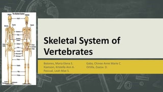
Skeletal System of
Vertebrates
Botones, Maria Elena S. Gaba, Chinee Anne Marie C.
Kiamzon, Kristelle Ann A. Ortilla, Zsazsa D.
Pascual, Leah Mae S.
SKELETAL SYSTEM
Skeletal System
 Most important organ system in the study of
vertebrate morphology.
- Provides reliable information about the
specific adaptations of vertebrates such as
posture and locomotor adaptations
- Tells about other organ systems
- Due to its hardness and durability, skeleton
becomes fossilized and the study of past
vertebrate life is mostly based on fossils
Functions
 Give shape to the body and support its
weight
 Offers a system of levers that aid
muscles to produce contraction
 Protects soft parts such as nerves, blood
vessels and other viscera
 Two types of skeleton
 Exoskeleton (protective structure on
the outside of the body)
 Endoskeleton (protective structure
on the inside of the body)
Endoskeleton
Exoskeleton
Functions
 Two separate components
 Cranial skeleton or the skull
 Postcranial skeleton (axial and
appendicular)
 Composed of mineralized connective
tissues ( dentin, enamel, cartilage and
mostly bones)
-mesenchyme differentiated into
scleroblasts which give rise to
osteoblasts, odontoblasts,
chondroblast and ameloblast.
Cartilage
- Forms an important part of the endoskeleton in all
vertebrates
- Cartilage is avascular tissue,
- Other types include the elastic cartilage in the external ear
and epiglottis, and fibrous cartilage in the intervertebral discs
and attachment of tendons and ligaments.
- Cartilage is a tough, elastic, fibrous connective tissue found in
various parts of the body, such as the joints, outer ear, and
larynx. A major constituent of the embryonic and young
vertebrate skeleton, it is converted largely to bone with
maturation.
Bones
 Comprise most of the endoskeleton in
higher vertebrates
 Contrary to cartilage, blood vessels and
nerves are present in bony tissues
passing through small Haversian canals.
 Haversian canals are located in the bone
tissue at the center of the compact bone
through which blood vessels, nerve
fibres and lymph vessels pass.
Bones
 Organic component is primarily collagen, which gives
bone great tensile strength
 Inorganic components of bone comprise 60% of the dry
weight
 Functions:
- Support and movement via attachments for soft tissue
and muscle
- Protects vital organs
- Major site for red marrow for production of blood cells
- Plays a role in metabolism of minerals such as calcium
and phosphorus
Bones
2 basic structural types
based on bone tissue
- Compact bone
- Spongy bone or
cancellous bone
Periosteum – dense layer
of connective tissue that
surrounds the bone
As to kind of bone tissue
Cranial Skeleton
Skeletal framework
of the head (skull)
3 embryonic
components:
1. Chondrocranium
2. Dermatocranium
3. Splanchnocranium
Chondrocranium
Composed of cartilage which contributes
to the base of the skull, includes the
sense capsule and in most vertebrates, is
replaced by bone
Primary brain case
Surrounds the brain and the special
sense organs
Chondrocranium Ossification Centers
 Occipital centers - cartilage surrounding
the foramen magnum may be replaced by
as many as four bones: one basioccipital, two
exoccipital, and one supraoccipital
 The six centers that we can see on diagram are:
 - Basioccipital bone;
 - Exoccipital bone (two centers);
 - Supraoccipital bone;
 - Interparietal bone (two centers).
Sphenoid centers from
basisphenoid bone,
presphenoid bone, and the
side walls above
basisphenoid and
presphenoid form
orbitosphenoid,
pleurosphenoid and
alisphenoid.
Ethmoid centers tend to remain
cartilaginous and form cribiform plate of
ethmoid and several ethmoturbinal bones
Otic centers – the cartilaginous otic
capsule is replaced in lower vertebrates by
several bones: prootic, opithotic, epiotic
Types of Skull Based on Fenestrae (Temporal Openings)
1. Anapsid skull
- The primitive skull, has no temporal fenestra,
possessed by turtles and other primitive reptiles.
2. Diapsid skull
- The diapsid skull has two temporal fenestrae,
possessed by most members of diapsida including
crocodiles, birds and lizards.
3. Euryapsid skull – this is a derived
diapsid
skull where the lower temporal
fenestra is lost
4. Synapsid skull – has one fenestra
located in
a different place than the euryapsid
skull
Dermatocranium
Composed of dermal bones that overlie the
chondrocranium and splanchnocranium
Forms the sides and roof of the skull
protecting the brain, it also forms most of
the bony lining of the roof of the mouth
and encases much of the
splanchnocranium
Completes the protective cover of the brain
and jaws
Parts of Dermatocranium
Modern fishes and amphibians have simple skull
and the number of dermal bones present is
reduced, some have tended to be lost or fused
In amniotes, dermal bones predominate, forming
most of the braincase and lower jaw; they are
divided into six series of bones.
Parts of Dermatocranium
1. Facial Series – encircles the external naris forming
the snout.
2. Orbital series – encircles the eye defining the orbit
3. Temporal series – lies behind the orbit completing
the posterior wall of the braincase
4. Vault series or roofing bones – located across the
top of skull covering the brain beneath
5. Palatal series – dermal bones of the primary palate
covering the roof of the mouth
6. Mandibular series – encases the Meckel’s cartilage
Splanchnocranium
 An ancient chordate structure associated with the filter feeding surfaces
 Arises from the neural crest cells departed from the sides of the neural tube and
migrate into the walls of the pharynx between successive pharyngeal slits
1. Meckel's Cartilage
2. Palatoquadrate
3. Rostrum
4. Labial Cartilage
5. Basihyal Cartilage
6. Ceratohyal Cartilage
7. Hyomandibular Cartilage
8. Ceratobranchial Cartilage
9. Basibranchial Cartilage
10. Hypobranchial Cartilage
11. A. Mandibular Arch
12. B. Hyoid Arch
13. C. Branchial Arch
Figure 2. Articulated chondrocranium and
splanchnocranium
Types of Jaw Attachments
1. Paleostylic – characteristic of Agnathans
- None of the arches attach directly to the skull
2. Euautostylic – the earliest jawed condition
- Found in Placoderms and Acanthodians
- The mandibular arch is suspended from the skull by itself without aid from the hyoid arch
3. Amphistylic – found in early sharks, some osteichthyians and crossopterygians
- Attached to the braincase through two primary articulations
- Anteriorly by a ligament connecting the palatoquadrate to the skull
- Posteriorly by the hyomandubula
4. Hyostylic – found in most modern bony fishes
- The mandibular arch is attached to the braincase primarily through the
hyomandibula with the aid of the sympletic bone
5. Metautostylic – found in most amphibians, reptiles and birds.
- Attached to the braincase directly through the quadrate bone
- Formed in the posterior part of the palatoquadrate
6. Craniostylic – found in mammals
- The entire upper jaw is a part of the braincase but the lower jaw called
dentary bone is suspended from the dermal squamosal bone of the
braincase
- The palatoquadrate and Meckel’s cartilages remain cartilaginous exceot at
their posterior ends which becomes the incus and malleus of the middle
ear respectively
Postcranial Skeleton
Axial
Appendicular
Function of body skeleton includes
- Protects the viscera
- Contributes to ventilation of the lungs
- Store for various minerals
- Provides rigidity to the body
- Provides series of firm and hinged segments needed for
locomotion in conjunction with the muscles
Axial Skeleton
Forms the main axis of the body
Composed of the notochord, vertebral
column, ribs, sternum and skull
Notochord
 The primitive axial skeleton,
replaced by the vertebral
column
 Unsegmented and composed
of dense fibrous connective
tissue
 The first skeletal element to
appear in the embryo of
chordates
Structure and Development of Vertebral Column
 The vertebral column is the
main axial support of vertebrates
 A vertebra is composed of a
centrum, one or two arches, and
various processes
 It protects the spinal cord and
provides rigidity to the body
Types of Vertebra Based on Centra
1. Aspondyly – no centra
2. Monospondyly – with only one centrum per
segment
- Stereospondyly – a monospondylous vertebra
in which the single centrum (intercentrum) is
separate
3. Diplospondyly – with two centra per segment
- Embolomerous – a diplospondylous vertebra in
which the approximate equal-sized centra are
separate
No centra
4. Polyspondyly – with five to six centra per segment
5. Aspidospondyly – the centra and spines are separate
- Rhachitomous – an aspidospondylous vertebra with
numerous separate parts that constitute each
vertebral segment
6. Holospondyly – the centra and spines are fused into
a single bone
-Lepospondyly - a holospondylous vertebra with a
husk-shaped centrum usually pierced by a
notochordal canal.
Figure 2.33. Comparison of vertebrae of primitive
tetrapods and modern amniotes. The rachitomous type
(shown also in cross section, X.S.) occurred in
crossopterygians and in the earliest amphibians. B is from
a labyrinthodont in the reptile line. B1 and B2 are from
other labyrinthodonts. Whether the modern amphibian
centrum represents a hypocentrum (diagonal lines) or a
pleurocentrum (stippled) is not certain. The unmarked part
of the vertebra is the neural arch. Adapted, with
permission, from Kent, G. C. 4th ed. Comparative
anatomy of the vertebrates. St. Louis: C. V. Mosby Co.;
1978. [134]
Types of Centra Based on Shapes
1. Amphicoelous
2. Procoelous
3. Opisthocoelous
4. Heterocoelous
5. Acoelous
Structure and Function of Ribs
 Series of cartilaginous or elongated bony
structures served as attachment for the
vertebrae extending into the body wall
- Provide sites for secure muscle
attachment and help suspend the body
- Form a protective case (rib cage) around
viscera
- In Amniotes, contributes to the breathing
mechanism
Types of Ribs
1. True ribs – meet ventrally with the
sternum, consist of two jointed segments
 Vertebral or costal rib (proximal segment)
 Sternal rib (distal segment)
 Joint between costal and sternal ribs allows
changes in chest shape during respiration
2. False ribs – articulate with each other but
not with the sternum
3. Floating ribs – do not articulate ventrally
Structure and Function of Sternum
 A midventral skeletal element that usually articulates with the more anterior
thoracic ribs and with the pectoral girdle
 Strictly a tetrapod structure and primarily, and amniote characteristic
- Strengthen the anterior part of the trunk and body wall
- Helps protect the thoracic viscera
- Accommodates muscles of the pectoral limbs
- In amniotes, helps in ventilating the lungs
 The sternum forms either paired or midventral primodia that are regarded as
new structures not derived from the pectoral girdle or ribs
Appendicular Skeleton
Structure and Evolution of Median Fins
Occur in all jawless vertebrates and
fishes:
 Dorsal fins - located along the
middorsal line.
Anal fins - located between anus and
tail
Caudal Fin
DORSAL and ANAL FINS
o Prevent the body from turning around the
vertical axis (yawing) and around the longitudinal
axis (rolling).
o In primitive vertebrates, each fin is supported
within the contour of the body by a series of rod-
like radials or pterygiophores.
o The exposed membrane of fins of CEPHALASPIDS
and some PLACODERMS are supported only by
dorsal scales.
CAUDAL FIN
Classified into four types depending on size and shape of the spine.
1. Diphycercal – if the spine is straight to the tip of the tail
with equal dorsal and ventral lobe of the tail. (ex.
Cyclostomes, pleuracanths, and some sarcopterygians)
2. Hypocercal – if the spine tilts downward with longer
ventral lobe than dorsal lobe.(ex.anaspids)
3. Heterocercal – if the spine tilts upward with longer dorsal
lobe than ventral lobe.(ex.cephalaspids, placoderms, most
chondrichthyes, and primitive osteichthyes)
4. Homocercal - if all the fin membrane is posterior to the
spine with equal dorsal and ventral lobe.(ex.all teleosts)
Structures and Evolution of Girdles
Girdles of fishes
o the pectoral girdle is older, larger and more complicated
than pelvic girdle.
-It includes one or more cartilage or replacement
bones and several dermal bones derived from ancestral
scales and armour plates.
o Placoderms cartilaginous fins was related to overlying
plates of dermal skeleton.
o Cartiliginous fishes has no dermal elements
Scapulocoracoid – the right and left halves fused in
the midline forming a U-shaped girdle
1.Ceratotrichia
2. Scapulocoracoid Bar
3. Propterygium
4. Mesopterygium
5. Metapterygium
A. Basal Pterygiophores
Girdle and Tetrapod
o BIRDS have a bladelike scapula that
is oriented parallel to the spine.
- with large anterior coracoid
that is articulated with the sternum
- the posterior coracoid has
been lost
- two clavicles fuse ventrally
forming the furcula or absent in
some birds.
Girdle and Tetrapod
o the only membrane bone retained Therian
Mammals is the clavicle
- The anterior coracoid is completely lost.
- the posterior coracoid fuses to the scapula
forming the coracoid process of the scapula
- the scapula is unique in having spine which
represents its anterior border
- the ventral end of the spine is continued as
the acromion process to articulate with the clavicle.
Girdle and Tetrapod
o the pelvic girdle of Tetrapods is much enlarged over that of fishes and
is relatively uniform in basic structure.
-each half of the pelvic girdle is a single cartilaginous unit in the embryo.
-three bones are constant in the adult:
 a dorsal ilium, which articulates with one or more sacral vertebrae
 an anterior pubis
A posterior ischium
-the bones of one side usually fuse in the adult forming the innominate
bone
-one or both of the ventral bones of the two sides usually articulates of
fuse across the midventral line, the contact is called pelvic symphysis
Girdle and tetrapod
 Primitive amphibians had a solid,
triangular shaped pelvic girdle with the
ilium forming the apex
- the pubis can be distinguished
from the ischium by having a obturator
foramen that accommodates a nerve.
 In FROG, the girdle has a long, anteriorly
inclined ilium and cartilaginous pubis.
Girdle and Tetrapod
 REPTILES has various shapes patterned after
the basic plan of LABYRINTHODONTS
-the contact with the spine is firmer
-the large pubo-ischiadic fenestrum is present
between the two ventral bones
 Birds have a large pelvic girdle that is firmly
attached to the synsacrum
-the long ilium extends both anterior and
posterior to the socket for the femur or
acetabulum: The pubis is turned backward below
the ischium and there is no symphysis
Girdle and Tetrapod
 Mammals have a long and expanded ilium
extending only forward from the
acetabulum
- the large obturator fenestrum
represents both the obturator foramen and
the pbo-isichiadic fenestrum of the ancestor.
- a symphysis is always present
- MONOTREMES and MARSUPIALS have
epipubic bones that articulate with the
pubic bones extending forward in the
ventral body wall.
MISCELLANEOUS BONES
 SESAMOID BONE – bones embedded in or interrupting
tendon
 the largest is patella or knee cap
 Baculum (os penis) – bone in the penis of carnivores,
bats, insectivores, rodents, and some primates
 Additional small bones are found in the different
structures among TETRAPODS:
 in the eyelids of CROCODILIANS
 in the crest of a BIRD
 in the snout of PIGS
 at the base of the external ear of some RODENTS Baculum of a dog’s penis
TYPES of LOCOMOTION IN MAMMALS
 posterior limbs provide rapid acceleration and often support the greater
part of the weight.
 Types of locomotion
used by Tetrapods:
- GRAVIPORTAL
- CURSORIAL
- VOLANT
- AERIAL
- SALTATORIAL
TYPES of LOCOMOTION in MAMMALS
- AQUATIC
- FOSSORIAL
- SCANSORIAL
- ARBOREAL
Comparison of Vertebrates
Vertebrates Type of Skull Type of Jaw Attachment
Fishes Euryapsid Paleostylic/Hyostylic/
Amphistylic
Amphibians Anapsid Metautostylic
Reptiles Anapsid Metautostylic
Birds Diapsid Metautostylic
Mammals Synapsid Craniostylic
Type of Skull and Jaw Attachment
Girdles and Fins/Limbs
Dorsal and Caudal
Short/long bones
Hindlimbs are larger than forelimbs
Slightly broader and segmented
Uniform and specialized limb structure
Centra and Vertebral Column
Vertebrates Types of Centra based on
shape
Regions of Vertebral Column
Fishes Amphicoelus Anterior and Posterior (2)
Amphibians Amphicoelus, Procoelus or
Opisthocoelous
there is little or no regional specialization
of the vertebral column
Reptiles Procoelus 4 or 5 distinct region
Birds Heterocoelus consists of vertebrae, and is divided into
three sections: cervical (11-25) (neck),
Synsacrum (fused vertebrae of the back,
also fused to the hips (pelvis)), and
pygostyle (tail).
Mammals Acocoelus 5 distinct regions: Cervical, Thoracic,
Lumbar, Sacral and Caudal
Trunk and tail
Ribs and Sternum
Vertebrates Ribs Sternum
Fishes Dorsal and ventral set Absent
Amphibians Dorsal andventral set Some
(In early amphibians it is absent
xiphisternum; Anurans
omosternum)
Reptiles Cervical ribs Some
Birds Uncinate processes (are
extensions of bone that project
caudally from the vertical
segment of each rib)
Present
(Large:Carina)
Mammals only have distinct ribs on the
thoracic vertebra, although
fixed cervical ribs are also
Present
(sternebrae: modified into
manubrium and xiphisternum)
Most do not have ribs
Flight muscle
Girdles and Fins/Limbs
Vertebrates Girdles Fins/Limbs
Cartilaginous Fishes Large pectoral girdle Fins
Bony Fishes Increased girdle Fins
Amphibians Long girdle Limbs
Reptiles Various shapes Limbs
Birds Large pelvic girdle Limbs
Mammals Long and expanded Limbs

Weitere ähnliche Inhalte

Was ist angesagt?

Comparative Anatomy - Integumentary System
Comparative Anatomy - Integumentary SystemComparative Anatomy - Integumentary System
Comparative Anatomy - Integumentary SystemEmsi Onairpic
 
Phylum chordata - zoology
Phylum chordata -  zoologyPhylum chordata -  zoology
Phylum chordata - zoologySijo A
 
Respiration in amphibians and reptiles
Respiration in amphibians and reptilesRespiration in amphibians and reptiles
Respiration in amphibians and reptilesDinDin Horneja
 
Classification of aves
Classification of avesClassification of aves
Classification of avesHabibur Rahman
 
Receptor organs in vertebrates
Receptor organs in vertebratesReceptor organs in vertebrates
Receptor organs in vertebratessunandakumre
 
Flight adaptations in birds
Flight adaptations in birdsFlight adaptations in birds
Flight adaptations in birdsGauri Haval
 
Comparative Anatomy - Nervous System
Comparative Anatomy - Nervous SystemComparative Anatomy - Nervous System
Comparative Anatomy - Nervous SystemEmsi Onairpic
 
Comparative Anatomy of the Limb
Comparative Anatomy of the LimbComparative Anatomy of the Limb
Comparative Anatomy of the LimbRayhan Shahrear
 
Comparative anatomy respiratory system
Comparative anatomy  respiratory systemComparative anatomy  respiratory system
Comparative anatomy respiratory systemMaria Isabel
 
Flight adaptations in birds
Flight adaptations in birdsFlight adaptations in birds
Flight adaptations in birdsnusratperween2
 
Comparative Anatomy - Respiratory System
Comparative Anatomy - Respiratory SystemComparative Anatomy - Respiratory System
Comparative Anatomy - Respiratory SystemEmsi Onairpic
 
Comparative account of axial and appendicular skeleton of amniots
Comparative account of axial and appendicular skeleton of amniotsComparative account of axial and appendicular skeleton of amniots
Comparative account of axial and appendicular skeleton of amniotsHabibur Rahman
 
Comparative anatomy (skeletal system)
Comparative anatomy (skeletal system)Comparative anatomy (skeletal system)
Comparative anatomy (skeletal system)Omer Rasool
 
Respiratory system in mammals
Respiratory system  in mammalsRespiratory system  in mammals
Respiratory system in mammalsDinDin Horneja
 

Was ist angesagt? (20)

Comparative Anatomy - Integumentary System
Comparative Anatomy - Integumentary SystemComparative Anatomy - Integumentary System
Comparative Anatomy - Integumentary System
 
Phylum chordata - zoology
Phylum chordata -  zoologyPhylum chordata -  zoology
Phylum chordata - zoology
 
Respiration in amphibians and reptiles
Respiration in amphibians and reptilesRespiration in amphibians and reptiles
Respiration in amphibians and reptiles
 
Classification of aves
Classification of avesClassification of aves
Classification of aves
 
Receptor organs in vertebrates
Receptor organs in vertebratesReceptor organs in vertebrates
Receptor organs in vertebrates
 
Urino genitial system of vertebrates
Urino genitial system of vertebratesUrino genitial system of vertebrates
Urino genitial system of vertebrates
 
Flight adaptations in birds
Flight adaptations in birdsFlight adaptations in birds
Flight adaptations in birds
 
Origin of chordata
Origin of chordataOrigin of chordata
Origin of chordata
 
Vertebrate respiratory system
Vertebrate respiratory systemVertebrate respiratory system
Vertebrate respiratory system
 
Comparative Anatomy - Nervous System
Comparative Anatomy - Nervous SystemComparative Anatomy - Nervous System
Comparative Anatomy - Nervous System
 
Comparative Anatomy of the Limb
Comparative Anatomy of the LimbComparative Anatomy of the Limb
Comparative Anatomy of the Limb
 
Comparative anatomy respiratory system
Comparative anatomy  respiratory systemComparative anatomy  respiratory system
Comparative anatomy respiratory system
 
Flight adaptations in birds
Flight adaptations in birdsFlight adaptations in birds
Flight adaptations in birds
 
Avian respiration
Avian respirationAvian respiration
Avian respiration
 
Comparative Anatomy - Respiratory System
Comparative Anatomy - Respiratory SystemComparative Anatomy - Respiratory System
Comparative Anatomy - Respiratory System
 
Jaw suspension in vertebrates [autosaved]
Jaw suspension in vertebrates [autosaved]Jaw suspension in vertebrates [autosaved]
Jaw suspension in vertebrates [autosaved]
 
Comparative account of axial and appendicular skeleton of amniots
Comparative account of axial and appendicular skeleton of amniotsComparative account of axial and appendicular skeleton of amniots
Comparative account of axial and appendicular skeleton of amniots
 
Comparative anatomy (skeletal system)
Comparative anatomy (skeletal system)Comparative anatomy (skeletal system)
Comparative anatomy (skeletal system)
 
Respiratory System
Respiratory SystemRespiratory System
Respiratory System
 
Respiratory system in mammals
Respiratory system  in mammalsRespiratory system  in mammals
Respiratory system in mammals
 

Andere mochten auch

Reptiles, Amphibians and fish ppt
Reptiles, Amphibians and fish pptReptiles, Amphibians and fish ppt
Reptiles, Amphibians and fish ppt123456mnbvcxz
 
Birds and mammals diagram
Birds and mammals diagramBirds and mammals diagram
Birds and mammals diagramandare2
 
VERTEBRATE COMPARATIVE ANATOMY OF MUSCULAR SYSTEM
VERTEBRATE COMPARATIVE ANATOMY OF MUSCULAR SYSTEMVERTEBRATE COMPARATIVE ANATOMY OF MUSCULAR SYSTEM
VERTEBRATE COMPARATIVE ANATOMY OF MUSCULAR SYSTEMWasiu Adeseji
 
Comparative anatomy integumentary system
Comparative anatomy integumentary systemComparative anatomy integumentary system
Comparative anatomy integumentary systemOmer Rasool
 
Activity 2 - Histology and Integument
Activity 2 - Histology and IntegumentActivity 2 - Histology and Integument
Activity 2 - Histology and Integumentmeleebirdsong
 

Andere mochten auch (6)

Reptiles, Amphibians and fish ppt
Reptiles, Amphibians and fish pptReptiles, Amphibians and fish ppt
Reptiles, Amphibians and fish ppt
 
Birds and mammals diagram
Birds and mammals diagramBirds and mammals diagram
Birds and mammals diagram
 
VERTEBRATE COMPARATIVE ANATOMY OF MUSCULAR SYSTEM
VERTEBRATE COMPARATIVE ANATOMY OF MUSCULAR SYSTEMVERTEBRATE COMPARATIVE ANATOMY OF MUSCULAR SYSTEM
VERTEBRATE COMPARATIVE ANATOMY OF MUSCULAR SYSTEM
 
Comparative anatomy integumentary system
Comparative anatomy integumentary systemComparative anatomy integumentary system
Comparative anatomy integumentary system
 
Activity 2 - Histology and Integument
Activity 2 - Histology and IntegumentActivity 2 - Histology and Integument
Activity 2 - Histology and Integument
 
Zoo 2
Zoo 2Zoo 2
Zoo 2
 

Ähnlich wie Comparative Anatomy - Skeletal System

Skeletal system
Skeletal systemSkeletal system
Skeletal systemkiesha26
 
The Skeletal Sytem
The Skeletal SytemThe Skeletal Sytem
The Skeletal SytemDoc Lorie B
 
Ms T.J Mkhize support in animal and plant
 Ms T.J Mkhize support in animal and plant Ms T.J Mkhize support in animal and plant
Ms T.J Mkhize support in animal and plantThokozile Mkhize
 
13 premedical motor_mechanism
13 premedical motor_mechanism13 premedical motor_mechanism
13 premedical motor_mechanismMesut Deniz
 
Osseous System Part I.pptx
Osseous System Part I.pptxOsseous System Part I.pptx
Osseous System Part I.pptxSwatiingle7
 
Hap osseous system
Hap osseous systemHap osseous system
Hap osseous systemKIRTI GUPTA
 
Bone/orthodontic courses by Indian dental academy
Bone/orthodontic courses by Indian dental academyBone/orthodontic courses by Indian dental academy
Bone/orthodontic courses by Indian dental academyIndian dental academy
 
General osteology
General osteologyGeneral osteology
General osteologyJamil Anwar
 
Skeletal system_presentation.pptx
Skeletal system_presentation.pptxSkeletal system_presentation.pptx
Skeletal system_presentation.pptxAchame
 
Skeletal system.pptx
Skeletal system.pptxSkeletal system.pptx
Skeletal system.pptxKeyaArere
 

Ähnlich wie Comparative Anatomy - Skeletal System (20)

Sistem rangka
Sistem rangkaSistem rangka
Sistem rangka
 
Ana-physi 4.pptx
Ana-physi 4.pptxAna-physi 4.pptx
Ana-physi 4.pptx
 
Skeletal system
Skeletal systemSkeletal system
Skeletal system
 
bone
bonebone
bone
 
Skeletal system
Skeletal  system Skeletal  system
Skeletal system
 
The Skeletal Sytem
The Skeletal SytemThe Skeletal Sytem
The Skeletal Sytem
 
Lp 5 bones 2008 no path
Lp 5 bones 2008 no pathLp 5 bones 2008 no path
Lp 5 bones 2008 no path
 
Ms T.J Mkhize support in animal and plant
 Ms T.J Mkhize support in animal and plant Ms T.J Mkhize support in animal and plant
Ms T.J Mkhize support in animal and plant
 
13 premedical motor_mechanism
13 premedical motor_mechanism13 premedical motor_mechanism
13 premedical motor_mechanism
 
skeletal system
skeletal systemskeletal system
skeletal system
 
Skeletal system
Skeletal systemSkeletal system
Skeletal system
 
Osseous System Part I.pptx
Osseous System Part I.pptxOsseous System Part I.pptx
Osseous System Part I.pptx
 
Bone/ dental implant courses
Bone/ dental implant coursesBone/ dental implant courses
Bone/ dental implant courses
 
Hap osseous system
Hap osseous systemHap osseous system
Hap osseous system
 
Bone/orthodontic courses by Indian dental academy
Bone/orthodontic courses by Indian dental academyBone/orthodontic courses by Indian dental academy
Bone/orthodontic courses by Indian dental academy
 
General osteology
General osteologyGeneral osteology
General osteology
 
Joko re modified
Joko re modifiedJoko re modified
Joko re modified
 
Skeletal system_presentation.pptx
Skeletal system_presentation.pptxSkeletal system_presentation.pptx
Skeletal system_presentation.pptx
 
Axial skeleton
Axial skeletonAxial skeleton
Axial skeleton
 
Skeletal system.pptx
Skeletal system.pptxSkeletal system.pptx
Skeletal system.pptx
 

Mehr von Emsi Onairpic

Histology - Specialized Connective Tissues (BONE)
Histology - Specialized Connective Tissues (BONE)Histology - Specialized Connective Tissues (BONE)
Histology - Specialized Connective Tissues (BONE)Emsi Onairpic
 
Kingdom Plantae (Concept Map)
Kingdom Plantae (Concept Map)Kingdom Plantae (Concept Map)
Kingdom Plantae (Concept Map)Emsi Onairpic
 
General Zoology Lab - Activity 9 Echinodermata
General Zoology Lab - Activity 9 EchinodermataGeneral Zoology Lab - Activity 9 Echinodermata
General Zoology Lab - Activity 9 EchinodermataEmsi Onairpic
 
General Zoology Lab - Activity 8 Arthropoda
General Zoology Lab - Activity 8 ArthropodaGeneral Zoology Lab - Activity 8 Arthropoda
General Zoology Lab - Activity 8 ArthropodaEmsi Onairpic
 
General Zoology Lab - Activity 6 Mollusca
General Zoology Lab - Activity 6 MolluscaGeneral Zoology Lab - Activity 6 Mollusca
General Zoology Lab - Activity 6 MolluscaEmsi Onairpic
 
General Zoology Lab - Activity 5 Nematoda
General Zoology Lab - Activity 5 NematodaGeneral Zoology Lab - Activity 5 Nematoda
General Zoology Lab - Activity 5 NematodaEmsi Onairpic
 
General Zoology Lab - Activity 4 Platyhelminthes
General Zoology Lab - Activity 4 PlatyhelminthesGeneral Zoology Lab - Activity 4 Platyhelminthes
General Zoology Lab - Activity 4 PlatyhelminthesEmsi Onairpic
 
General Zoology Lab - Activity 3 Cnidarians
General Zoology Lab - Activity 3 CnidariansGeneral Zoology Lab - Activity 3 Cnidarians
General Zoology Lab - Activity 3 CnidariansEmsi Onairpic
 
10 Medicinal Plants Found in DLSU-D Botanical Garden
10 Medicinal Plants Found in DLSU-D Botanical Garden10 Medicinal Plants Found in DLSU-D Botanical Garden
10 Medicinal Plants Found in DLSU-D Botanical GardenEmsi Onairpic
 
Ethnobotany - Plant Taxonomy Chichirica (Catharanthus roseus)
Ethnobotany - Plant Taxonomy Chichirica (Catharanthus roseus)Ethnobotany - Plant Taxonomy Chichirica (Catharanthus roseus)
Ethnobotany - Plant Taxonomy Chichirica (Catharanthus roseus)Emsi Onairpic
 
Taxonomy of "Bahay Kubo" plants
Taxonomy of "Bahay Kubo" plantsTaxonomy of "Bahay Kubo" plants
Taxonomy of "Bahay Kubo" plantsEmsi Onairpic
 
Supporting/Connective Tissues Reviewer
Supporting/Connective Tissues ReviewerSupporting/Connective Tissues Reviewer
Supporting/Connective Tissues ReviewerEmsi Onairpic
 
Physics (Sounds, Waves & Optics) Laboratory Apparatus & Equipment
Physics (Sounds, Waves & Optics) Laboratory Apparatus & EquipmentPhysics (Sounds, Waves & Optics) Laboratory Apparatus & Equipment
Physics (Sounds, Waves & Optics) Laboratory Apparatus & EquipmentEmsi Onairpic
 
MEDICINAL PLANTS AND OTHER FORMS OF TRADITIONAL MEDICINE USED BY THE RESIDENT...
MEDICINAL PLANTS AND OTHER FORMS OF TRADITIONAL MEDICINE USED BY THE RESIDENT...MEDICINAL PLANTS AND OTHER FORMS OF TRADITIONAL MEDICINE USED BY THE RESIDENT...
MEDICINAL PLANTS AND OTHER FORMS OF TRADITIONAL MEDICINE USED BY THE RESIDENT...Emsi Onairpic
 
10 DOH Approved Herbal Plants in the Philippines
10 DOH Approved Herbal Plants in the Philippines10 DOH Approved Herbal Plants in the Philippines
10 DOH Approved Herbal Plants in the PhilippinesEmsi Onairpic
 
Comparative Anatomy - Reproductive System
Comparative Anatomy - Reproductive SystemComparative Anatomy - Reproductive System
Comparative Anatomy - Reproductive SystemEmsi Onairpic
 
Comparative Anatomy - Excretory System
Comparative Anatomy - Excretory SystemComparative Anatomy - Excretory System
Comparative Anatomy - Excretory SystemEmsi Onairpic
 
Comparative Anatomy - Muscular System
Comparative Anatomy - Muscular SystemComparative Anatomy - Muscular System
Comparative Anatomy - Muscular SystemEmsi Onairpic
 

Mehr von Emsi Onairpic (18)

Histology - Specialized Connective Tissues (BONE)
Histology - Specialized Connective Tissues (BONE)Histology - Specialized Connective Tissues (BONE)
Histology - Specialized Connective Tissues (BONE)
 
Kingdom Plantae (Concept Map)
Kingdom Plantae (Concept Map)Kingdom Plantae (Concept Map)
Kingdom Plantae (Concept Map)
 
General Zoology Lab - Activity 9 Echinodermata
General Zoology Lab - Activity 9 EchinodermataGeneral Zoology Lab - Activity 9 Echinodermata
General Zoology Lab - Activity 9 Echinodermata
 
General Zoology Lab - Activity 8 Arthropoda
General Zoology Lab - Activity 8 ArthropodaGeneral Zoology Lab - Activity 8 Arthropoda
General Zoology Lab - Activity 8 Arthropoda
 
General Zoology Lab - Activity 6 Mollusca
General Zoology Lab - Activity 6 MolluscaGeneral Zoology Lab - Activity 6 Mollusca
General Zoology Lab - Activity 6 Mollusca
 
General Zoology Lab - Activity 5 Nematoda
General Zoology Lab - Activity 5 NematodaGeneral Zoology Lab - Activity 5 Nematoda
General Zoology Lab - Activity 5 Nematoda
 
General Zoology Lab - Activity 4 Platyhelminthes
General Zoology Lab - Activity 4 PlatyhelminthesGeneral Zoology Lab - Activity 4 Platyhelminthes
General Zoology Lab - Activity 4 Platyhelminthes
 
General Zoology Lab - Activity 3 Cnidarians
General Zoology Lab - Activity 3 CnidariansGeneral Zoology Lab - Activity 3 Cnidarians
General Zoology Lab - Activity 3 Cnidarians
 
10 Medicinal Plants Found in DLSU-D Botanical Garden
10 Medicinal Plants Found in DLSU-D Botanical Garden10 Medicinal Plants Found in DLSU-D Botanical Garden
10 Medicinal Plants Found in DLSU-D Botanical Garden
 
Ethnobotany - Plant Taxonomy Chichirica (Catharanthus roseus)
Ethnobotany - Plant Taxonomy Chichirica (Catharanthus roseus)Ethnobotany - Plant Taxonomy Chichirica (Catharanthus roseus)
Ethnobotany - Plant Taxonomy Chichirica (Catharanthus roseus)
 
Taxonomy of "Bahay Kubo" plants
Taxonomy of "Bahay Kubo" plantsTaxonomy of "Bahay Kubo" plants
Taxonomy of "Bahay Kubo" plants
 
Supporting/Connective Tissues Reviewer
Supporting/Connective Tissues ReviewerSupporting/Connective Tissues Reviewer
Supporting/Connective Tissues Reviewer
 
Physics (Sounds, Waves & Optics) Laboratory Apparatus & Equipment
Physics (Sounds, Waves & Optics) Laboratory Apparatus & EquipmentPhysics (Sounds, Waves & Optics) Laboratory Apparatus & Equipment
Physics (Sounds, Waves & Optics) Laboratory Apparatus & Equipment
 
MEDICINAL PLANTS AND OTHER FORMS OF TRADITIONAL MEDICINE USED BY THE RESIDENT...
MEDICINAL PLANTS AND OTHER FORMS OF TRADITIONAL MEDICINE USED BY THE RESIDENT...MEDICINAL PLANTS AND OTHER FORMS OF TRADITIONAL MEDICINE USED BY THE RESIDENT...
MEDICINAL PLANTS AND OTHER FORMS OF TRADITIONAL MEDICINE USED BY THE RESIDENT...
 
10 DOH Approved Herbal Plants in the Philippines
10 DOH Approved Herbal Plants in the Philippines10 DOH Approved Herbal Plants in the Philippines
10 DOH Approved Herbal Plants in the Philippines
 
Comparative Anatomy - Reproductive System
Comparative Anatomy - Reproductive SystemComparative Anatomy - Reproductive System
Comparative Anatomy - Reproductive System
 
Comparative Anatomy - Excretory System
Comparative Anatomy - Excretory SystemComparative Anatomy - Excretory System
Comparative Anatomy - Excretory System
 
Comparative Anatomy - Muscular System
Comparative Anatomy - Muscular SystemComparative Anatomy - Muscular System
Comparative Anatomy - Muscular System
 

Kürzlich hochgeladen

STOPPED FLOW METHOD & APPLICATION MURUGAVENI B.pptx
STOPPED FLOW METHOD & APPLICATION MURUGAVENI B.pptxSTOPPED FLOW METHOD & APPLICATION MURUGAVENI B.pptx
STOPPED FLOW METHOD & APPLICATION MURUGAVENI B.pptxMurugaveni B
 
Pests of safflower_Binomics_Identification_Dr.UPR.pdf
Pests of safflower_Binomics_Identification_Dr.UPR.pdfPests of safflower_Binomics_Identification_Dr.UPR.pdf
Pests of safflower_Binomics_Identification_Dr.UPR.pdfPirithiRaju
 
Topic 9- General Principles of International Law.pptx
Topic 9- General Principles of International Law.pptxTopic 9- General Principles of International Law.pptx
Topic 9- General Principles of International Law.pptxJorenAcuavera1
 
GenBio2 - Lesson 1 - Introduction to Genetics.pptx
GenBio2 - Lesson 1 - Introduction to Genetics.pptxGenBio2 - Lesson 1 - Introduction to Genetics.pptx
GenBio2 - Lesson 1 - Introduction to Genetics.pptxBerniceCayabyab1
 
Pests of soyabean_Binomics_IdentificationDr.UPR.pdf
Pests of soyabean_Binomics_IdentificationDr.UPR.pdfPests of soyabean_Binomics_IdentificationDr.UPR.pdf
Pests of soyabean_Binomics_IdentificationDr.UPR.pdfPirithiRaju
 
Call Girls In Nihal Vihar Delhi ❤️8860477959 Looking Escorts In 24/7 Delhi NCR
Call Girls In Nihal Vihar Delhi ❤️8860477959 Looking Escorts In 24/7 Delhi NCRCall Girls In Nihal Vihar Delhi ❤️8860477959 Looking Escorts In 24/7 Delhi NCR
Call Girls In Nihal Vihar Delhi ❤️8860477959 Looking Escorts In 24/7 Delhi NCRlizamodels9
 
Best Call Girls In Sector 29 Gurgaon❤️8860477959 EscorTs Service In 24/7 Delh...
Best Call Girls In Sector 29 Gurgaon❤️8860477959 EscorTs Service In 24/7 Delh...Best Call Girls In Sector 29 Gurgaon❤️8860477959 EscorTs Service In 24/7 Delh...
Best Call Girls In Sector 29 Gurgaon❤️8860477959 EscorTs Service In 24/7 Delh...lizamodels9
 
Carbon Dioxide Capture and Storage (CSS)
Carbon Dioxide Capture and Storage (CSS)Carbon Dioxide Capture and Storage (CSS)
Carbon Dioxide Capture and Storage (CSS)Tamer Koksalan, PhD
 
OECD bibliometric indicators: Selected highlights, April 2024
OECD bibliometric indicators: Selected highlights, April 2024OECD bibliometric indicators: Selected highlights, April 2024
OECD bibliometric indicators: Selected highlights, April 2024innovationoecd
 
Citronella presentation SlideShare mani upadhyay
Citronella presentation SlideShare mani upadhyayCitronella presentation SlideShare mani upadhyay
Citronella presentation SlideShare mani upadhyayupadhyaymani499
 
Harmful and Useful Microorganisms Presentation
Harmful and Useful Microorganisms PresentationHarmful and Useful Microorganisms Presentation
Harmful and Useful Microorganisms Presentationtahreemzahra82
 
Neurodevelopmental disorders according to the dsm 5 tr
Neurodevelopmental disorders according to the dsm 5 trNeurodevelopmental disorders according to the dsm 5 tr
Neurodevelopmental disorders according to the dsm 5 trssuser06f238
 
Radiation physics in Dental Radiology...
Radiation physics in Dental Radiology...Radiation physics in Dental Radiology...
Radiation physics in Dental Radiology...navyadasi1992
 
Pests of Blackgram, greengram, cowpea_Dr.UPR.pdf
Pests of Blackgram, greengram, cowpea_Dr.UPR.pdfPests of Blackgram, greengram, cowpea_Dr.UPR.pdf
Pests of Blackgram, greengram, cowpea_Dr.UPR.pdfPirithiRaju
 
Speech, hearing, noise, intelligibility.pptx
Speech, hearing, noise, intelligibility.pptxSpeech, hearing, noise, intelligibility.pptx
Speech, hearing, noise, intelligibility.pptxpriyankatabhane
 
basic entomology with insect anatomy and taxonomy
basic entomology with insect anatomy and taxonomybasic entomology with insect anatomy and taxonomy
basic entomology with insect anatomy and taxonomyDrAnita Sharma
 
preservation, maintanence and improvement of industrial organism.pptx
preservation, maintanence and improvement of industrial organism.pptxpreservation, maintanence and improvement of industrial organism.pptx
preservation, maintanence and improvement of industrial organism.pptxnoordubaliya2003
 
User Guide: Pulsar™ Weather Station (Columbia Weather Systems)
User Guide: Pulsar™ Weather Station (Columbia Weather Systems)User Guide: Pulsar™ Weather Station (Columbia Weather Systems)
User Guide: Pulsar™ Weather Station (Columbia Weather Systems)Columbia Weather Systems
 

Kürzlich hochgeladen (20)

STOPPED FLOW METHOD & APPLICATION MURUGAVENI B.pptx
STOPPED FLOW METHOD & APPLICATION MURUGAVENI B.pptxSTOPPED FLOW METHOD & APPLICATION MURUGAVENI B.pptx
STOPPED FLOW METHOD & APPLICATION MURUGAVENI B.pptx
 
Pests of safflower_Binomics_Identification_Dr.UPR.pdf
Pests of safflower_Binomics_Identification_Dr.UPR.pdfPests of safflower_Binomics_Identification_Dr.UPR.pdf
Pests of safflower_Binomics_Identification_Dr.UPR.pdf
 
Topic 9- General Principles of International Law.pptx
Topic 9- General Principles of International Law.pptxTopic 9- General Principles of International Law.pptx
Topic 9- General Principles of International Law.pptx
 
GenBio2 - Lesson 1 - Introduction to Genetics.pptx
GenBio2 - Lesson 1 - Introduction to Genetics.pptxGenBio2 - Lesson 1 - Introduction to Genetics.pptx
GenBio2 - Lesson 1 - Introduction to Genetics.pptx
 
Pests of soyabean_Binomics_IdentificationDr.UPR.pdf
Pests of soyabean_Binomics_IdentificationDr.UPR.pdfPests of soyabean_Binomics_IdentificationDr.UPR.pdf
Pests of soyabean_Binomics_IdentificationDr.UPR.pdf
 
Call Girls In Nihal Vihar Delhi ❤️8860477959 Looking Escorts In 24/7 Delhi NCR
Call Girls In Nihal Vihar Delhi ❤️8860477959 Looking Escorts In 24/7 Delhi NCRCall Girls In Nihal Vihar Delhi ❤️8860477959 Looking Escorts In 24/7 Delhi NCR
Call Girls In Nihal Vihar Delhi ❤️8860477959 Looking Escorts In 24/7 Delhi NCR
 
Best Call Girls In Sector 29 Gurgaon❤️8860477959 EscorTs Service In 24/7 Delh...
Best Call Girls In Sector 29 Gurgaon❤️8860477959 EscorTs Service In 24/7 Delh...Best Call Girls In Sector 29 Gurgaon❤️8860477959 EscorTs Service In 24/7 Delh...
Best Call Girls In Sector 29 Gurgaon❤️8860477959 EscorTs Service In 24/7 Delh...
 
Carbon Dioxide Capture and Storage (CSS)
Carbon Dioxide Capture and Storage (CSS)Carbon Dioxide Capture and Storage (CSS)
Carbon Dioxide Capture and Storage (CSS)
 
OECD bibliometric indicators: Selected highlights, April 2024
OECD bibliometric indicators: Selected highlights, April 2024OECD bibliometric indicators: Selected highlights, April 2024
OECD bibliometric indicators: Selected highlights, April 2024
 
Hot Sexy call girls in Moti Nagar,🔝 9953056974 🔝 escort Service
Hot Sexy call girls in  Moti Nagar,🔝 9953056974 🔝 escort ServiceHot Sexy call girls in  Moti Nagar,🔝 9953056974 🔝 escort Service
Hot Sexy call girls in Moti Nagar,🔝 9953056974 🔝 escort Service
 
Citronella presentation SlideShare mani upadhyay
Citronella presentation SlideShare mani upadhyayCitronella presentation SlideShare mani upadhyay
Citronella presentation SlideShare mani upadhyay
 
Volatile Oils Pharmacognosy And Phytochemistry -I
Volatile Oils Pharmacognosy And Phytochemistry -IVolatile Oils Pharmacognosy And Phytochemistry -I
Volatile Oils Pharmacognosy And Phytochemistry -I
 
Harmful and Useful Microorganisms Presentation
Harmful and Useful Microorganisms PresentationHarmful and Useful Microorganisms Presentation
Harmful and Useful Microorganisms Presentation
 
Neurodevelopmental disorders according to the dsm 5 tr
Neurodevelopmental disorders according to the dsm 5 trNeurodevelopmental disorders according to the dsm 5 tr
Neurodevelopmental disorders according to the dsm 5 tr
 
Radiation physics in Dental Radiology...
Radiation physics in Dental Radiology...Radiation physics in Dental Radiology...
Radiation physics in Dental Radiology...
 
Pests of Blackgram, greengram, cowpea_Dr.UPR.pdf
Pests of Blackgram, greengram, cowpea_Dr.UPR.pdfPests of Blackgram, greengram, cowpea_Dr.UPR.pdf
Pests of Blackgram, greengram, cowpea_Dr.UPR.pdf
 
Speech, hearing, noise, intelligibility.pptx
Speech, hearing, noise, intelligibility.pptxSpeech, hearing, noise, intelligibility.pptx
Speech, hearing, noise, intelligibility.pptx
 
basic entomology with insect anatomy and taxonomy
basic entomology with insect anatomy and taxonomybasic entomology with insect anatomy and taxonomy
basic entomology with insect anatomy and taxonomy
 
preservation, maintanence and improvement of industrial organism.pptx
preservation, maintanence and improvement of industrial organism.pptxpreservation, maintanence and improvement of industrial organism.pptx
preservation, maintanence and improvement of industrial organism.pptx
 
User Guide: Pulsar™ Weather Station (Columbia Weather Systems)
User Guide: Pulsar™ Weather Station (Columbia Weather Systems)User Guide: Pulsar™ Weather Station (Columbia Weather Systems)
User Guide: Pulsar™ Weather Station (Columbia Weather Systems)
 

Comparative Anatomy - Skeletal System

  • 1. Skeletal System of Vertebrates Botones, Maria Elena S. Gaba, Chinee Anne Marie C. Kiamzon, Kristelle Ann A. Ortilla, Zsazsa D. Pascual, Leah Mae S.
  • 3. Skeletal System  Most important organ system in the study of vertebrate morphology. - Provides reliable information about the specific adaptations of vertebrates such as posture and locomotor adaptations - Tells about other organ systems - Due to its hardness and durability, skeleton becomes fossilized and the study of past vertebrate life is mostly based on fossils
  • 4. Functions  Give shape to the body and support its weight  Offers a system of levers that aid muscles to produce contraction  Protects soft parts such as nerves, blood vessels and other viscera  Two types of skeleton  Exoskeleton (protective structure on the outside of the body)  Endoskeleton (protective structure on the inside of the body)
  • 7. Functions  Two separate components  Cranial skeleton or the skull  Postcranial skeleton (axial and appendicular)  Composed of mineralized connective tissues ( dentin, enamel, cartilage and mostly bones) -mesenchyme differentiated into scleroblasts which give rise to osteoblasts, odontoblasts, chondroblast and ameloblast.
  • 8. Cartilage - Forms an important part of the endoskeleton in all vertebrates - Cartilage is avascular tissue, - Other types include the elastic cartilage in the external ear and epiglottis, and fibrous cartilage in the intervertebral discs and attachment of tendons and ligaments. - Cartilage is a tough, elastic, fibrous connective tissue found in various parts of the body, such as the joints, outer ear, and larynx. A major constituent of the embryonic and young vertebrate skeleton, it is converted largely to bone with maturation.
  • 9. Bones  Comprise most of the endoskeleton in higher vertebrates  Contrary to cartilage, blood vessels and nerves are present in bony tissues passing through small Haversian canals.  Haversian canals are located in the bone tissue at the center of the compact bone through which blood vessels, nerve fibres and lymph vessels pass.
  • 10. Bones  Organic component is primarily collagen, which gives bone great tensile strength  Inorganic components of bone comprise 60% of the dry weight  Functions: - Support and movement via attachments for soft tissue and muscle - Protects vital organs - Major site for red marrow for production of blood cells - Plays a role in metabolism of minerals such as calcium and phosphorus
  • 11. Bones 2 basic structural types based on bone tissue - Compact bone - Spongy bone or cancellous bone Periosteum – dense layer of connective tissue that surrounds the bone
  • 12. As to kind of bone tissue
  • 13. Cranial Skeleton Skeletal framework of the head (skull) 3 embryonic components: 1. Chondrocranium 2. Dermatocranium 3. Splanchnocranium
  • 14. Chondrocranium Composed of cartilage which contributes to the base of the skull, includes the sense capsule and in most vertebrates, is replaced by bone Primary brain case Surrounds the brain and the special sense organs
  • 15. Chondrocranium Ossification Centers  Occipital centers - cartilage surrounding the foramen magnum may be replaced by as many as four bones: one basioccipital, two exoccipital, and one supraoccipital  The six centers that we can see on diagram are:  - Basioccipital bone;  - Exoccipital bone (two centers);  - Supraoccipital bone;  - Interparietal bone (two centers).
  • 16. Sphenoid centers from basisphenoid bone, presphenoid bone, and the side walls above basisphenoid and presphenoid form orbitosphenoid, pleurosphenoid and alisphenoid.
  • 17. Ethmoid centers tend to remain cartilaginous and form cribiform plate of ethmoid and several ethmoturbinal bones Otic centers – the cartilaginous otic capsule is replaced in lower vertebrates by several bones: prootic, opithotic, epiotic
  • 18. Types of Skull Based on Fenestrae (Temporal Openings) 1. Anapsid skull - The primitive skull, has no temporal fenestra, possessed by turtles and other primitive reptiles. 2. Diapsid skull - The diapsid skull has two temporal fenestrae, possessed by most members of diapsida including crocodiles, birds and lizards.
  • 19. 3. Euryapsid skull – this is a derived diapsid skull where the lower temporal fenestra is lost 4. Synapsid skull – has one fenestra located in a different place than the euryapsid skull
  • 20. Dermatocranium Composed of dermal bones that overlie the chondrocranium and splanchnocranium Forms the sides and roof of the skull protecting the brain, it also forms most of the bony lining of the roof of the mouth and encases much of the splanchnocranium Completes the protective cover of the brain and jaws
  • 21. Parts of Dermatocranium Modern fishes and amphibians have simple skull and the number of dermal bones present is reduced, some have tended to be lost or fused In amniotes, dermal bones predominate, forming most of the braincase and lower jaw; they are divided into six series of bones.
  • 22. Parts of Dermatocranium 1. Facial Series – encircles the external naris forming the snout. 2. Orbital series – encircles the eye defining the orbit 3. Temporal series – lies behind the orbit completing the posterior wall of the braincase 4. Vault series or roofing bones – located across the top of skull covering the brain beneath 5. Palatal series – dermal bones of the primary palate covering the roof of the mouth 6. Mandibular series – encases the Meckel’s cartilage
  • 23.
  • 24. Splanchnocranium  An ancient chordate structure associated with the filter feeding surfaces  Arises from the neural crest cells departed from the sides of the neural tube and migrate into the walls of the pharynx between successive pharyngeal slits 1. Meckel's Cartilage 2. Palatoquadrate 3. Rostrum 4. Labial Cartilage 5. Basihyal Cartilage 6. Ceratohyal Cartilage 7. Hyomandibular Cartilage 8. Ceratobranchial Cartilage 9. Basibranchial Cartilage 10. Hypobranchial Cartilage 11. A. Mandibular Arch 12. B. Hyoid Arch 13. C. Branchial Arch Figure 2. Articulated chondrocranium and splanchnocranium
  • 25. Types of Jaw Attachments 1. Paleostylic – characteristic of Agnathans - None of the arches attach directly to the skull 2. Euautostylic – the earliest jawed condition - Found in Placoderms and Acanthodians - The mandibular arch is suspended from the skull by itself without aid from the hyoid arch 3. Amphistylic – found in early sharks, some osteichthyians and crossopterygians - Attached to the braincase through two primary articulations - Anteriorly by a ligament connecting the palatoquadrate to the skull - Posteriorly by the hyomandubula
  • 26. 4. Hyostylic – found in most modern bony fishes - The mandibular arch is attached to the braincase primarily through the hyomandibula with the aid of the sympletic bone 5. Metautostylic – found in most amphibians, reptiles and birds. - Attached to the braincase directly through the quadrate bone - Formed in the posterior part of the palatoquadrate
  • 27. 6. Craniostylic – found in mammals - The entire upper jaw is a part of the braincase but the lower jaw called dentary bone is suspended from the dermal squamosal bone of the braincase - The palatoquadrate and Meckel’s cartilages remain cartilaginous exceot at their posterior ends which becomes the incus and malleus of the middle ear respectively
  • 28. Postcranial Skeleton Axial Appendicular Function of body skeleton includes - Protects the viscera - Contributes to ventilation of the lungs - Store for various minerals - Provides rigidity to the body - Provides series of firm and hinged segments needed for locomotion in conjunction with the muscles
  • 29. Axial Skeleton Forms the main axis of the body Composed of the notochord, vertebral column, ribs, sternum and skull
  • 30. Notochord  The primitive axial skeleton, replaced by the vertebral column  Unsegmented and composed of dense fibrous connective tissue  The first skeletal element to appear in the embryo of chordates
  • 31. Structure and Development of Vertebral Column  The vertebral column is the main axial support of vertebrates  A vertebra is composed of a centrum, one or two arches, and various processes  It protects the spinal cord and provides rigidity to the body
  • 32. Types of Vertebra Based on Centra 1. Aspondyly – no centra 2. Monospondyly – with only one centrum per segment - Stereospondyly – a monospondylous vertebra in which the single centrum (intercentrum) is separate 3. Diplospondyly – with two centra per segment - Embolomerous – a diplospondylous vertebra in which the approximate equal-sized centra are separate No centra
  • 33. 4. Polyspondyly – with five to six centra per segment 5. Aspidospondyly – the centra and spines are separate - Rhachitomous – an aspidospondylous vertebra with numerous separate parts that constitute each vertebral segment 6. Holospondyly – the centra and spines are fused into a single bone -Lepospondyly - a holospondylous vertebra with a husk-shaped centrum usually pierced by a notochordal canal.
  • 34. Figure 2.33. Comparison of vertebrae of primitive tetrapods and modern amniotes. The rachitomous type (shown also in cross section, X.S.) occurred in crossopterygians and in the earliest amphibians. B is from a labyrinthodont in the reptile line. B1 and B2 are from other labyrinthodonts. Whether the modern amphibian centrum represents a hypocentrum (diagonal lines) or a pleurocentrum (stippled) is not certain. The unmarked part of the vertebra is the neural arch. Adapted, with permission, from Kent, G. C. 4th ed. Comparative anatomy of the vertebrates. St. Louis: C. V. Mosby Co.; 1978. [134]
  • 35. Types of Centra Based on Shapes 1. Amphicoelous 2. Procoelous 3. Opisthocoelous 4. Heterocoelous 5. Acoelous
  • 36. Structure and Function of Ribs  Series of cartilaginous or elongated bony structures served as attachment for the vertebrae extending into the body wall - Provide sites for secure muscle attachment and help suspend the body - Form a protective case (rib cage) around viscera - In Amniotes, contributes to the breathing mechanism
  • 37. Types of Ribs 1. True ribs – meet ventrally with the sternum, consist of two jointed segments  Vertebral or costal rib (proximal segment)  Sternal rib (distal segment)  Joint between costal and sternal ribs allows changes in chest shape during respiration 2. False ribs – articulate with each other but not with the sternum 3. Floating ribs – do not articulate ventrally
  • 38. Structure and Function of Sternum  A midventral skeletal element that usually articulates with the more anterior thoracic ribs and with the pectoral girdle  Strictly a tetrapod structure and primarily, and amniote characteristic - Strengthen the anterior part of the trunk and body wall - Helps protect the thoracic viscera - Accommodates muscles of the pectoral limbs - In amniotes, helps in ventilating the lungs  The sternum forms either paired or midventral primodia that are regarded as new structures not derived from the pectoral girdle or ribs
  • 40. Structure and Evolution of Median Fins Occur in all jawless vertebrates and fishes:  Dorsal fins - located along the middorsal line. Anal fins - located between anus and tail Caudal Fin
  • 41. DORSAL and ANAL FINS o Prevent the body from turning around the vertical axis (yawing) and around the longitudinal axis (rolling). o In primitive vertebrates, each fin is supported within the contour of the body by a series of rod- like radials or pterygiophores. o The exposed membrane of fins of CEPHALASPIDS and some PLACODERMS are supported only by dorsal scales.
  • 42. CAUDAL FIN Classified into four types depending on size and shape of the spine. 1. Diphycercal – if the spine is straight to the tip of the tail with equal dorsal and ventral lobe of the tail. (ex. Cyclostomes, pleuracanths, and some sarcopterygians) 2. Hypocercal – if the spine tilts downward with longer ventral lobe than dorsal lobe.(ex.anaspids) 3. Heterocercal – if the spine tilts upward with longer dorsal lobe than ventral lobe.(ex.cephalaspids, placoderms, most chondrichthyes, and primitive osteichthyes) 4. Homocercal - if all the fin membrane is posterior to the spine with equal dorsal and ventral lobe.(ex.all teleosts)
  • 43. Structures and Evolution of Girdles Girdles of fishes o the pectoral girdle is older, larger and more complicated than pelvic girdle. -It includes one or more cartilage or replacement bones and several dermal bones derived from ancestral scales and armour plates. o Placoderms cartilaginous fins was related to overlying plates of dermal skeleton. o Cartiliginous fishes has no dermal elements Scapulocoracoid – the right and left halves fused in the midline forming a U-shaped girdle 1.Ceratotrichia 2. Scapulocoracoid Bar 3. Propterygium 4. Mesopterygium 5. Metapterygium A. Basal Pterygiophores
  • 44. Girdle and Tetrapod o BIRDS have a bladelike scapula that is oriented parallel to the spine. - with large anterior coracoid that is articulated with the sternum - the posterior coracoid has been lost - two clavicles fuse ventrally forming the furcula or absent in some birds.
  • 45. Girdle and Tetrapod o the only membrane bone retained Therian Mammals is the clavicle - The anterior coracoid is completely lost. - the posterior coracoid fuses to the scapula forming the coracoid process of the scapula - the scapula is unique in having spine which represents its anterior border - the ventral end of the spine is continued as the acromion process to articulate with the clavicle.
  • 46. Girdle and Tetrapod o the pelvic girdle of Tetrapods is much enlarged over that of fishes and is relatively uniform in basic structure. -each half of the pelvic girdle is a single cartilaginous unit in the embryo. -three bones are constant in the adult:  a dorsal ilium, which articulates with one or more sacral vertebrae  an anterior pubis A posterior ischium -the bones of one side usually fuse in the adult forming the innominate bone -one or both of the ventral bones of the two sides usually articulates of fuse across the midventral line, the contact is called pelvic symphysis
  • 47. Girdle and tetrapod  Primitive amphibians had a solid, triangular shaped pelvic girdle with the ilium forming the apex - the pubis can be distinguished from the ischium by having a obturator foramen that accommodates a nerve.  In FROG, the girdle has a long, anteriorly inclined ilium and cartilaginous pubis.
  • 48. Girdle and Tetrapod  REPTILES has various shapes patterned after the basic plan of LABYRINTHODONTS -the contact with the spine is firmer -the large pubo-ischiadic fenestrum is present between the two ventral bones  Birds have a large pelvic girdle that is firmly attached to the synsacrum -the long ilium extends both anterior and posterior to the socket for the femur or acetabulum: The pubis is turned backward below the ischium and there is no symphysis
  • 49. Girdle and Tetrapod  Mammals have a long and expanded ilium extending only forward from the acetabulum - the large obturator fenestrum represents both the obturator foramen and the pbo-isichiadic fenestrum of the ancestor. - a symphysis is always present - MONOTREMES and MARSUPIALS have epipubic bones that articulate with the pubic bones extending forward in the ventral body wall.
  • 50. MISCELLANEOUS BONES  SESAMOID BONE – bones embedded in or interrupting tendon  the largest is patella or knee cap  Baculum (os penis) – bone in the penis of carnivores, bats, insectivores, rodents, and some primates  Additional small bones are found in the different structures among TETRAPODS:  in the eyelids of CROCODILIANS  in the crest of a BIRD  in the snout of PIGS  at the base of the external ear of some RODENTS Baculum of a dog’s penis
  • 51. TYPES of LOCOMOTION IN MAMMALS  posterior limbs provide rapid acceleration and often support the greater part of the weight.  Types of locomotion used by Tetrapods: - GRAVIPORTAL - CURSORIAL - VOLANT - AERIAL - SALTATORIAL
  • 52. TYPES of LOCOMOTION in MAMMALS - AQUATIC - FOSSORIAL - SCANSORIAL - ARBOREAL
  • 53. Comparison of Vertebrates Vertebrates Type of Skull Type of Jaw Attachment Fishes Euryapsid Paleostylic/Hyostylic/ Amphistylic Amphibians Anapsid Metautostylic Reptiles Anapsid Metautostylic Birds Diapsid Metautostylic Mammals Synapsid Craniostylic Type of Skull and Jaw Attachment
  • 54. Girdles and Fins/Limbs Dorsal and Caudal Short/long bones Hindlimbs are larger than forelimbs Slightly broader and segmented Uniform and specialized limb structure
  • 55. Centra and Vertebral Column Vertebrates Types of Centra based on shape Regions of Vertebral Column Fishes Amphicoelus Anterior and Posterior (2) Amphibians Amphicoelus, Procoelus or Opisthocoelous there is little or no regional specialization of the vertebral column Reptiles Procoelus 4 or 5 distinct region Birds Heterocoelus consists of vertebrae, and is divided into three sections: cervical (11-25) (neck), Synsacrum (fused vertebrae of the back, also fused to the hips (pelvis)), and pygostyle (tail). Mammals Acocoelus 5 distinct regions: Cervical, Thoracic, Lumbar, Sacral and Caudal Trunk and tail
  • 56. Ribs and Sternum Vertebrates Ribs Sternum Fishes Dorsal and ventral set Absent Amphibians Dorsal andventral set Some (In early amphibians it is absent xiphisternum; Anurans omosternum) Reptiles Cervical ribs Some Birds Uncinate processes (are extensions of bone that project caudally from the vertical segment of each rib) Present (Large:Carina) Mammals only have distinct ribs on the thoracic vertebra, although fixed cervical ribs are also Present (sternebrae: modified into manubrium and xiphisternum) Most do not have ribs Flight muscle
  • 57. Girdles and Fins/Limbs Vertebrates Girdles Fins/Limbs Cartilaginous Fishes Large pectoral girdle Fins Bony Fishes Increased girdle Fins Amphibians Long girdle Limbs Reptiles Various shapes Limbs Birds Large pelvic girdle Limbs Mammals Long and expanded Limbs