Placenta
•Als PPTX, PDF herunterladen•
48 gefällt mir•2,473 views

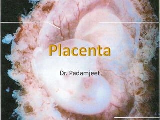
All eutherian mammals possess placenta. Human placenta is discoid, chorio-deciduate organ. Maternal and fetal tissue come in direct contact without rejection. It presents foetal and maternal surfaces and peripheral margins.

Empfohlen
Weitere ähnliche Inhalte
Was ist angesagt?
Was ist angesagt? (20)
Ähnlich wie Placenta
Ähnlich wie Placenta (20)
Kürzlich hochgeladen
Kürzlich hochgeladen (20)
Placenta
- 3. placenta • All eutherian mammals possess placenta. • Human placenta is discoid, chorio-deciduate organ. • Maternal and fetal tissue come in direct contact without rejection. • It presents foetal and maternal surfaces and peripheral margins. • Measurements – Diameter 15-20cm – Thickness 3cm (at center) – Weight 500gms. – Proprotional wt between placenta and foetus. • 1st month 6:1 • 4th month 1:1 • At birth 1:7 11/15/2017 dr.padam 3
- 4. placenta • The placenta is the primary site of nutrient and gas exchange between the mother and fetus. • The placenta is a fetomaternal organ that has two components: – A fetal part that develops from the chorionic sac – A maternal part that is derived from the endometrium 11/15/2017 dr.padam 4
- 5. placenta • It has two parts: – The fetal part of the placenta is formed by the villous chorion. The chorionic villi that arise from it project into the intervillous space containing maternal blood. – The maternal part of the placenta is formed by the decidua basalis, the part of the decidua related to the fetal component of the placenta. 11/15/2017 dr.padam 5
- 6. The Decidua • Decidua refers to the gravid endometrium, the functional layer of the endometrium in a pregnant woman that separates from the remainder of the uterus after parturition (childbirth). • The three regions of the decidua are named according to their relation to the implantation site – The decidua basalis is the part of the decidua deep to the conceptus that forms the maternal part of the placenta. – The decidua capsularis is the superficial part of the decidua overlying the conceptus. – The decidua parietalis is all the remaining parts of the decidua. 11/15/2017 dr.padam 6
- 7. Parts of Decidua 11/15/2017 dr.padam 7
- 8. Chorionic villi • It cover the entire chorionic sac until the beginning of the eighth week. • As this sac grows, the villi associated with the decidua capsularis are compressed, reducing the blood supply to them. These villi soon degenerate producing a relatively avascular bare area, the smooth chorion. • As these villi disappear, those associated with the decidua basalis rapidly increase in number, branch profusely, and enlarge. This bushy area of the chorionic sac is the villous chorion 11/15/2017 dr.padam 8
- 9. Maternal surface 15- 30 Polygonal area Peripheral margin Continuous With fetal membranes 11/15/2017 dr.padam 9
- 11. Structure of placenta • Chorionic plate (foetal side) • Basal plate(maternal side) • Stem villi extending between the plates • Intervilli spaces between the stem villi 11/15/2017 dr.padam 11
- 13. Chorionic plate • Composed of following structure from within outward( foetal to mother) – Primary mesoderm containing branches of umbilical vessels(foetal) – Cytotrophoblast – syncytiotrophoblast 11/15/2017 dr.padam 13
- 14. Basal plate • Consist of from outside inwards(mother to foetus) – Stratum spongiosum of decidua basalis – Outer layer of syncytiotrophoblast which undergoes fibrinoid degeneration (Nitabuch’s layer). – Outer shell of cytotrophoblast – Inner layer of syncytiotrophoblast; it also undergoes fibrinoid degeneration (Rohr’s stria). 11/15/2017 dr.padam 14
- 16. Constituents of Placental Membrane The placental membrane is made up of five layers. From the maternal side to fetal side these are: • Syncytiotrophoblast • Cytotrophoblast (up to 20 weeks) • Basement membrane of cytotrophoblast • Mesoderm in the core of villus • Endothelium and basement membrane of fetal capillaries. 11/15/2017 dr.padam 16
- 17. Placental circulation • The branch chorionic villi provide a large surface area for exchange across the very thin placental membrane ("barrier") interposed between the fetal and maternal circulations, consisting of extrafetal tissues. 11/15/2017 dr.padam 17
- 18. Fetal Placental Circulation • Poorly oxygenated blood leaves the fetus and passes through the umbilical arteries to the placenta. At the site of attachment of the umbilical cord to the placenta, these arteries divide into several radially disposed chorionic arteries that branch freely in the chorionic plate before entering the chorionic villi. The blood vessels form an extensive arteriocapillary-venous system within the chorionic villi. • There is normally no intermingling of fetal and maternal blood. The well-oxygenated fetal blood in the fetal capillaries passes into thin-walled veins that follow the chorionic arteries to the site of attachment of the umbilical cord. They converge here to form the umbilical vein. This large vessel carries oxygen-rich blood to the fetus. 11/15/2017 dr.padam 18
- 19. Maternal Placental Circulation • The blood flow from the spiral arteries is pulsatile and is propelled in the intervillous space and spurts toward the chorionic plate forming the "roof" of the intervillous space. • As the pressure dissipates, the blood flows slowly over the branch villi, allowing an exchange of metabolites and gases. • The blood eventually returns through the endometrial veins to the maternal circulation. • Reductions of uteroplacental circulation result in fetal hypoxia and intrauterine growth restriction (IUGR). • The intervillous space contains approximately 150 mL of blood that is replenished 3 to4 times per minute. • The intermittent contractions of the uterus during pregnancy decrease uteroplacental blood flow. 11/15/2017 dr.padam 19
- 22. According to the tissues forming the placental barrier, placenta may be classified phylogenetically as follows: (1) Epithelio-chorial (e. g. Pig) - Here endometrial epithelium remains intact, and the foetal and maternal tissues come in direct contact only. No part of the decidua is shed at full term. Hence, this type of placenta is called non- deciduate 11/15/2017 dr.padam 22
- 23. (2) Syndesmo-chorial (e. g: Bovine) Endometrial epithelium disappears, and the chorion is separated from the maternal blood by endometrial stroma and endothelium of maternal capillaries. 11/15/2017 dr.padam 23
- 24. (3) Endothelio-chorial (e. g. Dog) - Foetal chorion erodes the endometrial stroma upto the endothelium of the maternal vessels. 11/15/2017 dr.padam 24
- 25. • (4) Haemo-chorial (e. g. Man) - Here the endothelium of the maternal vessels disappears by the corrosive action of the chorion. Maternal blood directly comes in contact with the chorion and its villi 11/15/2017 dr.padam 25
- 26. (5) Haemo-endothelial (e.g. Rabbit) - this is one step more advanced in development than human placenta. The trophoblastic cells of the chorion degenerate to such an extent that only endotheliun of the foetal vessels intervenes between maternal and foetal blood. 11/15/2017 dr.padam 26
- 28. Placental macrophages (Hofbauer cells) are located close to trophoblast cells and fetal capillaries, which makes them ideal candidates for involvement in regulatory processes within the villous core. They have a role in production of various cytokines and prostaglandin (PG) synthesizing enzymes. 11/15/2017 dr.padam 28
- 29. Functions of the Placenta • The placenta has three main functions: – Metabolism (e.g., synthesis of glycogen) – Transport of gases and nutrients – Endocrine secretion (e.g., human chorionic gonadotropin hCG) 11/15/2017 dr.padam 29
- 33. Classification of Placenta : According to the attachment of the umbilical cord - (1) Battle-dore placenta, when the umbilical cord is attached close to the margin of placenta (2) Velamentous placenta, when the cord fails to reach the placenta and is attached to the foetal membrane close to the periphery of the organ. Battle-dore placenta Velamentous placenta 11/15/2017 dr.padam 33
- 34. According to the site of implantation: Placenta praevia - This condition takes place when the blastocyst is implanted in the lower part of the uterine cavity overlapping the internal os of the cervix. This produces serious haemorrhage before parturition. The placenta praevia may be central or marginal 11/15/2017 dr.padam 34
- 35. • Accessory placenta -. Sometimes an accessory lobe of placenta (Placenta succenturiata) is connected to the main mass by foetal membrane. • According to the degree of adhesion or penetration - (1) Placenta accreta, when it is adhered pathologically to the decidua basalis. (2) Placenta incerta, when it penetrates into the myometrium. (3) Placenta perceta, when it penetrates the entire uterine wall. 11/15/2017 dr.padam 35
- 36. • According to the shape- (1) Lobed placenta - It may exhibit two or more lobes. (2) Placenta membranacea - It is diffuse and thin, and the chorionic villi project from the entire blastocyst cavity. 11/15/2017 dr.padam 36 (3) Circumvallate - The peripheral margin of the placenta is surrounded by a sulcus and is overlapped by a circular fold of decidua. (4) fenestrated-
- 37. According to the distribution of the umbilical arteries - (1) Disperse type - The umbilical arteries divide in dichotomous manner and undergo successive reduction in calibre. (2) Magistral type- The arteries maintain almost a uniform calibre upto the periphery of the placenta and give off a number of smaller side branches. 11/15/2017 dr.padam 37