SlideShare ist ein Scribd-Unternehmen logo
1 von 2
Downloaden Sie, um offline zu lesen
ISSN 0975-8437                                        INTERNATIONAL JOURNAL OF DENTAL CLINICS 2011:3(1):115-116


  CASE REPORT

Kimura’s Disease: A case report
Shilpa.C.Patel, Vishal.R. Dave

Abstract
         Kimura’s disease is a rare inflammatory disorder of unknown cause, primarily seen in young
Asian males. The disease is characterized by a triad of painless subcutaneous masses in head and neck
region, blood and tissue eosinophilia and moderately elevated serum immunoglobulin E levels. Here, we
present a case report of this rare entity of unusual age presentation with classical histopathogical features.
Key Words: Kimura’s Disease; Eosinophilia; Parotid Gland; Lymphadenopathy
                              Received on: 22/09/2010 Accepted on: 06/12/2010

Introduction
          Kimura’s disease (KD) is a rare chronic           number. The histopathological feature of
angiolymphoid proliferative soft tissue disorder            excisional biopsy was matched with that of
of unknown origin.(1) It occurs most often in               incisional one. The patient was supplemented
young and middle aged asian male. It is most                with oral Dexamethasone for total duration of 6
common in head and neck region, with a                      weeks (0.5 mg twice daily for 1 week and then
predilection for preauricular area. Typical                 tapered over 5 weeks). The patient was
clinical presentations are painless subcutaneous            apparently normal without any local recurrence
masses, regional lymph node enlargement, blood              & systemic involvement within 6 month of
and tissue hyper eosinophilia, and markedly                 follow-up period.
elevated serum IgE levels.(1) According to the              Discussion
previous medical literatures, Kimura’s disease                        Kimura’s disease first described in1937
has a high recurrence rate so early & definitive            in Chinese literature by H.T.Kimm and C.Szeto
diagnosis of the disease is vital for effective             and they termed it as “eosinophilic hyperplastic
treatment plan.(1) Here we report a case of                 lymphogranuloma”.(2) The disease became
Kimura’s disease with clinico-pathological                  widely known as Kimura’s disease after Kimura
differential diagnosis.                                     and colleagues reported two cases of unusual
Case Report                                                 granulation combined with hyperplastic changes
          A 65 year old man reported to                     of lymphoid tissue.(1) The etiopathogenesis of
outpatient department with chief complaint of               KD remains unknown and it is considered
painless swelling on right side of face in parotid          nowadays as an allergic disease and it seems to
region since 4 months but it was painful on                 be a systemic immunological disorder.
mastication. Here we summarized details of                  Eosinophilia and increased serum IgE levels
present case in the following Table-1. With a               make KD be considered a CD4 (+) T helper 2
provisional diagnosis of salivary gland                     (Th2) allergic reactions. Th2 cells would produce
neoplasm, an incisional biopsy was performed.               interleukins (IL) IL-4, IL-5 and IL-13, which, in
The section stained with Haematoxylin & Eosin               turn, would act in B cells favoring the production
revealed unencapsulated ill-defined lesion with             of antigen-specific IgE. Th2 cell proliferation
lymphoid follicle formation, fibrosis and chronic           and the overexpression of cytokines would play
inflammatory cell infiltration with predominant             an essential role in the development of the
eosinophils (Figure 1a, 1b, 1c, 1d). Inter                  disease.(3)
follicular areas show increased number of                             It occurs predominantly as a unilateral
venules with endothelial cell proliferation. In             manifestation in the head and neck and it is
some areas lymphoid follicle were infiltrated by            frequently       associated     with        regional
eosinophils with resultant eosinophilic micro               lymphadenopathy with or without involvement
abscesses. With a diagnosis of Kimura’s disease,            of salivary glands.(4) In the present case, e
the patient was undergone superficial lobe                  presence unilateral parotid gland swelling with
parotidectomy with removal of multiple regional             regional lymphadenopathy led to the clinical
lymph nodes with vicinity.                                  diagnostic dilemma of other pathologic entities
          The excisional biopsy specimen                    affecting salivary glands & lymph nodes. The
received was firm nodular soft tissue measuring             differential diagnosis of Kimuras disease
approximately 4.2X2X1.5 cm3 and two in                      frequently     in    previous     literatures    are


©INTERNATIONAL JOURNAL OF DENTAL CLINICS VOLUME 3 ISSUE 1 JANUARY-MARCH 2011                                 115
ISSN 0975-8437                                                  INTERNATIONAL JOURNAL OF DENTAL CLINICS 2011:3(1):115-116


Lymphomas, salivary gland neoplasms, benign                              (AIL). Constant classical features of KD include
lymphoepithelial lesions (BLL / Mikulicz’s                               numerous       lymphoid      follicles,   mixed
disease), Angiolymphoid hyperplasia with                                 inflammatory infiltrate composed mainly of
eosinophilia (ALHE/ Epitheloid hemangioma)                               eosinophils and increased amount of post
and    angioimmunoblastic   lymphadenopathy                              capillary venules.(5)
Age/Sex    location           Physical findings        lymphadenopathy    Systemic involvement   Laboratory investigation
65/Male    Parotid (right)    3.5X3.5 cm firm,         Regional           No renal involvement   Eosinophilia (36%) Elevated
                              nontender & normal lymphadenopathy                                 erythrocyte sedimentation rate
                              overlying skin                                                     (ESR) Raised IgE levels
Table-1 clinical & investigation details of present case


Figure 1a. Lymphocytic infiltration with follicle formation and fibrosis .(4x), 1b. Well defined lymphoid follicle with
prominent germinal centre, (10x) 1c. Chronic Inflammatory cell infiltration with predominant eosinophils with
microabscesses.(40x), 1d. Interfollicular areas showing mixed inflammation with prominent eosinophils and
proliferation of venules.(40x), 1e. Interfollicular areas showing increased number of venules with endothelial cell
proliferation.(40x)
            Till date there is no definite treatment              Head, Department of Oral & Maxillofacial Pathology,
for KD is well established.(2) However the                        MGM Dental College & Hospital, Navi Mumbai, 2.
treatment plan should aim at preservation of vital                Dr. Vishal.R.Dave, M.S, Assistant Professor,
structures associated with the lesions & cosmetic                 Department of ENT, GCS Medical College,
                                                                  Ahmedabad, Gujarat, India.
rehabilitation, while preventing recurrences.                     References
Treatment options range from observation &                        1. Chung YG, Jee WH, Kang YK, Jung CK, Park
follow-up of mild & symptomatic cases to                                GS, Lee A, Bahk WJ, Cho HM, Park JW.
conservative surgical approach, medication &                            Kimura’s disease involving a long bone. Skeletal
radiotherapy in symptomatic & recurrent cases.                          Radiology 2010; 39(5):495-500.
If the lesion is primary, localized or present in                 2. Pitak-Arnnop P, Bellefqih S, Chaine A,
young age then surgical approach is more                                Dhanuthai K, Bertrand JC, Bertolus C. Head and
preferable. According to previous literature, only                      neck lesions of Kimura's disease: Exclusion of
surgical approach had high incidence of                                 human herpesvirus-8 and Epstein-Barr virus by in
                                                                        situ hybridisation and polymerase chain reaction.
recurrence.(1) If the lesion is recurrent with                          An immunohistochemical study. Journal of
systemic involvement, application of medication                         Cranio-Maxillofacial Surgery 2010; 38 (4):266-
like corticosteroid and immunosuppressive                               70.
agents have been shown to decrease size of the                    3. Briggs PL. Kimura disease is not angiolymphoid
lesion.(2) Irradiation should be considered in                          hyperplasia with eosinophilia: clinical and
patients resistant to the steroid or to prevent the                     pathological correlation with literature review
patient from deleterious effect of long term use                        and definition of diagnostic criteria. Anais
of steroid.(5)                                                          Brasileiros de Dermatologia 2006;81(2):167-73.
                                                                  4. Kung ITM, Gibson J, Bannatyne P. Kimura's
Conclusion
                                                                        Disease: A Clinico-Pathological Study Of 21
           In conclusion this paper draws attention                     Cases And Its Distinction From Angiolymphoid
on such a rare chronic inflammatory disease                             Hyperplasia           With          Eosinophils.
which mimics neoplastic conditions. This disease                        Pathology1984;16(1):39-44.
should be considered in differential diagnosis of                 5. Jani A, Coulson M. Kimura's disease. A typical
patients presented with head & neck mass and                            case of a rare disorder. Western Journal of
lymphadenopathy and investigated accordingly                            Medicine 1997; 166 (2):142-4.
as this disease has good prognosis. Knowledge                     Address for Correspondence
of signature features of Kimura’s disease put the                 Dr. Shilpa C Patel, M.D.S,
                                                                  801, Osho Kabir, Plot No 42, Sector 10,
physicians in a better position to evaluate its
                                                                  New Panvel, Maharashtra,India- 410206.
clinical outcome and optimal treatment regimen.                   Email: dr_shilpa0769@yahoo.com
Affiliations of Authors: 1. Dr. Shilpa.C.Patel,
M.D.S., Oral & Maxillofacial Pathology, Professor &
                           Source of Support: Nil,         Conflict of Interest: None Declared


©INTERNATIONAL JOURNAL OF DENTAL CLINICS VOLUME 3 ISSUE 1 JANUARY-MARCH 2011                                                  116

Weitere ähnliche Inhalte

Was ist angesagt?

Inflammatory effects of HLA-B27 as a Mechanism in Ankylosing Spondylitis
Inflammatory effects of HLA-B27 as a Mechanism in Ankylosing SpondylitisInflammatory effects of HLA-B27 as a Mechanism in Ankylosing Spondylitis
Inflammatory effects of HLA-B27 as a Mechanism in Ankylosing SpondylitisMeye1117
 
Infections of spine : Pyogenic and tuberculosis
Infections of spine : Pyogenic and tuberculosisInfections of spine : Pyogenic and tuberculosis
Infections of spine : Pyogenic and tuberculosisDr Shrikant Dhanani
 
General outline of musculoskeletal tuberculosis by dr ashutosh
General outline of musculoskeletal tuberculosis by dr ashutoshGeneral outline of musculoskeletal tuberculosis by dr ashutosh
General outline of musculoskeletal tuberculosis by dr ashutoshAshutosh Kumar
 
Acute transverse myelitis
Acute transverse myelitisAcute transverse myelitis
Acute transverse myelitisBakarAli3
 
Vertebral osteomyelitis
Vertebral osteomyelitisVertebral osteomyelitis
Vertebral osteomyelitisidchula
 
Discitis and osteomyelitis in Sickle cell disease
Discitis and osteomyelitis in Sickle cell diseaseDiscitis and osteomyelitis in Sickle cell disease
Discitis and osteomyelitis in Sickle cell diseaseJayanth Hiremagalur
 
Epidural abcess a case presentation
Epidural abcess a case presentationEpidural abcess a case presentation
Epidural abcess a case presentationRamy Mostafa
 
Spinal Epidural Abscess
Spinal Epidural Abscess Spinal Epidural Abscess
Spinal Epidural Abscess Ade Wijaya
 
Epidural abscess
Epidural abscessEpidural abscess
Epidural abscessyimsmart90
 
Analysis of lesion development pml
Analysis of lesion development pmlAnalysis of lesion development pml
Analysis of lesion development pmlClaudia Pfleger
 
Still’s Disease and Recurrent Complex Regional Pain Syndrome Type-I: The Firs...
Still’s Disease and Recurrent Complex Regional Pain Syndrome Type-I: The Firs...Still’s Disease and Recurrent Complex Regional Pain Syndrome Type-I: The Firs...
Still’s Disease and Recurrent Complex Regional Pain Syndrome Type-I: The Firs...Samantha Adcock
 
Surgical management of Tb spine
Surgical management of Tb spineSurgical management of Tb spine
Surgical management of Tb spineDrMdShafiulAlam
 
Other spine infections
Other spine infectionsOther spine infections
Other spine infectionsfarranajwa
 
Erythromelalgia: A New Password For Old Mystery
Erythromelalgia: A New Password For Old MysteryErythromelalgia: A New Password For Old Mystery
Erythromelalgia: A New Password For Old MysterySamir Mounir
 
tuberculosis of spine
tuberculosis of spinetuberculosis of spine
tuberculosis of spineHardik Pawar
 

Was ist angesagt? (20)

Inflammatory effects of HLA-B27 as a Mechanism in Ankylosing Spondylitis
Inflammatory effects of HLA-B27 as a Mechanism in Ankylosing SpondylitisInflammatory effects of HLA-B27 as a Mechanism in Ankylosing Spondylitis
Inflammatory effects of HLA-B27 as a Mechanism in Ankylosing Spondylitis
 
Infections of spine : Pyogenic and tuberculosis
Infections of spine : Pyogenic and tuberculosisInfections of spine : Pyogenic and tuberculosis
Infections of spine : Pyogenic and tuberculosis
 
Leri disease
Leri diseaseLeri disease
Leri disease
 
General outline of musculoskeletal tuberculosis by dr ashutosh
General outline of musculoskeletal tuberculosis by dr ashutoshGeneral outline of musculoskeletal tuberculosis by dr ashutosh
General outline of musculoskeletal tuberculosis by dr ashutosh
 
Acute transverse myelitis
Acute transverse myelitisAcute transverse myelitis
Acute transverse myelitis
 
Vertebral osteomyelitis
Vertebral osteomyelitisVertebral osteomyelitis
Vertebral osteomyelitis
 
Spondylodiscitis
SpondylodiscitisSpondylodiscitis
Spondylodiscitis
 
Discitis and osteomyelitis in Sickle cell disease
Discitis and osteomyelitis in Sickle cell diseaseDiscitis and osteomyelitis in Sickle cell disease
Discitis and osteomyelitis in Sickle cell disease
 
Epidural abcess a case presentation
Epidural abcess a case presentationEpidural abcess a case presentation
Epidural abcess a case presentation
 
Spinal Epidural Abscess
Spinal Epidural Abscess Spinal Epidural Abscess
Spinal Epidural Abscess
 
Epidural abscess
Epidural abscessEpidural abscess
Epidural abscess
 
Analysis of lesion development pml
Analysis of lesion development pmlAnalysis of lesion development pml
Analysis of lesion development pml
 
Still’s Disease and Recurrent Complex Regional Pain Syndrome Type-I: The Firs...
Still’s Disease and Recurrent Complex Regional Pain Syndrome Type-I: The Firs...Still’s Disease and Recurrent Complex Regional Pain Syndrome Type-I: The Firs...
Still’s Disease and Recurrent Complex Regional Pain Syndrome Type-I: The Firs...
 
Surgical management of Tb spine
Surgical management of Tb spineSurgical management of Tb spine
Surgical management of Tb spine
 
Other spine infections
Other spine infectionsOther spine infections
Other spine infections
 
Tb spine
Tb spineTb spine
Tb spine
 
Erythromelalgia: A New Password For Old Mystery
Erythromelalgia: A New Password For Old MysteryErythromelalgia: A New Password For Old Mystery
Erythromelalgia: A New Password For Old Mystery
 
Picture Medicine
Picture MedicinePicture Medicine
Picture Medicine
 
tuberculosis of spine
tuberculosis of spinetuberculosis of spine
tuberculosis of spine
 
Diskitis
DiskitisDiskitis
Diskitis
 

Andere mochten auch

Andere mochten auch (10)

Kikuchi fujimoto disease
Kikuchi fujimoto diseaseKikuchi fujimoto disease
Kikuchi fujimoto disease
 
Frontal sinus procedures
Frontal sinus proceduresFrontal sinus procedures
Frontal sinus procedures
 
Accustic neuroma 1
Accustic neuroma 1Accustic neuroma 1
Accustic neuroma 1
 
CP angle 360°
CP angle 360°CP angle 360°
CP angle 360°
 
Eosinophils in lymph node
Eosinophils in lymph nodeEosinophils in lymph node
Eosinophils in lymph node
 
surgical approaches to frontal sinus ppt
surgical approaches to frontal sinus pptsurgical approaches to frontal sinus ppt
surgical approaches to frontal sinus ppt
 
Acoustic neuroma
Acoustic neuromaAcoustic neuroma
Acoustic neuroma
 
Acoustic neuroma
Acoustic neuromaAcoustic neuroma
Acoustic neuroma
 
Acoustic neuroma
Acoustic neuromaAcoustic neuroma
Acoustic neuroma
 
Parotid salivary gland
Parotid salivary glandParotid salivary gland
Parotid salivary gland
 

Ähnlich wie Kimura’s disease a case report

50 1339
50 133950 1339
50 1339edcito
 
4th rd-template
4th rd-template4th rd-template
4th rd-templatemaha fahmy
 
HemangioEndotelioma epiteloide de la silla turca
HemangioEndotelioma epiteloide de la silla turcaHemangioEndotelioma epiteloide de la silla turca
HemangioEndotelioma epiteloide de la silla turcaCarlos Casallo
 
CHRONIC EOSINOPHILIC LEUKEMIA
CHRONIC EOSINOPHILIC LEUKEMIACHRONIC EOSINOPHILIC LEUKEMIA
CHRONIC EOSINOPHILIC LEUKEMIAnehaneemat
 
Atypical Presentation of Orbital Natural Killer cell Lymphoma | Crimson Publi...
Atypical Presentation of Orbital Natural Killer cell Lymphoma | Crimson Publi...Atypical Presentation of Orbital Natural Killer cell Lymphoma | Crimson Publi...
Atypical Presentation of Orbital Natural Killer cell Lymphoma | Crimson Publi...CrimsonpublishersMSOR
 
Crimson Publishers-Metastatic Atypical Fibroxanthoma: Case Report of an Uncom...
Crimson Publishers-Metastatic Atypical Fibroxanthoma: Case Report of an Uncom...Crimson Publishers-Metastatic Atypical Fibroxanthoma: Case Report of an Uncom...
Crimson Publishers-Metastatic Atypical Fibroxanthoma: Case Report of an Uncom...CromsonPublishersotolaryngology
 
Stevens-Johnson syndrome/ Toxic epidermal necrolysis emergency guidelines
Stevens-Johnson syndrome/ Toxic epidermal necrolysis emergency guidelinesStevens-Johnson syndrome/ Toxic epidermal necrolysis emergency guidelines
Stevens-Johnson syndrome/ Toxic epidermal necrolysis emergency guidelinesDaifallah Almansouri
 
Idiopathic Necrotizing Granulomas of the Thyroid: Report of a Rare Case
Idiopathic Necrotizing Granulomas of the Thyroid: Report of a Rare CaseIdiopathic Necrotizing Granulomas of the Thyroid: Report of a Rare Case
Idiopathic Necrotizing Granulomas of the Thyroid: Report of a Rare CaseCrimsonPublishersAICS
 
Fournier’s Gangrene in a 9 Yrs. Old Patient; A Rare Presentation in Paediatri...
Fournier’s Gangrene in a 9 Yrs. Old Patient; A Rare Presentation in Paediatri...Fournier’s Gangrene in a 9 Yrs. Old Patient; A Rare Presentation in Paediatri...
Fournier’s Gangrene in a 9 Yrs. Old Patient; A Rare Presentation in Paediatri...semualkaira
 
Sebaceous Carcinoma Masquerading As Chronic Blepharoconjunctivitis
Sebaceous Carcinoma Masquerading As Chronic BlepharoconjunctivitisSebaceous Carcinoma Masquerading As Chronic Blepharoconjunctivitis
Sebaceous Carcinoma Masquerading As Chronic BlepharoconjunctivitisNot Relevant
 
LEUKEMIA.pptx
LEUKEMIA.pptxLEUKEMIA.pptx
LEUKEMIA.pptxshiwani88
 

Ähnlich wie Kimura’s disease a case report (20)

Cervical lymphadenopathy
Cervical lymphadenopathyCervical lymphadenopathy
Cervical lymphadenopathy
 
50 1339
50 133950 1339
50 1339
 
Tropical
TropicalTropical
Tropical
 
4th rd-template
4th rd-template4th rd-template
4th rd-template
 
HemangioEndotelioma epiteloide de la silla turca
HemangioEndotelioma epiteloide de la silla turcaHemangioEndotelioma epiteloide de la silla turca
HemangioEndotelioma epiteloide de la silla turca
 
CHRONIC EOSINOPHILIC LEUKEMIA
CHRONIC EOSINOPHILIC LEUKEMIACHRONIC EOSINOPHILIC LEUKEMIA
CHRONIC EOSINOPHILIC LEUKEMIA
 
Eds 16 10
Eds 16 10Eds 16 10
Eds 16 10
 
Atypical Presentation of Orbital Natural Killer cell Lymphoma | Crimson Publi...
Atypical Presentation of Orbital Natural Killer cell Lymphoma | Crimson Publi...Atypical Presentation of Orbital Natural Killer cell Lymphoma | Crimson Publi...
Atypical Presentation of Orbital Natural Killer cell Lymphoma | Crimson Publi...
 
Crimson Publishers-Metastatic Atypical Fibroxanthoma: Case Report of an Uncom...
Crimson Publishers-Metastatic Atypical Fibroxanthoma: Case Report of an Uncom...Crimson Publishers-Metastatic Atypical Fibroxanthoma: Case Report of an Uncom...
Crimson Publishers-Metastatic Atypical Fibroxanthoma: Case Report of an Uncom...
 
Acute generalized exanthematous pustulosis (AGEP)
Acute generalized exanthematous pustulosis (AGEP)Acute generalized exanthematous pustulosis (AGEP)
Acute generalized exanthematous pustulosis (AGEP)
 
Scientifi c Journal of Clinical Research in Dermatology
Scientifi c Journal of Clinical Research in DermatologyScientifi c Journal of Clinical Research in Dermatology
Scientifi c Journal of Clinical Research in Dermatology
 
Stevens-Johnson syndrome/ Toxic epidermal necrolysis emergency guidelines
Stevens-Johnson syndrome/ Toxic epidermal necrolysis emergency guidelinesStevens-Johnson syndrome/ Toxic epidermal necrolysis emergency guidelines
Stevens-Johnson syndrome/ Toxic epidermal necrolysis emergency guidelines
 
Journal of Pathology & Microbiology
Journal of Pathology & MicrobiologyJournal of Pathology & Microbiology
Journal of Pathology & Microbiology
 
Idiopathic Necrotizing Granulomas of the Thyroid: Report of a Rare Case
Idiopathic Necrotizing Granulomas of the Thyroid: Report of a Rare CaseIdiopathic Necrotizing Granulomas of the Thyroid: Report of a Rare Case
Idiopathic Necrotizing Granulomas of the Thyroid: Report of a Rare Case
 
Fournier’s Gangrene in a 9 Yrs. Old Patient; A Rare Presentation in Paediatri...
Fournier’s Gangrene in a 9 Yrs. Old Patient; A Rare Presentation in Paediatri...Fournier’s Gangrene in a 9 Yrs. Old Patient; A Rare Presentation in Paediatri...
Fournier’s Gangrene in a 9 Yrs. Old Patient; A Rare Presentation in Paediatri...
 
Fournier’s Gangrene in a 9 Yrs. Old Patient; A Rare Presentation in Paediatri...
Fournier’s Gangrene in a 9 Yrs. Old Patient; A Rare Presentation in Paediatri...Fournier’s Gangrene in a 9 Yrs. Old Patient; A Rare Presentation in Paediatri...
Fournier’s Gangrene in a 9 Yrs. Old Patient; A Rare Presentation in Paediatri...
 
Apla
AplaApla
Apla
 
A Descriptive Study of Differentially Placed Hydatid cysts
A Descriptive Study of Differentially Placed Hydatid cysts A Descriptive Study of Differentially Placed Hydatid cysts
A Descriptive Study of Differentially Placed Hydatid cysts
 
Sebaceous Carcinoma Masquerading As Chronic Blepharoconjunctivitis
Sebaceous Carcinoma Masquerading As Chronic BlepharoconjunctivitisSebaceous Carcinoma Masquerading As Chronic Blepharoconjunctivitis
Sebaceous Carcinoma Masquerading As Chronic Blepharoconjunctivitis
 
LEUKEMIA.pptx
LEUKEMIA.pptxLEUKEMIA.pptx
LEUKEMIA.pptx
 

Mehr von Dr Amolkumar W Diwan

Acquired a amyloidosis from injection drug use presenting with atraumatic spl...
Acquired a amyloidosis from injection drug use presenting with atraumatic spl...Acquired a amyloidosis from injection drug use presenting with atraumatic spl...
Acquired a amyloidosis from injection drug use presenting with atraumatic spl...Dr Amolkumar W Diwan
 
Naco guidelines for hiv aids management
Naco guidelines for hiv aids managementNaco guidelines for hiv aids management
Naco guidelines for hiv aids managementDr Amolkumar W Diwan
 
Traction bronchiectasis bronchiectasis notes
Traction bronchiectasis bronchiectasis notesTraction bronchiectasis bronchiectasis notes
Traction bronchiectasis bronchiectasis notesDr Amolkumar W Diwan
 
Naco guidelines for hiv aids management
Naco guidelines for hiv aids managementNaco guidelines for hiv aids management
Naco guidelines for hiv aids managementDr Amolkumar W Diwan
 
Acquired a amyloidosis from injection drug use presenting with atraumatic spl...
Acquired a amyloidosis from injection drug use presenting with atraumatic spl...Acquired a amyloidosis from injection drug use presenting with atraumatic spl...
Acquired a amyloidosis from injection drug use presenting with atraumatic spl...Dr Amolkumar W Diwan
 

Mehr von Dr Amolkumar W Diwan (20)

Diabetes mellitus amol
Diabetes mellitus amolDiabetes mellitus amol
Diabetes mellitus amol
 
Incremental shuttle walking test
Incremental shuttle walking testIncremental shuttle walking test
Incremental shuttle walking test
 
Pulmonary rehab clinic protocol
Pulmonary rehab clinic protocolPulmonary rehab clinic protocol
Pulmonary rehab clinic protocol
 
Copd cipladoc
Copd cipladocCopd cipladoc
Copd cipladoc
 
Acquired a amyloidosis from injection drug use presenting with atraumatic spl...
Acquired a amyloidosis from injection drug use presenting with atraumatic spl...Acquired a amyloidosis from injection drug use presenting with atraumatic spl...
Acquired a amyloidosis from injection drug use presenting with atraumatic spl...
 
Anatomic variation
Anatomic variationAnatomic variation
Anatomic variation
 
Chest and tuberculosis
Chest and tuberculosisChest and tuberculosis
Chest and tuberculosis
 
Md & ms curriculum
Md &  ms curriculumMd &  ms curriculum
Md & ms curriculum
 
T beff
T beffT beff
T beff
 
Steroidsfleishman
SteroidsfleishmanSteroidsfleishman
Steroidsfleishman
 
Intrapleural stk
Intrapleural stkIntrapleural stk
Intrapleural stk
 
Guide to pleural_thickening
Guide to pleural_thickeningGuide to pleural_thickening
Guide to pleural_thickening
 
Naco guidelines for hiv aids management
Naco guidelines for hiv aids managementNaco guidelines for hiv aids management
Naco guidelines for hiv aids management
 
Traction bronchiectasis bronchiectasis notes
Traction bronchiectasis bronchiectasis notesTraction bronchiectasis bronchiectasis notes
Traction bronchiectasis bronchiectasis notes
 
Sgrq c%20 manual%202008
Sgrq c%20 manual%202008Sgrq c%20 manual%202008
Sgrq c%20 manual%202008
 
Guide to pleural_thickening
Guide to pleural_thickeningGuide to pleural_thickening
Guide to pleural_thickening
 
Publication guidelines
Publication guidelinesPublication guidelines
Publication guidelines
 
Naco guidelines for hiv aids management
Naco guidelines for hiv aids managementNaco guidelines for hiv aids management
Naco guidelines for hiv aids management
 
Copd cipladoc
Copd cipladocCopd cipladoc
Copd cipladoc
 
Acquired a amyloidosis from injection drug use presenting with atraumatic spl...
Acquired a amyloidosis from injection drug use presenting with atraumatic spl...Acquired a amyloidosis from injection drug use presenting with atraumatic spl...
Acquired a amyloidosis from injection drug use presenting with atraumatic spl...
 

Kimura’s disease a case report

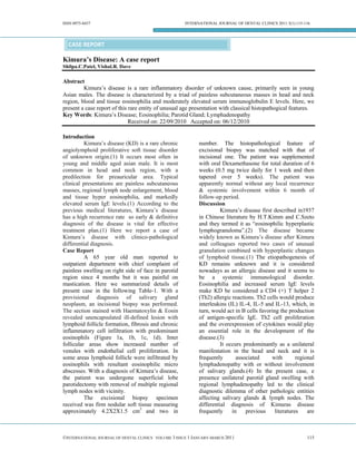
  • 1. ISSN 0975-8437 INTERNATIONAL JOURNAL OF DENTAL CLINICS 2011:3(1):115-116 CASE REPORT Kimura’s Disease: A case report Shilpa.C.Patel, Vishal.R. Dave Abstract Kimura’s disease is a rare inflammatory disorder of unknown cause, primarily seen in young Asian males. The disease is characterized by a triad of painless subcutaneous masses in head and neck region, blood and tissue eosinophilia and moderately elevated serum immunoglobulin E levels. Here, we present a case report of this rare entity of unusual age presentation with classical histopathogical features. Key Words: Kimura’s Disease; Eosinophilia; Parotid Gland; Lymphadenopathy Received on: 22/09/2010 Accepted on: 06/12/2010 Introduction Kimura’s disease (KD) is a rare chronic number. The histopathological feature of angiolymphoid proliferative soft tissue disorder excisional biopsy was matched with that of of unknown origin.(1) It occurs most often in incisional one. The patient was supplemented young and middle aged asian male. It is most with oral Dexamethasone for total duration of 6 common in head and neck region, with a weeks (0.5 mg twice daily for 1 week and then predilection for preauricular area. Typical tapered over 5 weeks). The patient was clinical presentations are painless subcutaneous apparently normal without any local recurrence masses, regional lymph node enlargement, blood & systemic involvement within 6 month of and tissue hyper eosinophilia, and markedly follow-up period. elevated serum IgE levels.(1) According to the Discussion previous medical literatures, Kimura’s disease Kimura’s disease first described in1937 has a high recurrence rate so early & definitive in Chinese literature by H.T.Kimm and C.Szeto diagnosis of the disease is vital for effective and they termed it as “eosinophilic hyperplastic treatment plan.(1) Here we report a case of lymphogranuloma”.(2) The disease became Kimura’s disease with clinico-pathological widely known as Kimura’s disease after Kimura differential diagnosis. and colleagues reported two cases of unusual Case Report granulation combined with hyperplastic changes A 65 year old man reported to of lymphoid tissue.(1) The etiopathogenesis of outpatient department with chief complaint of KD remains unknown and it is considered painless swelling on right side of face in parotid nowadays as an allergic disease and it seems to region since 4 months but it was painful on be a systemic immunological disorder. mastication. Here we summarized details of Eosinophilia and increased serum IgE levels present case in the following Table-1. With a make KD be considered a CD4 (+) T helper 2 provisional diagnosis of salivary gland (Th2) allergic reactions. Th2 cells would produce neoplasm, an incisional biopsy was performed. interleukins (IL) IL-4, IL-5 and IL-13, which, in The section stained with Haematoxylin & Eosin turn, would act in B cells favoring the production revealed unencapsulated ill-defined lesion with of antigen-specific IgE. Th2 cell proliferation lymphoid follicle formation, fibrosis and chronic and the overexpression of cytokines would play inflammatory cell infiltration with predominant an essential role in the development of the eosinophils (Figure 1a, 1b, 1c, 1d). Inter disease.(3) follicular areas show increased number of It occurs predominantly as a unilateral venules with endothelial cell proliferation. In manifestation in the head and neck and it is some areas lymphoid follicle were infiltrated by frequently associated with regional eosinophils with resultant eosinophilic micro lymphadenopathy with or without involvement abscesses. With a diagnosis of Kimura’s disease, of salivary glands.(4) In the present case, e the patient was undergone superficial lobe presence unilateral parotid gland swelling with parotidectomy with removal of multiple regional regional lymphadenopathy led to the clinical lymph nodes with vicinity. diagnostic dilemma of other pathologic entities The excisional biopsy specimen affecting salivary glands & lymph nodes. The received was firm nodular soft tissue measuring differential diagnosis of Kimuras disease approximately 4.2X2X1.5 cm3 and two in frequently in previous literatures are ©INTERNATIONAL JOURNAL OF DENTAL CLINICS VOLUME 3 ISSUE 1 JANUARY-MARCH 2011 115
  • 2. ISSN 0975-8437 INTERNATIONAL JOURNAL OF DENTAL CLINICS 2011:3(1):115-116 Lymphomas, salivary gland neoplasms, benign (AIL). Constant classical features of KD include lymphoepithelial lesions (BLL / Mikulicz’s numerous lymphoid follicles, mixed disease), Angiolymphoid hyperplasia with inflammatory infiltrate composed mainly of eosinophilia (ALHE/ Epitheloid hemangioma) eosinophils and increased amount of post and angioimmunoblastic lymphadenopathy capillary venules.(5) Age/Sex location Physical findings lymphadenopathy Systemic involvement Laboratory investigation 65/Male Parotid (right) 3.5X3.5 cm firm, Regional No renal involvement Eosinophilia (36%) Elevated nontender & normal lymphadenopathy erythrocyte sedimentation rate overlying skin (ESR) Raised IgE levels Table-1 clinical & investigation details of present case Figure 1a. Lymphocytic infiltration with follicle formation and fibrosis .(4x), 1b. Well defined lymphoid follicle with prominent germinal centre, (10x) 1c. Chronic Inflammatory cell infiltration with predominant eosinophils with microabscesses.(40x), 1d. Interfollicular areas showing mixed inflammation with prominent eosinophils and proliferation of venules.(40x), 1e. Interfollicular areas showing increased number of venules with endothelial cell proliferation.(40x) Till date there is no definite treatment Head, Department of Oral & Maxillofacial Pathology, for KD is well established.(2) However the MGM Dental College & Hospital, Navi Mumbai, 2. treatment plan should aim at preservation of vital Dr. Vishal.R.Dave, M.S, Assistant Professor, structures associated with the lesions & cosmetic Department of ENT, GCS Medical College, Ahmedabad, Gujarat, India. rehabilitation, while preventing recurrences. References Treatment options range from observation & 1. Chung YG, Jee WH, Kang YK, Jung CK, Park follow-up of mild & symptomatic cases to GS, Lee A, Bahk WJ, Cho HM, Park JW. conservative surgical approach, medication & Kimura’s disease involving a long bone. Skeletal radiotherapy in symptomatic & recurrent cases. Radiology 2010; 39(5):495-500. If the lesion is primary, localized or present in 2. Pitak-Arnnop P, Bellefqih S, Chaine A, young age then surgical approach is more Dhanuthai K, Bertrand JC, Bertolus C. Head and preferable. According to previous literature, only neck lesions of Kimura's disease: Exclusion of surgical approach had high incidence of human herpesvirus-8 and Epstein-Barr virus by in situ hybridisation and polymerase chain reaction. recurrence.(1) If the lesion is recurrent with An immunohistochemical study. Journal of systemic involvement, application of medication Cranio-Maxillofacial Surgery 2010; 38 (4):266- like corticosteroid and immunosuppressive 70. agents have been shown to decrease size of the 3. Briggs PL. Kimura disease is not angiolymphoid lesion.(2) Irradiation should be considered in hyperplasia with eosinophilia: clinical and patients resistant to the steroid or to prevent the pathological correlation with literature review patient from deleterious effect of long term use and definition of diagnostic criteria. Anais of steroid.(5) Brasileiros de Dermatologia 2006;81(2):167-73. 4. Kung ITM, Gibson J, Bannatyne P. Kimura's Conclusion Disease: A Clinico-Pathological Study Of 21 In conclusion this paper draws attention Cases And Its Distinction From Angiolymphoid on such a rare chronic inflammatory disease Hyperplasia With Eosinophils. which mimics neoplastic conditions. This disease Pathology1984;16(1):39-44. should be considered in differential diagnosis of 5. Jani A, Coulson M. Kimura's disease. A typical patients presented with head & neck mass and case of a rare disorder. Western Journal of lymphadenopathy and investigated accordingly Medicine 1997; 166 (2):142-4. as this disease has good prognosis. Knowledge Address for Correspondence of signature features of Kimura’s disease put the Dr. Shilpa C Patel, M.D.S, 801, Osho Kabir, Plot No 42, Sector 10, physicians in a better position to evaluate its New Panvel, Maharashtra,India- 410206. clinical outcome and optimal treatment regimen. Email: dr_shilpa0769@yahoo.com Affiliations of Authors: 1. Dr. Shilpa.C.Patel, M.D.S., Oral & Maxillofacial Pathology, Professor & Source of Support: Nil, Conflict of Interest: None Declared ©INTERNATIONAL JOURNAL OF DENTAL CLINICS VOLUME 3 ISSUE 1 JANUARY-MARCH 2011 116