SlideShare a Scribd company logo
1 of 84
JOINT PATHOLOGY
Aim
To discuss pathogenesis, morphology and
clinical course of common diseases of joint
Osteoarthritis
rheumatoid arthritis
gouty arthritis
infectious arthritis
OBJECTIVE
Students should be able to:
• Define and identify osteoarthritis, rheumatoid arthritis,
gouty arthritis and infectious arthritis
• Explain the pathogenesis of osteoarthritis, rheumatoid
arthritis, gouty arthritis and infectious arthritis
• Describe the morphology of osteoarthritis, rheumatoid
arthritis, gouty arthritis and infectious arthritis
• Outline the clinical course of osteoarthritis,
rheumatoid arthritis, gouty arthritis and infectious
arthritis
SYNOVIAL JOINTS
Normal articular cartilage performs two functions:
(1) Along with the synovial fluid, it provides virtually
friction-free movement within the joint
(2) in weight-bearing joints, it spreads the load across the
joint surface
These functions require the cartilage to be elastic and to
have high tensile strength provided by proteoglycans and
type II collagen produced by chondrocytes
articular cartilage constantly undergoes matrix degradation
and replacement
Normal chondrocyte function is critical to maintain cartilage
synthesis and degradation
JOINT DISEASES
Joints are subject to a wide variety of disorders
 Degeneration
 Infections
 immune-mediated injury
 metabolic derangements
 neoplasms
JOINT DISEASES
ARTHRITIS
– Degenerative (OSTEOARTHRITIS)
– Rheumatoid
– Infectious: Supp., TB, Lyme, Viral
– Gout (URATE)
– Pseugout (PYROPHOSPHATE)
TUMORS
– Ganglion (Synovial Cyst)
– Giant Cell Tumor (Pigmented VilloNodular Synovitis[PVNS])
– Synovial Sarcoma
OSTEOARTHRITIS
(degenerative joint disease)
characterized by
degeneration of cartilage that results in
structural and functional failure of
synovial joints
an intrinsic disease of cartilage in which
chondrocytes respond to biochemical &
mechanical stresses resulting in breakdown of the
matrix
OSTEOARTHRITIS
Primary osteoarthritis
Onset - insidiously with age and
without apparent initiating cause
Most commonly affected joints:
joints of the hands, knees, hips, and spine
Secondary osteoarthritis
 strikes in youth
 predisposing conditions
- previous trauma
- developmental deformity
- systemic disease : diabetes, ochronosis,
hemochromatosis
- marked obesity
OSTEOARTHRITIS
PATHOGENESIS
Risk Factors
A wear-and-tear phenomenon
 Mechanical stresses
 Aging
 Genetic factors : polymorphisms and mutations
in genes encoding components of the matrix and
signaling molecules
sustained high estrogen levels
increased bone density
Imbalance in the expression, activity, and signalling
of cytokines and growth factors involved in synthesis
and degradation of matrix
PATHOGENESIS
Aging
Mechanical Stress
Degradation and loss of matrix
 degenerating cartilage containing more water and less
proteoglycan
 diminished type II collagen network
MORPHOLOGY
early changes in osteoarthritis include
alterations in the composition and structure of
the matrix
Microscopic :
vertical and horizontal fibrillation and
cracking of the matrix
occur as the superficial layers of the cartilage
are degraded
MORPHOLOGY
fibrillation of the articular cartilage
Gross morphology
Chondromalacia: at early stage - a soft granular-
appearing articular cartilage surface
bone eburnation: Eventually full-thickness portions
of the cartilage are lost and the subchondral bone plate
is exposed and is smoothened and burnished by
friction, giving it the appearance of polished ivory
 underlying cancellous bone becomes sclerotic and
thickened
 subchondral cyst: fracture gaps allow synovial fluid
to be forced into the subchondral regions to form
fibrous walled cysts
MORPHOLOGY
residual articular cartilage
subchondral cyst
eburnated articular surface
exposing subchondral
bone
Advanced subchondral degenerative
cyst in femoral head
joint mice: Small fractures can dislodge pieces
of cartilage and subchondral bone into the
joint, forming loose bodies
MORPHOLOGY
Osteophytes: Mushroom-shaped bony outgrowths
develop at the margins of the articular surface
• pannus: a fibrous synovial pannus covers the
peripheral portions of the articular surface in severe
disease
MORPHOLOGY
The synovium is
usually only mildly
congested and
fibrotic, and may
have scattered
chronic
inflammatory cells
Clinical Course
insidious disease
predominantly affecting patients beginning in
their 50s and 60s
Characteristic symptoms and signs
1. deep, aching pain exacerbated by use
2. morning stiffness
3. crepitus (grating or popping sensation in the
joint)
4. limitation in range of movement
Clinical Course
5. Osteophyte impingement on spinal foramina
can cause nerve root compression with
radicular pain, muscle spasms,muscle atrophy,
and neurologic deficits
6. Heberden nodes in the fingers, representing
prominent osteophytes at the distal
interphalangeal joints are characteristic in
women
7. significant joint deformity
1. Hips
2. Knees
3. lower lumbar
4. cervical vertebrae
5. proximal and distal
interphalangeal joints of
the fingers
6. first carpometacarpal
joints
7. first tarsometatarsal joints
of the feet are commonly
involved
RHEUMATOID ARTHRITIS
 systemic chronic inflammatory disorder that
 affect many tissues and organs—skin, blood
vessels, heart, lungs, and muscles but principally
attacks the joints
 causing a non suppurative proliferative synovitis
 progresses to destruction of the articular
cartilage and underlying bone resulting ankylosis
of the joints
RHEUMATOID ARTHRITIS
Pathogenesis
RA is an autoimmune disease involving complex
interactions of
- genetic risk factors
- environment
- immune system
genetic and environmental factors contribute to
the breakdown of tolerance to self-antigens
Pathogenesis
Genetic susceptibility
50% of the risk of developing RA is related to
genetic factors
Specific HLA-DRB1 alleles have been shown to
be associated with rheumatoid arthritis
Another gene associated with rheumatoid
arthritis is PTPN22 gene which encodes a
tyrosine phosphatase that is postulated to
inhibit T cell activation
RHEUMATOID ARTHRITIS
Pathogenesis
Environmental factors:
Many infectious agents whose antigens may
activate T or B cells have been considered
Inflammatory and environmental insults such as
smoking and infections may induce the
citrullination of some self proteins
creating new epitopes that trigger autoimmune
reactions
RHEUMATOID ARTHRITIS
Pathogenesis
Environmental factors:
Cyclic citrullinated peptides are derived from
proteins in which arginine residues are
converted to citrulline residues post
translationally
70% of patients the blood contains anti-CCP
antibody (antibodies against cyclic citrullinated
peptides) which may be produced during
inflammation
RHEUMATOID ARTHRITIS
Pathogenesis
Autoimmunity
Pathologic changes are caused mainly by
antibodies against self-antigens & cytokine-
mediated inflammation (with CD4+ T cells
princinpal source of the cytokines)
In RA, antibodies to citrullinated fibrinogen, type II
collagen, α-enolase, and vimentin
immune complexes that deposit in the joints
These antibodies are a diagnostic marker for the disease
and may be involved in tissue injury
RHEUMATOID ARTHRITIS
About 80% of individuals with rheumatoid arthritis
have
autoantibodies to the Fc portion of autologous
IgG (rheumatoid factors)
Rheumatoid factor, however, is not present in some
individuals with the disease (seronegative)
and is sometimes found in other disease states and
even in otherwise healthy people
Pathogenesis
RHEUMATOID ARTHRITIS
disease is initiated in a genetically predisposed
person by activation of CD4+ helper T cells
responding to some arthritogenic agent, possibly
microbial,or to a self-antigen such as CCP
IL-1, IL-8, TNF, IL-6, IL-17, У interferon
Morphology
RA typically manifests as symmetric arthritis
principally affecting
small joints of the hands and feet, ankles, knees,
wrists, elbows & shoulders
Most often - proximal interphalangeal &
- metacarpophalangeal joints
distal interphalangeal joints are spared
Axial involvement, when it occurs is limited to the
upper cervical spine
RHEUMATOID ARTHRITIS
Morphology
Joints
Gross
synovium becomes
 Thickened
 Transforming thin, smooth synovial membrane is
into lush, edematous, frondlike (villous)
projections
RHEUMATOID ARTHRITIS
Morphology
Affected joints show chronic papillary synovitis
The characteristic histologic features include
1. synovial cell hyperplasia and proliferation
2. infiltration of synovial stroma by a dense perivascular
inflammatory infiltrate composed of lymphoid aggregates
(mostly CD4+ helper T cells), B cells, plasma cells, dendritic cells,
and macrophages
3. increased vascularity due to vasodilation and
angiogenesis - with superficial hemosiderin deposits
4. aggregation of organizing fibrin - covering portions of the
synovium and floating in the joint space as rice bodies
RHEUMATOID ARTHRITIS
5. accumulation of neutrophils - in the synovial fluid and
along the surface of synovium but usually not deep in the
synovial stroma
6. osteoclastic activity in underlying bone - allowing the
synovium to penetrate into the bone and cause and periarticular
bone erosion s, subchondral cysts, and osteoporosis
7. pannus formation
- pannus is a mass of synovium and synovial stroma consisting of
proliferating synovial lining cells admixed with inflammatory cells,
granulation tissue, and fibrous connective tissue
- overgrowth of this tissue is so exuberant that thin, smooth
synovial membrane is transformed into lush, edematous, frondlike
(villous) projections
In time, after the cartilage has been destroyed,
the pannus bridges the apposing bones to form
a fibrous ankylosis which eventually ossifies and
results in bony ankylosis
Inflammation in the tendons, ligaments, and
occasionally the adjacent skeletal muscle
frequently accompanies the arthritis
Skin
Rheumatoid nodules are firm, non tender, and round to
oval, in the skin arise in the subcutaneous tissue
the most common cutaneous lesion
in approximately 25% of affected individuals
Sites:
arise in regions of the skin that are subjected to pressure,
including the ulnar aspect of the forearm, elbows,
occiput, and lumbosacral area
Less commonly
they form in the lungs, spleen, pericardium, myocardium,
heart valves, aorta, and other viscera
Microscopically
A central zone of fibrinoid necrosis
surrounded by
a prominent rim of epithelioid histiocytes
(activated macrophages) and
numerous lymphocytes and plasma cells
The rheumatoid “nodule” shows “palisading”
fibroblasts
HANDSWRISTELBOWS
Blood Vessels
 with severe erosive disease
 rheumatoid nodules
 high titers of rheumatoid factor
are at risk of developing vasculitic syndromes
Rheumatoid vasculitis is a potentially catastrophic
complication of rheumatoid arthritis,
particularly when it affects vital organs
Frequently, segments of small arteries such as
vasa nervorum and digital arteries are
obstructed by an obliterating endarteritis
resulting in peripheral neuropathy, ulcers, and
gangrene
Leukocytoclastic venulitis produces purpura,
cutaneous ulcers, and nail bed infarction
Clinical Course
Symmetric polyarticular arthritis
Constitutional symptoms (weakness, malaise,
and low-grade fever)
Subcutaneous rheumatoid nodules
Vasculitic involvement of the extremities may
give rise to Raynaud phenomenon and chronic
leg ulcers
RHEUMATOID ARTHRITIS
Symmetric polyarticular arthritis
The arthritis first appears insidiously with
aching and stiffness of the joints, particularly in the
morning
As the disease advances the joints become
- swollen, painful
- Limited movement (minimal or no range of motion)
- complete ankylosis
characteristic deformities
- radial deviation of the wrist
- ulnar deviation of the fingers
- flexion-hyperextension abnormalities of the fingers (swan
neck, boutonnière)
Large synovial cysts
Hand, acute rheumatoid arthritis
This is a classic presentation of acute rheumatoid arthritis
It is polyarthritic, affects the proximal interphalangeal joints
has produced a fusiform swelling of the soft tissue
The lesion is usually symmetric
Chronic RA
The radiographic hallmarks are
 joint effusions
juxta-articular osteopenia
erosions and narrowing of the joint space
loss of articular cartilage
Diffuse osteopenia
Narrow joint spaces
Periarticular bony erosions
Ulnar drift of the fingers
The disease course may be slow or rapid, and
fluctuates over the years, with the greatest
damage occurring in the first 4 or 5 years
Approximately 20% of affected individuals enjoy
periods of partial or complete remission
Juvenile Rheumatoid Arthritis
 A group of multifactorial disorders with
environmental and genetic components
 unknown etiology
 classified according to their presentation into
oligoarthritis, polyarthritis & systemic (Still’s
disease) variants
 Large joints often are affected
 begin before the age of 16 years and persist for
more than 6 weeks
 Extraarticular inflammatory manifestations
such as uveitis also may be present
GOUT
Crystal-Induced Arthritis
 affects about 1% of the population & shows a
predeliction for males
 caused by excessive amounts of uric acid within
tissues and body fluids
 Monosodium urate crystals precipitate from
supersaturated body fluids and induce an acute
inflammatory reaction
GOUT
Definition
Gout is marked by
 transient attacks of acute arthritis initiated by
crystallization of monosodium urate within and
around joints
 sometime accompanied by the formation of
large crystalline aggregates called tophi, and
eventual permanent joint deformity
Gout is divided into primary and secondary forms
Primary gout - 90% of cases
designates cases in which the basic cause is
unknown or (less commonly) in which the disorder
is due to an inborn metabolic defect that causes
hyperuricemia
Secondary gout - 10% of cases
cause of the hyperuricemia is known, but gout is
not necessarily the main or even dominant clinical
disorder
GOUT
PATHOGENESIS
 elevated level of uric acid is an essential
component of gout, not all such persons develop
gout, and genetic and environmental factors also
contribute to its pathogenesis
 elevated uric acid levels can result from
overproduction or reduced excretion of uric
acid, or both
GOUT
Uric acid synthesis -end product of purine
catabolism
increased urate synthesis typically reflects some
abnormality in purine nucleotide production
synthesis of purine nucleotides involves two
different but interlinked pathways:
the de novo - involved in the synthesis of purine
nucleotides from nonpurine precursors.
salvage pathways - involved in the synthesis of
purine nucleotides from free purine bases, derived
from dietary intake and by catabolizing nucleic
acids and purine nucleotides
GOUT
PATHOGENESIS
Uric acid excretion
Circulating uric acid is freely filtered by the
glomerulus and virtually completely resorbed in
the proximal tubules of the kidney
A small fraction of the resorbed uric acid is
subsequently secreted by the distal nephron and
excreted in the urine
GOUT
PATHOGENESIS
Primary gout - cause of excessive uric acid biosynthesis
is unknown in most cases
 rare patients have identifiable enzymatic defects [e.g.
complete lack of HGPRT in Lesch-Nyhan syndrome]
Secondary gout - hyperuricemia can be caused by
 increased urate production[e.g.rapid cell lysis during
chemotherapy for lymphoma or leukemia)
 decreased excretion [chronic renal insufficiency or
reduced renal excretion caused by drugs such as
thiazide diuretics]
GOUT
increased levels of uric acid in the blood & body fluids ( synovium)
precipitation of monosodium urate crystals
chain of events that culminate in joint injury
Activation of complement system leading to
production of chemotactic and pro-inflammatory mediators
Phagocytosis of crystal by macrophages, stimulates the
production of the cytokine IL-1
local accumulation of neutrophils & macrophages in the joints &
synovial membranes
release of chemokines, cytokines, toxic free radicals, leukotriene B &
destructive lysosomal enzymes from activated inflammatory cells
Cytokines activate synovial cells & cartilage cells to release proteases
MORPHOLOGY
Major morphologic manifestations of gout are
acute arthritis
chronic tophaceous arthritis
tophi in various sites
gouty nephropathy
GOUT
MORPHOLOGY
Acute arthritis : characterized by
 a dense neutrophilic infiltrate permeating the
synovium and synovial fluid
 presence of long, slender, needle-shaped monosodium
urate crystals in the cytoplasm of the neutrophils & in
small clusters in the synovium
 synovium is edematous and congested & contains
scattered mononuclear inflammatory cells
when the episode of crystallization abates and the crystals
resolubilize, the attack remits
GOUT
MORPHOLOGY
Chronic tophaceous arthritis - evolves from
 Repetitive precipitation of urate crystals during acute
attacks
 urates can heavily encrust the articular surfaces and
form visible deposits in the synovium
 synovium becomes hyperplastic, fibrotic, and thickened
by inflammatory cells, forming a pannus that destroys
the underlying cartilage, leading to juxtaarticular bone
erosions
 In severe cases, fibrous or bony ankylosis ensues,
resulting in loss of joint function
GOUT
MORPHOLOGY - Tophi in various sites
Tophi are pathognomonic for gout
 formed by large aggregations of urate crystals
surrounded by an intense inflammatory
reaction of lymphocytes, macrophages, &
foreign-body giant cells, attempting to engulf
the masses of crystals
 Sites - articular cartilage of joints
- periarticular ligaments, tendons, & soft tissues
- ear lobes, nasal cartilages, skin of the fingertips
[Superficial tophi can lead to large ulcerations of the overlying
skin]
GOUT
MORPHOLOGY
Gouty nephropathy
 refers to renal complications associated with
urate deposition, variously forming medullary
tophi, intratubular precipitations, or free uric
acid crystals and renal calculi
 Secondary complications such as pyelonephritis
can occur, especially when there is urinary
obstruction
GOUT
GOUT
Clinical Features
more common in men than in women
not usually cause symptoms before the age of 30
Risk factors :
- obesity
- excess alcohol intake
- consumption of purine-rich foods
- diabetes
- metabolic syndrome
- renal failure
GOUT
Clinical Features
Four stages are classically recognized:
(1)asymptomatic hyperuricemia:
around puberty in males and after menopause in
women
(2) acute gouty arthritis
(3) “intercritical”gout (asymptomatic intercritical period)
(4) chronic tophaceous gout
GOUT
(2) acute gouty arthritis :
after a long interval of years appear as
sudden onset, excruciating joint pain with
localized erythema & warmth mostly
monoarticular
50% occur in the first metatarsophalangeal joint
(great toe) & 90% in ankle, heel, or wrist
last for hours to weeks & gradually completely
resolves, and the patient enters an
asymptomatic intercritical period
GOUT
GOUT
(4) chronic tophaceous gout
At this stage, radiographs show characteristic
- juxtaarticular bone erosion caused by the
crystal deposits and loss of the joint space
Progression leads to chronic gouty nephropathy
- renal colic associated with the passage of
gravel and stones
Pseudogout
calcium pyrophosphate crystal deposition disease
(also known as chondrocalcinosis )
 crystal deposits first appear in structures composed of
cartilage such as menisci, intervertebral discs, and
articular surfaces
 When the deposits enlarge enough, they may rupture,
inducing an inflammatory reaction
 typically first occurs in persons 50 years of age or older
becoming more common with increasing age, and
eventually reaching a prevalence of 30% to 60% in those
age 85
 no gender or race predilection
Infectious Arthritis
 Mode of entry: hematogenous dissemination &
direct inoculation or by contiguous spread from
osteomyelitis or a soft tissue abscess
 Infectious arthritis is serious because it can
cause rapid joint destruction and permanent
deformities
Suppurative Arthritis
Aetiology (causative agent )
 In children < 2 years of age - Haemophilus influenzae
 In older children and adults - S. aureus & gonococcus
 Patients with sickle cell disease - Salmonella infection
at any age
 Both genders are affected equally except for
gonococcal arthritis, which occurs mainly in sexually
active women
 In this group, those with deficiency of complement
proteins (C5, C6, C7) are particularly susceptible to
disseminated gonococcal infections
Infectious Arthritis
Suppurative Arthritis
Clinical presentation
 sudden onset with cardinal signs of acute
inflammation
 Fever, leukocytosis, and elevated ESR
 involves only a single joint—usually the knee—
followed in order by hip, shoulder, elbow, wrist,
and sternoclavicular joints
 Joint aspiration typically yields a purulent fluid
in which the causal agent can be identified
Infectious Arthritis
Lyme Arthritis
Aetiology - spirochete Borrelia burgdorferi
Mode of transmission : arthropod-borne disease
deer ticks of the Ixodes ricinus complexin
Clinical course - involves multiple organ systems
and in its classic form progresses through three
successive stages
Stage 1 - multiply at the site of the tick bite and cause
an expanding area of redness, often with an indurated or
pale center (erythema chronicum migrans) with fever
&lymphadenopathy last for few weeks’time
Infectious Arthritis
Stage 2 - early disseminated stage ,spread
hematogenously &cause secondary annular skin
lesions, lymphadenopathy, migratory joint and
muscle pain, cardiac arrhythmias, and meningitis,
often with cranial nerve involvement
- Diagnostically useful antibodies (usually both IgM
and IgG) against Borrelia antigens appear in the
serum at this stage of the disease
Infectious Arthritis
Stage 3 - late disseminated stage, which occurs 2
or 3 years after the initial bite
chronic arthritis, with severe damage to large
joints, and an encephalitis that ranges in
severity from mild to debilitating disease
 The arthritis may be caused by immune
responses against Borrelia antigens that cross-
react with proteins in the joints
disease tends to be migratory, with remissions
&relapses
 involves mainly large joints, especially the
knees, shoulders, elbows, and ankles, in
descending order of frequency
Infectious Arthritis
Morphology
Histologic examination reveals a
Chronic papillary synovitis with synoviocyte
hyperplasia fibrin deposition, mononuclear cell
infiltrates, onion-skin thickening of arterial
walls
Infectious Arthritis
Joint pathology

More Related Content

What's hot (20)

Seronegative arthropathies
Seronegative arthropathiesSeronegative arthropathies
Seronegative arthropathies
 
Crystal Arthritis Amh
Crystal Arthritis AmhCrystal Arthritis Amh
Crystal Arthritis Amh
 
Granulomatous inflammation
Granulomatous inflammation Granulomatous inflammation
Granulomatous inflammation
 
Chronic inflammation
Chronic inflammationChronic inflammation
Chronic inflammation
 
Bone tumors
Bone tumorsBone tumors
Bone tumors
 
Pseudogout
PseudogoutPseudogout
Pseudogout
 
Osteomyelitis
OsteomyelitisOsteomyelitis
Osteomyelitis
 
Osteosarcoma ppt
Osteosarcoma pptOsteosarcoma ppt
Osteosarcoma ppt
 
CHRONIC OSTEOMYELITIS
CHRONIC  OSTEOMYELITISCHRONIC  OSTEOMYELITIS
CHRONIC OSTEOMYELITIS
 
Rickets & osteomalacia
Rickets & osteomalaciaRickets & osteomalacia
Rickets & osteomalacia
 
Clinical approach to Arthritis
Clinical approach to ArthritisClinical approach to Arthritis
Clinical approach to Arthritis
 
AMYLOIDOSIS
AMYLOIDOSISAMYLOIDOSIS
AMYLOIDOSIS
 
Crystal arthropathies gout &amp; pseudogout
Crystal arthropathies  gout &amp; pseudogoutCrystal arthropathies  gout &amp; pseudogout
Crystal arthropathies gout &amp; pseudogout
 
Leukocytosis. Leukopenia. Leukosis
Leukocytosis. Leukopenia. LeukosisLeukocytosis. Leukopenia. Leukosis
Leukocytosis. Leukopenia. Leukosis
 
Pathophysiology of Rheumatoid Arthritis
Pathophysiology of  Rheumatoid ArthritisPathophysiology of  Rheumatoid Arthritis
Pathophysiology of Rheumatoid Arthritis
 
SERO-NEGATIVE ARTHRITIS
SERO-NEGATIVE ARTHRITISSERO-NEGATIVE ARTHRITIS
SERO-NEGATIVE ARTHRITIS
 
Spondyloarthritis a brief
Spondyloarthritis a briefSpondyloarthritis a brief
Spondyloarthritis a brief
 
Infarct
InfarctInfarct
Infarct
 
Central nervous system pathology
Central nervous system pathology Central nervous system pathology
Central nervous system pathology
 
Synovial fluid
Synovial fluidSynovial fluid
Synovial fluid
 

Viewers also liked

Viewers also liked (8)

Clinical Presentation on Rheumatoid Arthritis (Amavata)
Clinical Presentation on Rheumatoid Arthritis (Amavata)Clinical Presentation on Rheumatoid Arthritis (Amavata)
Clinical Presentation on Rheumatoid Arthritis (Amavata)
 
Osteoarthritis
OsteoarthritisOsteoarthritis
Osteoarthritis
 
Osteoarthritis.ppt
Osteoarthritis.pptOsteoarthritis.ppt
Osteoarthritis.ppt
 
Pathology of Arthritis
Pathology of ArthritisPathology of Arthritis
Pathology of Arthritis
 
Osteoarthritis
OsteoarthritisOsteoarthritis
Osteoarthritis
 
Osteoarthritis Presentation
Osteoarthritis PresentationOsteoarthritis Presentation
Osteoarthritis Presentation
 
Osteoarthritis lecture for UG
Osteoarthritis lecture for UGOsteoarthritis lecture for UG
Osteoarthritis lecture for UG
 
Osteoarthritis ppt
Osteoarthritis pptOsteoarthritis ppt
Osteoarthritis ppt
 

Similar to Joint pathology

RHEUMATOID ARTHRITIS BY DR BASHIR AHMED DAR ASSOCIATE PROFESSOR MEDICINE SOPO...
RHEUMATOID ARTHRITIS BY DR BASHIR AHMED DAR ASSOCIATE PROFESSOR MEDICINE SOPO...RHEUMATOID ARTHRITIS BY DR BASHIR AHMED DAR ASSOCIATE PROFESSOR MEDICINE SOPO...
RHEUMATOID ARTHRITIS BY DR BASHIR AHMED DAR ASSOCIATE PROFESSOR MEDICINE SOPO...Prof Dr Bashir Ahmed Dar
 
TREATMENT OF RHEUMATOID ARTHRITIS BY DR BASHIR AHMED DAR ASSOCIATE PROFESSOR ...
TREATMENT OF RHEUMATOID ARTHRITIS BY DR BASHIR AHMED DAR ASSOCIATE PROFESSOR ...TREATMENT OF RHEUMATOID ARTHRITIS BY DR BASHIR AHMED DAR ASSOCIATE PROFESSOR ...
TREATMENT OF RHEUMATOID ARTHRITIS BY DR BASHIR AHMED DAR ASSOCIATE PROFESSOR ...Prof Dr Bashir Ahmed Dar
 
RHEUMATOID ARTHRITIS BY DR BASHIR AHMED DAR ASSOCIATE PROFESSOR MEDICINE SOPO...
RHEUMATOID ARTHRITIS BY DR BASHIR AHMED DAR ASSOCIATE PROFESSOR MEDICINE SOPO...RHEUMATOID ARTHRITIS BY DR BASHIR AHMED DAR ASSOCIATE PROFESSOR MEDICINE SOPO...
RHEUMATOID ARTHRITIS BY DR BASHIR AHMED DAR ASSOCIATE PROFESSOR MEDICINE SOPO...Prof Dr Bashir Ahmed Dar
 
rheumatoid arthritis details ins and outs
rheumatoid arthritis details ins and outsrheumatoid arthritis details ins and outs
rheumatoid arthritis details ins and outsBosan Khalid
 
Musculoskeletal System III__Physioterapy_Spring_ 2018.ppt
Musculoskeletal System III__Physioterapy_Spring_ 2018.pptMusculoskeletal System III__Physioterapy_Spring_ 2018.ppt
Musculoskeletal System III__Physioterapy_Spring_ 2018.pptManal445405
 
Chapter 20 Rheumatologic Disorders
Chapter 20 Rheumatologic Disorders Chapter 20 Rheumatologic Disorders
Chapter 20 Rheumatologic Disorders MeganSimpson27
 
Rheumatoid arthitis
Rheumatoid arthitis Rheumatoid arthitis
Rheumatoid arthitis SUDHEER BABU
 
Osteoarth & Rheumatoid arthritis
Osteoarth & Rheumatoid arthritisOsteoarth & Rheumatoid arthritis
Osteoarth & Rheumatoid arthritisKemUnited
 
Rhematoid arthiritis
Rhematoid arthiritisRhematoid arthiritis
Rhematoid arthiritisjasleenbrar03
 
Arthiritis and related Diseases-1.pptx
Arthiritis and related Diseases-1.pptxArthiritis and related Diseases-1.pptx
Arthiritis and related Diseases-1.pptxAshu Bansal
 
14-180530185343.pdf
14-180530185343.pdf14-180530185343.pdf
14-180530185343.pdfBucky10
 

Similar to Joint pathology (20)

osteoarthritis.pptx
osteoarthritis.pptxosteoarthritis.pptx
osteoarthritis.pptx
 
Osteomyelitis
OsteomyelitisOsteomyelitis
Osteomyelitis
 
RHEUMATOID ARTHRITIS BY DR BASHIR AHMED DAR ASSOCIATE PROFESSOR MEDICINE SOPO...
RHEUMATOID ARTHRITIS BY DR BASHIR AHMED DAR ASSOCIATE PROFESSOR MEDICINE SOPO...RHEUMATOID ARTHRITIS BY DR BASHIR AHMED DAR ASSOCIATE PROFESSOR MEDICINE SOPO...
RHEUMATOID ARTHRITIS BY DR BASHIR AHMED DAR ASSOCIATE PROFESSOR MEDICINE SOPO...
 
Arthritis
ArthritisArthritis
Arthritis
 
Arthritis
ArthritisArthritis
Arthritis
 
JIA.pptx
JIA.pptxJIA.pptx
JIA.pptx
 
Lec arthritis
Lec arthritisLec arthritis
Lec arthritis
 
TREATMENT OF RHEUMATOID ARTHRITIS BY DR BASHIR AHMED DAR ASSOCIATE PROFESSOR ...
TREATMENT OF RHEUMATOID ARTHRITIS BY DR BASHIR AHMED DAR ASSOCIATE PROFESSOR ...TREATMENT OF RHEUMATOID ARTHRITIS BY DR BASHIR AHMED DAR ASSOCIATE PROFESSOR ...
TREATMENT OF RHEUMATOID ARTHRITIS BY DR BASHIR AHMED DAR ASSOCIATE PROFESSOR ...
 
RHEUMATOID ARTHRITIS BY DR BASHIR AHMED DAR ASSOCIATE PROFESSOR MEDICINE SOPO...
RHEUMATOID ARTHRITIS BY DR BASHIR AHMED DAR ASSOCIATE PROFESSOR MEDICINE SOPO...RHEUMATOID ARTHRITIS BY DR BASHIR AHMED DAR ASSOCIATE PROFESSOR MEDICINE SOPO...
RHEUMATOID ARTHRITIS BY DR BASHIR AHMED DAR ASSOCIATE PROFESSOR MEDICINE SOPO...
 
rheumatoid arthritis details ins and outs
rheumatoid arthritis details ins and outsrheumatoid arthritis details ins and outs
rheumatoid arthritis details ins and outs
 
Musculoskeletal System III__Physioterapy_Spring_ 2018.ppt
Musculoskeletal System III__Physioterapy_Spring_ 2018.pptMusculoskeletal System III__Physioterapy_Spring_ 2018.ppt
Musculoskeletal System III__Physioterapy_Spring_ 2018.ppt
 
Chapter 20 Rheumatologic Disorders
Chapter 20 Rheumatologic Disorders Chapter 20 Rheumatologic Disorders
Chapter 20 Rheumatologic Disorders
 
Rheumatoid arthitis
Rheumatoid arthitis Rheumatoid arthitis
Rheumatoid arthitis
 
Rheumatology
RheumatologyRheumatology
Rheumatology
 
Osteoarth & Rheumatoid arthritis
Osteoarth & Rheumatoid arthritisOsteoarth & Rheumatoid arthritis
Osteoarth & Rheumatoid arthritis
 
Rheumatoid arthritis
Rheumatoid arthritisRheumatoid arthritis
Rheumatoid arthritis
 
Rhematoid arthiritis
Rhematoid arthiritisRhematoid arthiritis
Rhematoid arthiritis
 
Arthiritis and related Diseases-1.pptx
Arthiritis and related Diseases-1.pptxArthiritis and related Diseases-1.pptx
Arthiritis and related Diseases-1.pptx
 
mss(0).pptx
mss(0).pptxmss(0).pptx
mss(0).pptx
 
14-180530185343.pdf
14-180530185343.pdf14-180530185343.pdf
14-180530185343.pdf
 

More from ayeayetun08

Cardiac hyprtrophy and heart failure
Cardiac hyprtrophy and heart failureCardiac hyprtrophy and heart failure
Cardiac hyprtrophy and heart failureayeayetun08
 
Wbc disoders practical
Wbc disoders practicalWbc disoders practical
Wbc disoders practicalayeayetun08
 
Blood film examination
Blood film examinationBlood film examination
Blood film examinationayeayetun08
 
Pathology of cervix &uterus
Pathology of cervix &uterusPathology of cervix &uterus
Pathology of cervix &uterusayeayetun08
 
Lecture 28. common repratory pathological condirtion part 3
Lecture 28. common repratory pathological condirtion part 3Lecture 28. common repratory pathological condirtion part 3
Lecture 28. common repratory pathological condirtion part 3ayeayetun08
 
Tumors of kidney & urinary tract 2012
Tumors of kidney & urinary tract 2012Tumors of kidney & urinary tract 2012
Tumors of kidney & urinary tract 2012ayeayetun08
 
Lecture 50 chronic inflammation.ppt 4.11.11
Lecture 50  chronic inflammation.ppt 4.11.11Lecture 50  chronic inflammation.ppt 4.11.11
Lecture 50 chronic inflammation.ppt 4.11.11ayeayetun08
 

More from ayeayetun08 (10)

Cardiac hyprtrophy and heart failure
Cardiac hyprtrophy and heart failureCardiac hyprtrophy and heart failure
Cardiac hyprtrophy and heart failure
 
Wbc disoders practical
Wbc disoders practicalWbc disoders practical
Wbc disoders practical
 
Blood film examination
Blood film examinationBlood film examination
Blood film examination
 
Bm examination
Bm examinationBm examination
Bm examination
 
Pathology of cervix &uterus
Pathology of cervix &uterusPathology of cervix &uterus
Pathology of cervix &uterus
 
Cns tumors
Cns tumorsCns tumors
Cns tumors
 
Tumor immunity
Tumor immunityTumor immunity
Tumor immunity
 
Lecture 28. common repratory pathological condirtion part 3
Lecture 28. common repratory pathological condirtion part 3Lecture 28. common repratory pathological condirtion part 3
Lecture 28. common repratory pathological condirtion part 3
 
Tumors of kidney & urinary tract 2012
Tumors of kidney & urinary tract 2012Tumors of kidney & urinary tract 2012
Tumors of kidney & urinary tract 2012
 
Lecture 50 chronic inflammation.ppt 4.11.11
Lecture 50  chronic inflammation.ppt 4.11.11Lecture 50  chronic inflammation.ppt 4.11.11
Lecture 50 chronic inflammation.ppt 4.11.11
 

Recently uploaded

PNEUMOTHORAX AND ITS MANAGEMENTS.pdf
PNEUMOTHORAX   AND  ITS  MANAGEMENTS.pdfPNEUMOTHORAX   AND  ITS  MANAGEMENTS.pdf
PNEUMOTHORAX AND ITS MANAGEMENTS.pdfDolisha Warbi
 
Statistical modeling in pharmaceutical research and development.
Statistical modeling in pharmaceutical research and development.Statistical modeling in pharmaceutical research and development.
Statistical modeling in pharmaceutical research and development.ANJALI
 
Biomechanics- Shoulder Joint!!!!!!!!!!!!
Biomechanics- Shoulder Joint!!!!!!!!!!!!Biomechanics- Shoulder Joint!!!!!!!!!!!!
Biomechanics- Shoulder Joint!!!!!!!!!!!!ibtesaam huma
 
Music Therapy's Impact in Palliative Care| IAPCON2024| Dr. Tara Rajendran
Music Therapy's Impact in Palliative Care| IAPCON2024| Dr. Tara RajendranMusic Therapy's Impact in Palliative Care| IAPCON2024| Dr. Tara Rajendran
Music Therapy's Impact in Palliative Care| IAPCON2024| Dr. Tara RajendranTara Rajendran
 
Introduction to Sports Injuries by- Dr. Anjali Rai
Introduction to Sports Injuries by- Dr. Anjali RaiIntroduction to Sports Injuries by- Dr. Anjali Rai
Introduction to Sports Injuries by- Dr. Anjali RaiGoogle
 
call girls in munirka DELHI 🔝 >༒9540349809 🔝 genuine Escort Service 🔝✔️✔️
call girls in munirka  DELHI 🔝 >༒9540349809 🔝 genuine Escort Service 🔝✔️✔️call girls in munirka  DELHI 🔝 >༒9540349809 🔝 genuine Escort Service 🔝✔️✔️
call girls in munirka DELHI 🔝 >༒9540349809 🔝 genuine Escort Service 🔝✔️✔️saminamagar
 
Hematology and Immunology - Leukocytes Functions
Hematology and Immunology - Leukocytes FunctionsHematology and Immunology - Leukocytes Functions
Hematology and Immunology - Leukocytes FunctionsMedicoseAcademics
 
Informed Consent Empowering Healthcare Decision-Making.pptx
Informed Consent Empowering Healthcare Decision-Making.pptxInformed Consent Empowering Healthcare Decision-Making.pptx
Informed Consent Empowering Healthcare Decision-Making.pptxSasikiranMarri
 
97111 47426 Call Girls In Delhi MUNIRKAA
97111 47426 Call Girls In Delhi MUNIRKAA97111 47426 Call Girls In Delhi MUNIRKAA
97111 47426 Call Girls In Delhi MUNIRKAAjennyeacort
 
History and Development of Pharmacovigilence.pdf
History and Development of Pharmacovigilence.pdfHistory and Development of Pharmacovigilence.pdf
History and Development of Pharmacovigilence.pdfSasikiranMarri
 
epilepsy and status epilepticus for undergraduate.pptx
epilepsy and status epilepticus  for undergraduate.pptxepilepsy and status epilepticus  for undergraduate.pptx
epilepsy and status epilepticus for undergraduate.pptxMohamed Rizk Khodair
 
Primary headache and facial pain. (2024)
Primary headache and facial pain. (2024)Primary headache and facial pain. (2024)
Primary headache and facial pain. (2024)Mohamed Rizk Khodair
 
PULMONARY EDEMA AND ITS MANAGEMENT.pdf
PULMONARY EDEMA AND  ITS  MANAGEMENT.pdfPULMONARY EDEMA AND  ITS  MANAGEMENT.pdf
PULMONARY EDEMA AND ITS MANAGEMENT.pdfDolisha Warbi
 
LUNG TUMORS AND ITS CLASSIFICATIONS.pdf
LUNG TUMORS AND ITS  CLASSIFICATIONS.pdfLUNG TUMORS AND ITS  CLASSIFICATIONS.pdf
LUNG TUMORS AND ITS CLASSIFICATIONS.pdfDolisha Warbi
 
PERFECT BUT PAINFUL TKR -ROLE OF SYNOVECTOMY.pptx
PERFECT BUT PAINFUL TKR -ROLE OF SYNOVECTOMY.pptxPERFECT BUT PAINFUL TKR -ROLE OF SYNOVECTOMY.pptx
PERFECT BUT PAINFUL TKR -ROLE OF SYNOVECTOMY.pptxdrashraf369
 
Big Data Analysis Suggests COVID Vaccination Increases Excess Mortality Of ...
Big Data Analysis Suggests COVID  Vaccination Increases Excess Mortality Of  ...Big Data Analysis Suggests COVID  Vaccination Increases Excess Mortality Of  ...
Big Data Analysis Suggests COVID Vaccination Increases Excess Mortality Of ...sdateam0
 
Culture and Health Disorders Social change.pptx
Culture and Health Disorders Social change.pptxCulture and Health Disorders Social change.pptx
Culture and Health Disorders Social change.pptxDr. Dheeraj Kumar
 
COVID-19 (NOVEL CORONA VIRUS DISEASE PANDEMIC ).pptx
COVID-19  (NOVEL CORONA  VIRUS DISEASE PANDEMIC ).pptxCOVID-19  (NOVEL CORONA  VIRUS DISEASE PANDEMIC ).pptx
COVID-19 (NOVEL CORONA VIRUS DISEASE PANDEMIC ).pptxBibekananda shah
 
Measurement of Radiation and Dosimetric Procedure.pptx
Measurement of Radiation and Dosimetric Procedure.pptxMeasurement of Radiation and Dosimetric Procedure.pptx
Measurement of Radiation and Dosimetric Procedure.pptxDr. Dheeraj Kumar
 
The next social challenge to public health: the information environment.pptx
The next social challenge to public health:  the information environment.pptxThe next social challenge to public health:  the information environment.pptx
The next social challenge to public health: the information environment.pptxTina Purnat
 

Recently uploaded (20)
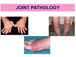
PNEUMOTHORAX AND ITS MANAGEMENTS.pdf
PNEUMOTHORAX   AND  ITS  MANAGEMENTS.pdfPNEUMOTHORAX   AND  ITS  MANAGEMENTS.pdf
PNEUMOTHORAX AND ITS MANAGEMENTS.pdf
 
Statistical modeling in pharmaceutical research and development.
Statistical modeling in pharmaceutical research and development.Statistical modeling in pharmaceutical research and development.
Statistical modeling in pharmaceutical research and development.
 
Biomechanics- Shoulder Joint!!!!!!!!!!!!
Biomechanics- Shoulder Joint!!!!!!!!!!!!Biomechanics- Shoulder Joint!!!!!!!!!!!!
Biomechanics- Shoulder Joint!!!!!!!!!!!!
 
Music Therapy's Impact in Palliative Care| IAPCON2024| Dr. Tara Rajendran
Music Therapy's Impact in Palliative Care| IAPCON2024| Dr. Tara RajendranMusic Therapy's Impact in Palliative Care| IAPCON2024| Dr. Tara Rajendran
Music Therapy's Impact in Palliative Care| IAPCON2024| Dr. Tara Rajendran
 
Introduction to Sports Injuries by- Dr. Anjali Rai
Introduction to Sports Injuries by- Dr. Anjali RaiIntroduction to Sports Injuries by- Dr. Anjali Rai
Introduction to Sports Injuries by- Dr. Anjali Rai
 
call girls in munirka DELHI 🔝 >༒9540349809 🔝 genuine Escort Service 🔝✔️✔️
call girls in munirka  DELHI 🔝 >༒9540349809 🔝 genuine Escort Service 🔝✔️✔️call girls in munirka  DELHI 🔝 >༒9540349809 🔝 genuine Escort Service 🔝✔️✔️
call girls in munirka DELHI 🔝 >༒9540349809 🔝 genuine Escort Service 🔝✔️✔️
 
Hematology and Immunology - Leukocytes Functions
Hematology and Immunology - Leukocytes FunctionsHematology and Immunology - Leukocytes Functions
Hematology and Immunology - Leukocytes Functions
 
Informed Consent Empowering Healthcare Decision-Making.pptx
Informed Consent Empowering Healthcare Decision-Making.pptxInformed Consent Empowering Healthcare Decision-Making.pptx
Informed Consent Empowering Healthcare Decision-Making.pptx
 
97111 47426 Call Girls In Delhi MUNIRKAA
97111 47426 Call Girls In Delhi MUNIRKAA97111 47426 Call Girls In Delhi MUNIRKAA
97111 47426 Call Girls In Delhi MUNIRKAA
 
History and Development of Pharmacovigilence.pdf
History and Development of Pharmacovigilence.pdfHistory and Development of Pharmacovigilence.pdf
History and Development of Pharmacovigilence.pdf
 
epilepsy and status epilepticus for undergraduate.pptx
epilepsy and status epilepticus  for undergraduate.pptxepilepsy and status epilepticus  for undergraduate.pptx
epilepsy and status epilepticus for undergraduate.pptx
 
Primary headache and facial pain. (2024)
Primary headache and facial pain. (2024)Primary headache and facial pain. (2024)
Primary headache and facial pain. (2024)
 
PULMONARY EDEMA AND ITS MANAGEMENT.pdf
PULMONARY EDEMA AND  ITS  MANAGEMENT.pdfPULMONARY EDEMA AND  ITS  MANAGEMENT.pdf
PULMONARY EDEMA AND ITS MANAGEMENT.pdf
 
LUNG TUMORS AND ITS CLASSIFICATIONS.pdf
LUNG TUMORS AND ITS  CLASSIFICATIONS.pdfLUNG TUMORS AND ITS  CLASSIFICATIONS.pdf
LUNG TUMORS AND ITS CLASSIFICATIONS.pdf
 
PERFECT BUT PAINFUL TKR -ROLE OF SYNOVECTOMY.pptx
PERFECT BUT PAINFUL TKR -ROLE OF SYNOVECTOMY.pptxPERFECT BUT PAINFUL TKR -ROLE OF SYNOVECTOMY.pptx
PERFECT BUT PAINFUL TKR -ROLE OF SYNOVECTOMY.pptx
 
Big Data Analysis Suggests COVID Vaccination Increases Excess Mortality Of ...
Big Data Analysis Suggests COVID  Vaccination Increases Excess Mortality Of  ...Big Data Analysis Suggests COVID  Vaccination Increases Excess Mortality Of  ...
Big Data Analysis Suggests COVID Vaccination Increases Excess Mortality Of ...
 
Culture and Health Disorders Social change.pptx
Culture and Health Disorders Social change.pptxCulture and Health Disorders Social change.pptx
Culture and Health Disorders Social change.pptx
 
COVID-19 (NOVEL CORONA VIRUS DISEASE PANDEMIC ).pptx
COVID-19  (NOVEL CORONA  VIRUS DISEASE PANDEMIC ).pptxCOVID-19  (NOVEL CORONA  VIRUS DISEASE PANDEMIC ).pptx
COVID-19 (NOVEL CORONA VIRUS DISEASE PANDEMIC ).pptx
 
Measurement of Radiation and Dosimetric Procedure.pptx
Measurement of Radiation and Dosimetric Procedure.pptxMeasurement of Radiation and Dosimetric Procedure.pptx
Measurement of Radiation and Dosimetric Procedure.pptx
 
The next social challenge to public health: the information environment.pptx
The next social challenge to public health:  the information environment.pptxThe next social challenge to public health:  the information environment.pptx
The next social challenge to public health: the information environment.pptx
 

Joint pathology

  • 2. Aim To discuss pathogenesis, morphology and clinical course of common diseases of joint Osteoarthritis rheumatoid arthritis gouty arthritis infectious arthritis
  • 3. OBJECTIVE Students should be able to: • Define and identify osteoarthritis, rheumatoid arthritis, gouty arthritis and infectious arthritis • Explain the pathogenesis of osteoarthritis, rheumatoid arthritis, gouty arthritis and infectious arthritis • Describe the morphology of osteoarthritis, rheumatoid arthritis, gouty arthritis and infectious arthritis • Outline the clinical course of osteoarthritis, rheumatoid arthritis, gouty arthritis and infectious arthritis
  • 5.
  • 6. Normal articular cartilage performs two functions: (1) Along with the synovial fluid, it provides virtually friction-free movement within the joint (2) in weight-bearing joints, it spreads the load across the joint surface These functions require the cartilage to be elastic and to have high tensile strength provided by proteoglycans and type II collagen produced by chondrocytes articular cartilage constantly undergoes matrix degradation and replacement Normal chondrocyte function is critical to maintain cartilage synthesis and degradation
  • 7. JOINT DISEASES Joints are subject to a wide variety of disorders  Degeneration  Infections  immune-mediated injury  metabolic derangements  neoplasms
  • 8. JOINT DISEASES ARTHRITIS – Degenerative (OSTEOARTHRITIS) – Rheumatoid – Infectious: Supp., TB, Lyme, Viral – Gout (URATE) – Pseugout (PYROPHOSPHATE) TUMORS – Ganglion (Synovial Cyst) – Giant Cell Tumor (Pigmented VilloNodular Synovitis[PVNS]) – Synovial Sarcoma
  • 9. OSTEOARTHRITIS (degenerative joint disease) characterized by degeneration of cartilage that results in structural and functional failure of synovial joints an intrinsic disease of cartilage in which chondrocytes respond to biochemical & mechanical stresses resulting in breakdown of the matrix
  • 10. OSTEOARTHRITIS Primary osteoarthritis Onset - insidiously with age and without apparent initiating cause Most commonly affected joints: joints of the hands, knees, hips, and spine
  • 11. Secondary osteoarthritis  strikes in youth  predisposing conditions - previous trauma - developmental deformity - systemic disease : diabetes, ochronosis, hemochromatosis - marked obesity OSTEOARTHRITIS
  • 12. PATHOGENESIS Risk Factors A wear-and-tear phenomenon  Mechanical stresses  Aging  Genetic factors : polymorphisms and mutations in genes encoding components of the matrix and signaling molecules sustained high estrogen levels increased bone density
  • 13. Imbalance in the expression, activity, and signalling of cytokines and growth factors involved in synthesis and degradation of matrix PATHOGENESIS Aging Mechanical Stress Degradation and loss of matrix  degenerating cartilage containing more water and less proteoglycan  diminished type II collagen network
  • 14. MORPHOLOGY early changes in osteoarthritis include alterations in the composition and structure of the matrix Microscopic : vertical and horizontal fibrillation and cracking of the matrix occur as the superficial layers of the cartilage are degraded
  • 15. MORPHOLOGY fibrillation of the articular cartilage
  • 16. Gross morphology Chondromalacia: at early stage - a soft granular- appearing articular cartilage surface bone eburnation: Eventually full-thickness portions of the cartilage are lost and the subchondral bone plate is exposed and is smoothened and burnished by friction, giving it the appearance of polished ivory  underlying cancellous bone becomes sclerotic and thickened  subchondral cyst: fracture gaps allow synovial fluid to be forced into the subchondral regions to form fibrous walled cysts MORPHOLOGY
  • 17. residual articular cartilage subchondral cyst eburnated articular surface exposing subchondral bone Advanced subchondral degenerative cyst in femoral head
  • 18. joint mice: Small fractures can dislodge pieces of cartilage and subchondral bone into the joint, forming loose bodies MORPHOLOGY
  • 19. Osteophytes: Mushroom-shaped bony outgrowths develop at the margins of the articular surface • pannus: a fibrous synovial pannus covers the peripheral portions of the articular surface in severe disease MORPHOLOGY The synovium is usually only mildly congested and fibrotic, and may have scattered chronic inflammatory cells
  • 20.
  • 21. Clinical Course insidious disease predominantly affecting patients beginning in their 50s and 60s Characteristic symptoms and signs 1. deep, aching pain exacerbated by use 2. morning stiffness 3. crepitus (grating or popping sensation in the joint) 4. limitation in range of movement
  • 22. Clinical Course 5. Osteophyte impingement on spinal foramina can cause nerve root compression with radicular pain, muscle spasms,muscle atrophy, and neurologic deficits 6. Heberden nodes in the fingers, representing prominent osteophytes at the distal interphalangeal joints are characteristic in women 7. significant joint deformity
  • 23. 1. Hips 2. Knees 3. lower lumbar 4. cervical vertebrae 5. proximal and distal interphalangeal joints of the fingers 6. first carpometacarpal joints 7. first tarsometatarsal joints of the feet are commonly involved
  • 24. RHEUMATOID ARTHRITIS  systemic chronic inflammatory disorder that  affect many tissues and organs—skin, blood vessels, heart, lungs, and muscles but principally attacks the joints  causing a non suppurative proliferative synovitis  progresses to destruction of the articular cartilage and underlying bone resulting ankylosis of the joints
  • 25. RHEUMATOID ARTHRITIS Pathogenesis RA is an autoimmune disease involving complex interactions of - genetic risk factors - environment - immune system genetic and environmental factors contribute to the breakdown of tolerance to self-antigens
  • 26. Pathogenesis Genetic susceptibility 50% of the risk of developing RA is related to genetic factors Specific HLA-DRB1 alleles have been shown to be associated with rheumatoid arthritis Another gene associated with rheumatoid arthritis is PTPN22 gene which encodes a tyrosine phosphatase that is postulated to inhibit T cell activation RHEUMATOID ARTHRITIS
  • 27. Pathogenesis Environmental factors: Many infectious agents whose antigens may activate T or B cells have been considered Inflammatory and environmental insults such as smoking and infections may induce the citrullination of some self proteins creating new epitopes that trigger autoimmune reactions RHEUMATOID ARTHRITIS
  • 28. Pathogenesis Environmental factors: Cyclic citrullinated peptides are derived from proteins in which arginine residues are converted to citrulline residues post translationally 70% of patients the blood contains anti-CCP antibody (antibodies against cyclic citrullinated peptides) which may be produced during inflammation RHEUMATOID ARTHRITIS
  • 29. Pathogenesis Autoimmunity Pathologic changes are caused mainly by antibodies against self-antigens & cytokine- mediated inflammation (with CD4+ T cells princinpal source of the cytokines) In RA, antibodies to citrullinated fibrinogen, type II collagen, α-enolase, and vimentin immune complexes that deposit in the joints These antibodies are a diagnostic marker for the disease and may be involved in tissue injury RHEUMATOID ARTHRITIS
  • 30. About 80% of individuals with rheumatoid arthritis have autoantibodies to the Fc portion of autologous IgG (rheumatoid factors) Rheumatoid factor, however, is not present in some individuals with the disease (seronegative) and is sometimes found in other disease states and even in otherwise healthy people
  • 31. Pathogenesis RHEUMATOID ARTHRITIS disease is initiated in a genetically predisposed person by activation of CD4+ helper T cells responding to some arthritogenic agent, possibly microbial,or to a self-antigen such as CCP
  • 32. IL-1, IL-8, TNF, IL-6, IL-17, У interferon
  • 33. IL-1, IL-8, TNF, IL-6, IL-17, У interferon
  • 34.
  • 35. Morphology RA typically manifests as symmetric arthritis principally affecting small joints of the hands and feet, ankles, knees, wrists, elbows & shoulders Most often - proximal interphalangeal & - metacarpophalangeal joints distal interphalangeal joints are spared Axial involvement, when it occurs is limited to the upper cervical spine RHEUMATOID ARTHRITIS
  • 36. Morphology Joints Gross synovium becomes  Thickened  Transforming thin, smooth synovial membrane is into lush, edematous, frondlike (villous) projections RHEUMATOID ARTHRITIS
  • 37. Morphology Affected joints show chronic papillary synovitis The characteristic histologic features include 1. synovial cell hyperplasia and proliferation 2. infiltration of synovial stroma by a dense perivascular inflammatory infiltrate composed of lymphoid aggregates (mostly CD4+ helper T cells), B cells, plasma cells, dendritic cells, and macrophages 3. increased vascularity due to vasodilation and angiogenesis - with superficial hemosiderin deposits 4. aggregation of organizing fibrin - covering portions of the synovium and floating in the joint space as rice bodies RHEUMATOID ARTHRITIS
  • 38. 5. accumulation of neutrophils - in the synovial fluid and along the surface of synovium but usually not deep in the synovial stroma 6. osteoclastic activity in underlying bone - allowing the synovium to penetrate into the bone and cause and periarticular bone erosion s, subchondral cysts, and osteoporosis 7. pannus formation - pannus is a mass of synovium and synovial stroma consisting of proliferating synovial lining cells admixed with inflammatory cells, granulation tissue, and fibrous connective tissue - overgrowth of this tissue is so exuberant that thin, smooth synovial membrane is transformed into lush, edematous, frondlike (villous) projections
  • 39. In time, after the cartilage has been destroyed, the pannus bridges the apposing bones to form a fibrous ankylosis which eventually ossifies and results in bony ankylosis Inflammation in the tendons, ligaments, and occasionally the adjacent skeletal muscle frequently accompanies the arthritis
  • 40. Skin Rheumatoid nodules are firm, non tender, and round to oval, in the skin arise in the subcutaneous tissue the most common cutaneous lesion in approximately 25% of affected individuals Sites: arise in regions of the skin that are subjected to pressure, including the ulnar aspect of the forearm, elbows, occiput, and lumbosacral area Less commonly they form in the lungs, spleen, pericardium, myocardium, heart valves, aorta, and other viscera
  • 41. Microscopically A central zone of fibrinoid necrosis surrounded by a prominent rim of epithelioid histiocytes (activated macrophages) and numerous lymphocytes and plasma cells
  • 42. The rheumatoid “nodule” shows “palisading” fibroblasts HANDSWRISTELBOWS
  • 43.
  • 44. Blood Vessels  with severe erosive disease  rheumatoid nodules  high titers of rheumatoid factor are at risk of developing vasculitic syndromes Rheumatoid vasculitis is a potentially catastrophic complication of rheumatoid arthritis, particularly when it affects vital organs
  • 45. Frequently, segments of small arteries such as vasa nervorum and digital arteries are obstructed by an obliterating endarteritis resulting in peripheral neuropathy, ulcers, and gangrene Leukocytoclastic venulitis produces purpura, cutaneous ulcers, and nail bed infarction
  • 46. Clinical Course Symmetric polyarticular arthritis Constitutional symptoms (weakness, malaise, and low-grade fever) Subcutaneous rheumatoid nodules Vasculitic involvement of the extremities may give rise to Raynaud phenomenon and chronic leg ulcers RHEUMATOID ARTHRITIS
  • 47. Symmetric polyarticular arthritis The arthritis first appears insidiously with aching and stiffness of the joints, particularly in the morning As the disease advances the joints become - swollen, painful - Limited movement (minimal or no range of motion) - complete ankylosis characteristic deformities - radial deviation of the wrist - ulnar deviation of the fingers - flexion-hyperextension abnormalities of the fingers (swan neck, boutonnière) Large synovial cysts
  • 48. Hand, acute rheumatoid arthritis This is a classic presentation of acute rheumatoid arthritis It is polyarthritic, affects the proximal interphalangeal joints has produced a fusiform swelling of the soft tissue The lesion is usually symmetric
  • 49.
  • 51. The radiographic hallmarks are  joint effusions juxta-articular osteopenia erosions and narrowing of the joint space loss of articular cartilage
  • 52. Diffuse osteopenia Narrow joint spaces Periarticular bony erosions Ulnar drift of the fingers
  • 53. The disease course may be slow or rapid, and fluctuates over the years, with the greatest damage occurring in the first 4 or 5 years Approximately 20% of affected individuals enjoy periods of partial or complete remission
  • 54. Juvenile Rheumatoid Arthritis  A group of multifactorial disorders with environmental and genetic components  unknown etiology  classified according to their presentation into oligoarthritis, polyarthritis & systemic (Still’s disease) variants  Large joints often are affected  begin before the age of 16 years and persist for more than 6 weeks  Extraarticular inflammatory manifestations such as uveitis also may be present
  • 55. GOUT Crystal-Induced Arthritis  affects about 1% of the population & shows a predeliction for males  caused by excessive amounts of uric acid within tissues and body fluids  Monosodium urate crystals precipitate from supersaturated body fluids and induce an acute inflammatory reaction
  • 56. GOUT Definition Gout is marked by  transient attacks of acute arthritis initiated by crystallization of monosodium urate within and around joints  sometime accompanied by the formation of large crystalline aggregates called tophi, and eventual permanent joint deformity
  • 57. Gout is divided into primary and secondary forms Primary gout - 90% of cases designates cases in which the basic cause is unknown or (less commonly) in which the disorder is due to an inborn metabolic defect that causes hyperuricemia Secondary gout - 10% of cases cause of the hyperuricemia is known, but gout is not necessarily the main or even dominant clinical disorder GOUT
  • 58.
  • 59. PATHOGENESIS  elevated level of uric acid is an essential component of gout, not all such persons develop gout, and genetic and environmental factors also contribute to its pathogenesis  elevated uric acid levels can result from overproduction or reduced excretion of uric acid, or both GOUT
  • 60. Uric acid synthesis -end product of purine catabolism increased urate synthesis typically reflects some abnormality in purine nucleotide production synthesis of purine nucleotides involves two different but interlinked pathways: the de novo - involved in the synthesis of purine nucleotides from nonpurine precursors. salvage pathways - involved in the synthesis of purine nucleotides from free purine bases, derived from dietary intake and by catabolizing nucleic acids and purine nucleotides GOUT
  • 61. PATHOGENESIS Uric acid excretion Circulating uric acid is freely filtered by the glomerulus and virtually completely resorbed in the proximal tubules of the kidney A small fraction of the resorbed uric acid is subsequently secreted by the distal nephron and excreted in the urine GOUT
  • 62. PATHOGENESIS Primary gout - cause of excessive uric acid biosynthesis is unknown in most cases  rare patients have identifiable enzymatic defects [e.g. complete lack of HGPRT in Lesch-Nyhan syndrome] Secondary gout - hyperuricemia can be caused by  increased urate production[e.g.rapid cell lysis during chemotherapy for lymphoma or leukemia)  decreased excretion [chronic renal insufficiency or reduced renal excretion caused by drugs such as thiazide diuretics] GOUT
  • 63. increased levels of uric acid in the blood & body fluids ( synovium) precipitation of monosodium urate crystals chain of events that culminate in joint injury Activation of complement system leading to production of chemotactic and pro-inflammatory mediators Phagocytosis of crystal by macrophages, stimulates the production of the cytokine IL-1 local accumulation of neutrophils & macrophages in the joints & synovial membranes release of chemokines, cytokines, toxic free radicals, leukotriene B & destructive lysosomal enzymes from activated inflammatory cells Cytokines activate synovial cells & cartilage cells to release proteases
  • 64.
  • 65. MORPHOLOGY Major morphologic manifestations of gout are acute arthritis chronic tophaceous arthritis tophi in various sites gouty nephropathy GOUT
  • 66. MORPHOLOGY Acute arthritis : characterized by  a dense neutrophilic infiltrate permeating the synovium and synovial fluid  presence of long, slender, needle-shaped monosodium urate crystals in the cytoplasm of the neutrophils & in small clusters in the synovium  synovium is edematous and congested & contains scattered mononuclear inflammatory cells when the episode of crystallization abates and the crystals resolubilize, the attack remits GOUT
  • 67. MORPHOLOGY Chronic tophaceous arthritis - evolves from  Repetitive precipitation of urate crystals during acute attacks  urates can heavily encrust the articular surfaces and form visible deposits in the synovium  synovium becomes hyperplastic, fibrotic, and thickened by inflammatory cells, forming a pannus that destroys the underlying cartilage, leading to juxtaarticular bone erosions  In severe cases, fibrous or bony ankylosis ensues, resulting in loss of joint function GOUT
  • 68. MORPHOLOGY - Tophi in various sites Tophi are pathognomonic for gout  formed by large aggregations of urate crystals surrounded by an intense inflammatory reaction of lymphocytes, macrophages, & foreign-body giant cells, attempting to engulf the masses of crystals  Sites - articular cartilage of joints - periarticular ligaments, tendons, & soft tissues - ear lobes, nasal cartilages, skin of the fingertips [Superficial tophi can lead to large ulcerations of the overlying skin] GOUT
  • 69. MORPHOLOGY Gouty nephropathy  refers to renal complications associated with urate deposition, variously forming medullary tophi, intratubular precipitations, or free uric acid crystals and renal calculi  Secondary complications such as pyelonephritis can occur, especially when there is urinary obstruction GOUT
  • 70. GOUT
  • 71. Clinical Features more common in men than in women not usually cause symptoms before the age of 30 Risk factors : - obesity - excess alcohol intake - consumption of purine-rich foods - diabetes - metabolic syndrome - renal failure GOUT
  • 72. Clinical Features Four stages are classically recognized: (1)asymptomatic hyperuricemia: around puberty in males and after menopause in women (2) acute gouty arthritis (3) “intercritical”gout (asymptomatic intercritical period) (4) chronic tophaceous gout GOUT
  • 73. (2) acute gouty arthritis : after a long interval of years appear as sudden onset, excruciating joint pain with localized erythema & warmth mostly monoarticular 50% occur in the first metatarsophalangeal joint (great toe) & 90% in ankle, heel, or wrist last for hours to weeks & gradually completely resolves, and the patient enters an asymptomatic intercritical period GOUT
  • 74. GOUT (4) chronic tophaceous gout At this stage, radiographs show characteristic - juxtaarticular bone erosion caused by the crystal deposits and loss of the joint space Progression leads to chronic gouty nephropathy - renal colic associated with the passage of gravel and stones
  • 75. Pseudogout calcium pyrophosphate crystal deposition disease (also known as chondrocalcinosis )  crystal deposits first appear in structures composed of cartilage such as menisci, intervertebral discs, and articular surfaces  When the deposits enlarge enough, they may rupture, inducing an inflammatory reaction  typically first occurs in persons 50 years of age or older becoming more common with increasing age, and eventually reaching a prevalence of 30% to 60% in those age 85  no gender or race predilection
  • 76. Infectious Arthritis  Mode of entry: hematogenous dissemination & direct inoculation or by contiguous spread from osteomyelitis or a soft tissue abscess  Infectious arthritis is serious because it can cause rapid joint destruction and permanent deformities
  • 77. Suppurative Arthritis Aetiology (causative agent )  In children < 2 years of age - Haemophilus influenzae  In older children and adults - S. aureus & gonococcus  Patients with sickle cell disease - Salmonella infection at any age  Both genders are affected equally except for gonococcal arthritis, which occurs mainly in sexually active women  In this group, those with deficiency of complement proteins (C5, C6, C7) are particularly susceptible to disseminated gonococcal infections Infectious Arthritis
  • 78. Suppurative Arthritis Clinical presentation  sudden onset with cardinal signs of acute inflammation  Fever, leukocytosis, and elevated ESR  involves only a single joint—usually the knee— followed in order by hip, shoulder, elbow, wrist, and sternoclavicular joints  Joint aspiration typically yields a purulent fluid in which the causal agent can be identified Infectious Arthritis
  • 79. Lyme Arthritis Aetiology - spirochete Borrelia burgdorferi Mode of transmission : arthropod-borne disease deer ticks of the Ixodes ricinus complexin Clinical course - involves multiple organ systems and in its classic form progresses through three successive stages Stage 1 - multiply at the site of the tick bite and cause an expanding area of redness, often with an indurated or pale center (erythema chronicum migrans) with fever &lymphadenopathy last for few weeks’time Infectious Arthritis
  • 80. Stage 2 - early disseminated stage ,spread hematogenously &cause secondary annular skin lesions, lymphadenopathy, migratory joint and muscle pain, cardiac arrhythmias, and meningitis, often with cranial nerve involvement - Diagnostically useful antibodies (usually both IgM and IgG) against Borrelia antigens appear in the serum at this stage of the disease Infectious Arthritis
  • 81. Infectious Arthritis Stage 3 - late disseminated stage, which occurs 2 or 3 years after the initial bite chronic arthritis, with severe damage to large joints, and an encephalitis that ranges in severity from mild to debilitating disease  The arthritis may be caused by immune responses against Borrelia antigens that cross- react with proteins in the joints disease tends to be migratory, with remissions &relapses
  • 82.  involves mainly large joints, especially the knees, shoulders, elbows, and ankles, in descending order of frequency Infectious Arthritis
  • 83. Morphology Histologic examination reveals a Chronic papillary synovitis with synoviocyte hyperplasia fibrin deposition, mononuclear cell infiltrates, onion-skin thickening of arterial walls Infectious Arthritis

Editor's Notes

  1. Although the term osteoarthritis implies an inflammatory disease,it is considered to be an intrinsic disease of cartilage in which chondrocytes respond to biochemical and mechanical stresses resulting in breakdown of the matrix
  2. Rheumatoid nodules can be associated with the joint or in far off places like lung. Remember it is a SYSTEMIC auto-immune disease!
  3. ULNAR DEVIATION of MP joints is MOST consistent reliable finding. Bouchard:Rheumatoid (PIP)::Heberden:Degenerative (DIP)
  4. Uric acid - an end product of purine metabolism
  5. Uric acid - an end product of purine metabolism