1. Apollo BGS Hospitals, Mysore, Karnataka.
Dr Srinath S ( consultant Laparoscopic & GI Surgeon)
Dr Ravindranath S.M ( consultant Laparoscopic & GI Surgeon)
*Dr Vijaya Lakshmi L , Dr Kiran Kumar G, Dr Amarnath Reddy E, Dr Chetan S
(DNB General Surgery Residents)
Venous aneurysm is a rare cause of a neck mass.
Aneurysmal dilatations in cervical veins are due to low
pressure in the vena cava system. Among neck veins,
involvement of the external jugular vein is uncommon. The
cause remains unknown. Diagnosis is suggested by clinical
features and can be confirmed by noninvasive radiology.
Important complications are pulmonary embolism,
thrombophlebitis, rupture and thrombus formation.
1. Fishmen.G, D Rowe A, Singhal V congenital internal
and external jugular venous aneurysm in a child Br
J plastic surgery 2004; 57:165-7
2. Sami Karapolat multiple aneurysms of left external
jugular vein- sag 35-1-7-0411-4
3. Nenad S. iilijevski, Sandra Radak jugular vein
aneurysm posted 05/26/2009, vascular. 2008;16(5):
291-294
4. International Journal of Otolaryngology vol 2009,
article ID 535617, 4 pages, doi: 10.1155/2009/535617
5. M A Kaygin External Jugular vein aneurysm – a rare
cause of neck swelling 2008 issn = 0256-
4947,Year=2008
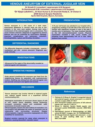
Under general anesthesia the aneurysm was freed from the
neighbouring tissues by separation with supraclavicular
incision. Swelling was arising from left external jugular vein
and saccular aneurysm was ligated and excised.
Venous aneurysm may involve internal or external jugular
vein, anterior jugular branch or a superficial cervical
communicating vein.
Histological examination of aneurysm shows layers of wall
of vein, elastic tissue dysplasia, intimal thickening,
increased connective tissue and endothelial cells,
prominence of smooth muscle cells.
Ultrasonography remains the best method of evaluation. It is
non-invasive and delineates the extent of aneurysm and its
relationship with surrounding structures. Doppler
ultrasonography, magnetic resonance angiography are other
modalities for assessment.
Surgical excision reserved for cases where aneurysm is
extensive, causing discomfort, cosmetic reason
A 35 year old female was admitted to our hospital with a
progressive swelling in the left side of the neck since 15
months with significant increase in size in the past 2
months prior to admission. The mass localized laterally
to the left sternocleidomastoid muscle. Physical
examination revealed a soft, round, mobile, non
pulsating mass approximately 8×6 cm in size. Other
systems were normal.
INTRODUCTION PRESENTATION
DISCUSSION
References
VENOUS ANEURYSM OF EXTERNAL JUGULAR VEIN
OPERATIVE FINDINGS
INVESTIGATIONS
Ultrasound of the region of the abnormality revealed an
external jugular vein aneurysm .
DIFFERENTIAL DIAGNOSIS
The differential diagnosis included a laryngocele, superior
mediastinal cyst and venous aneurysm. Diagnosis was
made clinically.