
Empfohlen
Weitere ähnliche Inhalte
Was ist angesagt?
Was ist angesagt? (20)
Ähnlich wie cardiac action potential
Ähnlich wie cardiac action potential (20)
Mehr von Anirudh Allam
Kürzlich hochgeladen
Kürzlich hochgeladen (20)
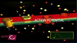
cardiac action potential
- 2. DEFINATION • Short term change in the electrical potential on the surface of a cell in response to stimulation and then leads to transmission of electrical impulse that travels across the cell membrane.
- 3. TERMS EXPLAINED • RESTING MEMBRANE POTENTIAL : It is the relative static membrane potential of quiescent cells . • THRESHOLD POTENTIAL : it is the critical level to which the membrane potential must be depolarized to initiate action potential .
- 4. WHAT MAKES IONS TO MOVE ACROSS? • Nernst equation • EK =RT/ZF ln [K]2 / [K]1 • Where, • T is temperature [370 C] • R is the gas constant • F is the Faraday constant • Z is the valence of ion [1] • [K]2 and [K]1 are the final concentrations of potassium in compartments 2 and 1, • respectively. [150mmol, 5 mmol] • EK is the equilibrium potential for potassium [-90mV] • At equilibrium potential net diffusion is 0 • All ions try to reach equilibrium i.e., tries to drive the membrane potential towards its equilibrium potential • At RMP, membrane is permeable mostly to potassium , hence RMP is close to the EK
- 6. CARDIAC ACTION POTENTIAL PHASES • FIVE PHASES : one phase of depolarization three phases of repolarization one phase of resting membrane potential diastolic depolarization
- 7. • Phase 0: the upstroke or rapid depolarization • Phase 1: early rapid repolarization • Phase 2: plateau phase • Phase 3: final rapid repolarization • Phase 4: resting membrane potential and diastolic depolarization
- 9. PHASE 0: THE UPSTROKE OR RAPID DEPOLARIZATION • Increased inward sodium currents (INa) • The rate at which depolarization occurs during phase 0 (Vmax) is reasonable approximation of the rate and magnitude of Na entry into the cell and when the equilibrium potential for Na (ENa 60mV) is reached, Na no longer enters the cell. • The membrane conductance of Na during phase 0 is hypothetically regulated by two types of gates— m and h
- 10. • Three ‘m’ (activation) gates on the extracellular side • One ‘h’ (inactivation) gate on the intracellular side of the membrane, which modulate Na passage through the sodium channels • When the membrane is in a resting polarized state, the ‘m’ gates are almost completely closed and ‘h’ gate is open and hence, no Na can enter the cell • Depolarization of the membrane opens the ‘m’ gates and closes the ‘h’ gate, ‘m’ gates opening faster than the ‘h’ gate closing i.e. activation of the Na channels proceeds faster than inactivation can occur and Na flows through for about 1–2 msec when both gates are simultaneously open
- 12. PHASE 1: EARLY RAPID REPOLARIZATION • It is partly owing to the inactivation of inward current ( I Na) and the activation of a transient outward current (Ito) carried through K channels. • Phase 1 is well defined in Purkinje fibers and some muscle fibers but is indistinct and not separated in SA and AV nodes.
- 13. PHASE 2: PLATEAU PHASE • The membrane voltage remains zero for more than 100 ms • Plateau phase is due to: Fall of K conductance Slow inward current (I Si) through Ca channels Small inward Cl (I Cl) flux through Cl channel.
- 14. PHASE 3: FINAL RAPID REPOLARIZATION • Final phase of rapid repolarization is due to: Time dependent inactivation of slow inward currents ISi and ICl so that intracellular movement of positive charges decreases Activation of outward K current (I k) • The net membrane current becomes more outward and the membrane potential shifts in a negative direction
- 15. PHASE 4: (A) THE RESTING MEMBRANE POTENTIAL • It is - 50 to - 95 mV depending on the cell type • The resting negative membrane potential is mainly due to inward K current (IKi ) • During diastole, the cell membrane is quite permeable to K and relatively impermeable to Na and the sodium pump (Na, K ATPase) pumps three Na out of the cell and two K into the cell which results in high intracellular K (150 mM) and low intracellular Na (15 mM)
- 16. • The Ca2 contributes little to the resting membrane potential although changes in the Ca2 concentration can affect the permeability of the cell membrane to other ions. • An increase in Ca2 increases the K conductance. Besides under normal conditions, one internal Ca2 is exchanged for three external Na by the Na/Ca2 exchanger.
- 17. PHASE 4: (B) DIASTOLIC DEPOLARIZATION • The membrane potential of the atrial and ventricular muscle cells remains steady throughout the diastole • In SA node, distal portion of AV node, muscles of mitral and tricuspid valves and Purkinje fibers the resting membrane potential does not remain constant in diastole but gradually depolarizes and when it reaches the threshold potential, it produces spontaneous action potential. • This property possessed by the spontaneously discharging cells is known as phase 4 diastolic depolarization and automaticity results when it leads to the initiation of action potential
- 18. IONIC BASIS OF ACTION POTENTIAL OF PACEMAKER CELLS • Phase 4: Pacemaker Potential: • Opening of voltage-gated Sodium channels called Funny channels (If or f channels ). • Closure of voltage-gated Potassium channels. • Opening of Voltage-gated Transient-type Calcium (T-type Ca2+ channels) channels . • Phase 0: The Rising Phase or Depolarization: • Opening of Long-lasting voltage-gated Calcium channels (L-type Ca2+ channels). • Large influx of Calcium. • Phase 3: The Falling Phase or Repolarization: • Opening of voltage-gated Potassium channels • Closing of L-type Ca channels. • Potassium Efflux.