SlideShare ist ein Scribd-Unternehmen logo
1 von 46
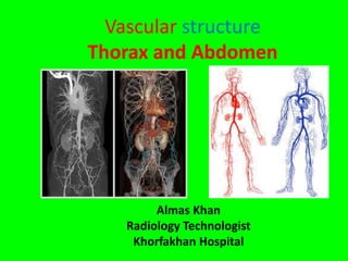
Vascular structure
Thorax and Abdomen
Almas Khan
Radiology Technologist
Khorfakhan Hospital
Vascular structure Thorax and
Abdomen
Talk plan:-
 Vascular system and Circulations
 Co relation with Anatomical landmark
 Common Indications.
Components of circulatory system
Circulatory system
Cardiovascular Lymphatic system
Cardio Vascular Lymph
(heart) (vessels) Lymph vessels
Lymphatic nodules
Pulmonary Systemic
( lung) ( body)
The circulatory system consists of cardiovascular and lymphatic components
.
The cardiovascular portion includes heart, blood , and vessels that
Transport the blood.
Blood vessels
• Arteries: carry blood away
from the heart
Arterioles – smallest
arteries , carrying blood
to tissue.
• Veins: carry blood back to
the heart
• Venules – smallest vein ,
returning blood from tissue.
• Capillaries: microscopic
blood vessels .
Aorta
Four segments of Aorta
1) Aortic bulb ( root )
2) Ascending aorta
3) Ascending arch
4) Descending aorta.
Branches of Arch of Aorta :-
1) Brachiocephalic artery
2 ) Left common carotid artery
3) Left subclavian artery
Vascular structure Head and neck
Thoracic Aorta and major branches
• The thoracic aorta begins at the level of vertebra T5 and
continue through to the diaphragm at the level of T12.
• Initially traveling within the mediastinum to the left of
vertebral column , as it passes through the thoracic region.
Thoracic aorta branches :
Visceral branches : Supply blood primarily to visceral organs of
thorax
• Bronchial artery
• Esophageal artery
• Pericardial artery
• Mediastinal artery
Parietal (somatic ) branches : blood to the muscles of thoracic
cavity and vertebral column
• Intercostal artery
• Superior phrenic artery.
Thoracic aorta branches :
• Each bronchial artery supplies systemic blood to the lung and
visceral pleura.
• Each pericardial artery supplies blood to the pericardium.
• Esophageal artery provides blood to the esophagus
• Mediastinal artery provides blood to the mediastinum.
• Each intercostal artery provides blood to the muscles of the
thoracic cavity and vertebral column.
• Superior phrenic artery provides blood to the superior surface
of the diaphram.
Abdominal Aorta and Major branches
• After crossing through the diaphragm at the aortic hiatus, the
thoracic aorta is called abdominal aorta.
• It formally ends at the level of L4 vertebra.
• It bifurcates to form the common iliac arteries.
• Abdomen aorta gives rise to several important branches.
• Celiac trunk:-
Also called celiac artery
a major branch of abdomen
aorta , gives rise to the gastric
artery , splenic artery , and
common hepatic artery that
forms the hepatic artery to the
liver
The right artery to the
stomach , cystic artery to the
gall bladder.
CHA: common hepatic artery; CT: celiac trunk; GDA:
gastroduodenal artery; LGA: left gastric artery; LHA: left
hepatic artery; RHA: right hepatic artery; SA: splenic
artery; SMA: superior mesenteric artery
Abdomen aorta Branches…..
• Left gastric artery : Branch of
celiac trunk ; supply blood to
stomach.
• Splenic artery : Branch of celiac
trunk ; supply blood to the
spleen.
• Common hepatic artery (CHA) :
Br of celiac trunk that forms the
hepatic artery, right gastric artery
and cystic artery.
• Hepatic artery proper : Br of CHA
; supplies systemic blood to the
liver.
• Right gastric artery : Br of CHA ;
supplies blood to the stomach.
• Cystic artery : Br of CHA ; supplies
blood to the gall bladder .
• Superior mesenteric artery : Br of
abdominal aorta ; suppilies blood
to the small intestine (duodenum,
jejunum, ileum) , pancreases and
a majority of the large intestine.
• Inferior meseneteric artery:
supplies blood to the distal
segment of the large intestine
and rectum.
• Inferior phrenic artery : supplies
blood to the inferior surface of
diaphragm.
• Adrenal artery: Br of
abdominal aorta ; supplies
blood to the adrenal
(suprarenal ) glands.
• Renal artery : Branches
approximately 2.5 cm inferior
to the superior mesenteric
arteries and supplies a kidney.
The right renal artery is longer
than the left since the aorta lies
to the left of vertebral column.
• Gonadal artery : Supplies blood
to the gonads ,or reproductive
organs.
• Lumbar arteries : Supply blood to
the lumbar region, the abdomen
wall , and the spinal cord.
• The aorta divided at the level of
vertebra L4 into left and right
Common iliac artery. But
continues as a small vessels , the
median sacral artery in to
sacrum.
• The common iliac arteries provides
blood to the pelvic region and lower
limbs
• They split into external and internal
iliac arteries.
• Internal iliac arteries: Supplies blood
to the urinary bladder , wall of pelvis
, external genitalia , medial portion of
the femoral region.
• External iliac artery: It leaves the
body cavity and become a femoral
artery, supplies blood to lower limbs
Thoracic vein
• The right atrium receives all of the systemic venous return.
• The major veins the chest are the superior vena cava , azygos and
pulmonary artery.
• Most of blood flow into either superior vena cava or inferior vena cava.
• Superior vena cava : Large systemic vein , blood from most areas superior
to the diaphragm , empties in to right atrium .
• Azygos vein : return blood
from posterior thoracic wall
to superior vena cava .
• The superior and inferior
pulmonary veins return
oxygenated blood from the
lungs to the left atrium.
• Inferior vena cava return
blood from the abdomen
and lower limbs to the right
atrium.
Abdominal veins
• Inferior vena cava : parallel the
abdominal aorta. Receives blood
from abdominal veins.
• This veins include the right and left
common iliacs , internal iliacs
external iliacs , renal veins and
hepatic portal systems.
• Superior and inferior mesenteric
veins return blood from the small
and large intestine through the
hepatic portal vein.
• Liver drains to each hepatic vein and
directly in inferior vena cava.
Tributaries Of The Inferior Vena Cava
Right
suprarenal vein
Right
gonadal vein
Hepatic veins
Inferior vena cava
External iliac
vein
Inferior phrenic
vein
Renal veins
Lumbar veins
Left ascending
lumbar vein
Left gonadal
vein
Common iliac
vein
Internal iliac
vein
Left suprarenal
vein
Major Branches Of The Abdominal Aorta
Adrenal
gland
Celiac trunk
Kidney
Abdominal
aorta
Diaphragm
Suprarenal
artery
Renal artery
Superior
mesenteric artery
Gonadal
artery
Inferior
mesenteric artery
Common iliac
artery
Hepatic portal system :
• Hepatic portal system :
• Contains substance absorbed
by the stomach and intestines
• Delivers these compounds to
the liver for
• Storage
• Metabolic conversion
• Excretion
• Nutrients from the digestive
tract enter the hepatic portal
vein
Tributaries of the
Hepatic Portal Vein
Inferior mesenteric vein:
drains part of large intestine
Splenic vein:
drains spleen, part of stomach, and pancreas
Superior mesenteric vein:
drains part of stomach, small intestine, and part of large
intestine
Left and right gastric veins:
drains part of stomach
Cystic vein:
drains gallbladder
Veins Of The Hepatic Portal System
Hepatic veins
Liver
Spleen
Gastric veins
Inferior vena cava
Splenic vein
Inferior
mesenteric vein
Superior
mesenteric vein
Large intestine
Hepatic portal vein
Small intestine
Rectum
The Hepatic Portal System
• A specialized part of the vascular circuit
• Picks up digested nutrients
• Delivers nutrients to the liver for processing
CT Angiogram
CT Angiography is a medical exam that combines a CT scan with
an injection of a contrast material to produce pictures of blood
vessels and tissue in a part of body.
CT Angiography to helps diagnose a narrowing or obstruction of
the arteries, an aneurysm, deep vein thrombosis, pulmonary
embolism, or other vascular condition.
MDCT angiography findings of the most common aortic diseases:
1. Congenital disorders
2. Inflammatory pathology
3. Aortic aneurysm
4. Complications of aortic aneurysm
- Rupture
- Impeding rupture
5. Acute aortic syndrome
- Aortic dissection
- Intramural hematoma
- Penetrating atherosclerotic ulcer
6. Traumatic injuries
7. Postsurgical complications
Locator position for Angiograms
Example of the regions of interest (ROIs) drawn on
the aorta (arrow) to obtain the CT values. ROI 1: Arch
of aorta at Th 7 level, ROI 2: abdominal aorta at L 1
level, and ROI 3: S 1 level.
CT Pulmonary Angiogram (CTA)
• localize the pulmonary trunk, which is
just at or below the level of the
bifurcation of the trachea (white lines).
• The resulting axial CT localization
image is shown on the right. An
elliptical ROI is placed in the right
pulmonary artery to measure the time
for contrast to reach this area from the
injection site.
CT Pulmonary Angiogram (CTA)
A picture of a heart model that displays the right atrium
(RA) that receives deoxygenated blood from the superior
vena cava (SVC). The blood then flows to the right
ventricle (RV)) and to the pulmonary trunk (PT). These
structures are also seen on the coronal CT image on the
right. Also labeled are the aorta (A) and left ventricle (LV).
CT Pulmonary Angiogram (CTA)
• These two axial CT images show
profound pulmonary emboli. On the
left the embolus almost completely
blocks the right pulmonary artery
(yellow arrow). Right image show an
extensive saddle embolus forming in
both pulmonary arteries and
becoming extensive. Both of these
types of pulmonary emboli are life
threatening and require immediate
medical attention.
Left- axial CT image demonstrate the
anatomical presentations of the
pulmonary trunk when normal in size,
caliber, and pressure (yellow arrow), and
on the right an example of a large
widened pulmonary trunk (pink arrow)
due to pulmonary hypertension.
MIP- and VRT-images:
steno-obstructive disease
Calcifications Aneurysms
CT Angiography
Aorta bolus tracking
time
CT Pulmonary study timing
• Post Inj delay : 7sec
• Post threshold : Automatic minimum
• Scan start time : 12 seconds
• HU :120 /150
• Locator : Just below the bifurcation
CT Abdomen bolus tracking timing
• Post Inj delay : 12
• Arterial : 18 to 20 seconds
• Portal venous phase : 70 sec
• Equilibrium : 120 sec
• CT KUB with Contrast
• Scan to start 5 to 10 mts from the start of Inj
• Post Inj delay : 8 sec
• Post threshold : 5 sec
• Scan start time : 15 sec
• HU : 120/150
• Locator : Arch of
aorta
Case Studies of
Radiology Department Khorfakhan Hospital
CONCLUSION
 We are able to understand how the circulation of blood is
organized in the thorax and Abdomen.
 Helps to identify vessels after injection of contrast.
 A good understanding of anatomy is essential while performing
angiography.
Normal and abnormal distribution of circulatory system
 Co relation with Anatomical landmark helps to identify vascular
structures of Thorax and Abdomen precisely .
Thanks for the motivation and guidance provided by our
Radiologists with out which we would not have achieved good
results.
Congrats and expecting continued best support from all my
colleagues.
Thank you

Weitere ähnliche Inhalte

Was ist angesagt?

Arterial supply of the Abdomen : Abdominal Aorta
Arterial supply of the Abdomen : Abdominal AortaArterial supply of the Abdomen : Abdominal Aorta
Arterial supply of the Abdomen : Abdominal AortaBitew Mekonnen
 
Vena cava anatomy and variants
Vena cava anatomy and variantsVena cava anatomy and variants
Vena cava anatomy and variantsMilan Silwal
 
Anatomy of the heart
Anatomy of the heartAnatomy of the heart
Anatomy of the heartAhmed Hammad
 
Anatomy of Blood vessels of abdomen pelvic cavities. Portacaval & Cavacaval A...
Anatomy of Blood vessels of abdomen pelvic cavities. Portacaval & Cavacaval A...Anatomy of Blood vessels of abdomen pelvic cavities. Portacaval & Cavacaval A...
Anatomy of Blood vessels of abdomen pelvic cavities. Portacaval & Cavacaval A...Eneutron
 
Mesentery, jejunum, ileum & superior mesenteric artery -PDF Lecture Notes -Dr...
Mesentery, jejunum, ileum & superior mesenteric artery -PDF Lecture Notes -Dr...Mesentery, jejunum, ileum & superior mesenteric artery -PDF Lecture Notes -Dr...
Mesentery, jejunum, ileum & superior mesenteric artery -PDF Lecture Notes -Dr...MUGUNTHAN Dr.Mugunthan
 
Presentation1.pptx, radiological anatomy of the abdomen and pelvis.
Presentation1.pptx, radiological anatomy of the abdomen and pelvis.Presentation1.pptx, radiological anatomy of the abdomen and pelvis.
Presentation1.pptx, radiological anatomy of the abdomen and pelvis.Abdellah Nazeer
 
Anatomy of the oesophagus
Anatomy of the oesophagusAnatomy of the oesophagus
Anatomy of the oesophagusGeorge Owusu
 
Great vessels artery
Great vessels arteryGreat vessels artery
Great vessels arteryRohit Paswan
 
Azygos system of veins
Azygos system of veinsAzygos system of veins
Azygos system of veinsIdris Siddiqui
 
Development of superior venacava and azygous vein
Development of superior venacava and azygous veinDevelopment of superior venacava and azygous vein
Development of superior venacava and azygous veinanuppslides
 

Was ist angesagt? (20)

Arterial supply of the Abdomen : Abdominal Aorta
Arterial supply of the Abdomen : Abdominal AortaArterial supply of the Abdomen : Abdominal Aorta
Arterial supply of the Abdomen : Abdominal Aorta
 
Mediastinum
Mediastinum Mediastinum
Mediastinum
 
Vena cava anatomy and variants
Vena cava anatomy and variantsVena cava anatomy and variants
Vena cava anatomy and variants
 
Anatomy of the heart
Anatomy of the heartAnatomy of the heart
Anatomy of the heart
 
Abdominal arota
Abdominal arotaAbdominal arota
Abdominal arota
 
Anatomy of the thorax
Anatomy of the thoraxAnatomy of the thorax
Anatomy of the thorax
 
Anatomy of Blood vessels of abdomen pelvic cavities. Portacaval & Cavacaval A...
Anatomy of Blood vessels of abdomen pelvic cavities. Portacaval & Cavacaval A...Anatomy of Blood vessels of abdomen pelvic cavities. Portacaval & Cavacaval A...
Anatomy of Blood vessels of abdomen pelvic cavities. Portacaval & Cavacaval A...
 
Mesentery, jejunum, ileum & superior mesenteric artery -PDF Lecture Notes -Dr...
Mesentery, jejunum, ileum & superior mesenteric artery -PDF Lecture Notes -Dr...Mesentery, jejunum, ileum & superior mesenteric artery -PDF Lecture Notes -Dr...
Mesentery, jejunum, ileum & superior mesenteric artery -PDF Lecture Notes -Dr...
 
Mediastinum
MediastinumMediastinum
Mediastinum
 
The pulmonary trunk
The pulmonary trunkThe pulmonary trunk
The pulmonary trunk
 
Blood supply of abdomen
Blood supply of abdomenBlood supply of abdomen
Blood supply of abdomen
 
Ductus deference
Ductus deferenceDuctus deference
Ductus deference
 
Presentation1.pptx, radiological anatomy of the abdomen and pelvis.
Presentation1.pptx, radiological anatomy of the abdomen and pelvis.Presentation1.pptx, radiological anatomy of the abdomen and pelvis.
Presentation1.pptx, radiological anatomy of the abdomen and pelvis.
 
Anatomy of the oesophagus
Anatomy of the oesophagusAnatomy of the oesophagus
Anatomy of the oesophagus
 
Great vessels artery
Great vessels arteryGreat vessels artery
Great vessels artery
 
Azygos system of veins
Azygos system of veinsAzygos system of veins
Azygos system of veins
 
Right ventricle
Right ventricleRight ventricle
Right ventricle
 
2 the heart
2 the heart2 the heart
2 the heart
 
Development of superior venacava and azygous vein
Development of superior venacava and azygous veinDevelopment of superior venacava and azygous vein
Development of superior venacava and azygous vein
 
Azygos system
Azygos systemAzygos system
Azygos system
 

Andere mochten auch

ARTERIAL AND VENOUS SUPPLY OF BRAIN
ARTERIAL AND VENOUS SUPPLY OF BRAIN ARTERIAL AND VENOUS SUPPLY OF BRAIN
ARTERIAL AND VENOUS SUPPLY OF BRAIN Battulga Munkhtsetseg
 
Presentation1.pptx, radiological vascular anatomy of the upper and lower limbs.
Presentation1.pptx, radiological vascular anatomy of the upper and lower limbs.Presentation1.pptx, radiological vascular anatomy of the upper and lower limbs.
Presentation1.pptx, radiological vascular anatomy of the upper and lower limbs.Abdellah Nazeer
 
Legg calve perthes
Legg calve perthesLegg calve perthes
Legg calve perthesAsish Rajak
 
Upper limbs arteries and veins
Upper limbs  arteries and veinsUpper limbs  arteries and veins
Upper limbs arteries and veinsLorenas13
 
Arterial Supply of Upper Limb
Arterial Supply of Upper LimbArterial Supply of Upper Limb
Arterial Supply of Upper LimbAsish Rajak
 
Vascular brain anatomy for Radiology by Dr Soumitra Halder
Vascular brain anatomy for Radiology by Dr Soumitra HalderVascular brain anatomy for Radiology by Dr Soumitra Halder
Vascular brain anatomy for Radiology by Dr Soumitra HalderSoumitra Halder
 
Neck triangles anatomy
Neck triangles anatomyNeck triangles anatomy
Neck triangles anatomyJamil Anwar
 

Andere mochten auch (9)

Axilla & brachial plexus
Axilla & brachial plexusAxilla & brachial plexus
Axilla & brachial plexus
 
ARTERIAL AND VENOUS SUPPLY OF BRAIN
ARTERIAL AND VENOUS SUPPLY OF BRAIN ARTERIAL AND VENOUS SUPPLY OF BRAIN
ARTERIAL AND VENOUS SUPPLY OF BRAIN
 
Presentation1.pptx, radiological vascular anatomy of the upper and lower limbs.
Presentation1.pptx, radiological vascular anatomy of the upper and lower limbs.Presentation1.pptx, radiological vascular anatomy of the upper and lower limbs.
Presentation1.pptx, radiological vascular anatomy of the upper and lower limbs.
 
Legg calve perthes
Legg calve perthesLegg calve perthes
Legg calve perthes
 
Celiac trunk
Celiac trunk Celiac trunk
Celiac trunk
 
Upper limbs arteries and veins
Upper limbs  arteries and veinsUpper limbs  arteries and veins
Upper limbs arteries and veins
 
Arterial Supply of Upper Limb
Arterial Supply of Upper LimbArterial Supply of Upper Limb
Arterial Supply of Upper Limb
 
Vascular brain anatomy for Radiology by Dr Soumitra Halder
Vascular brain anatomy for Radiology by Dr Soumitra HalderVascular brain anatomy for Radiology by Dr Soumitra Halder
Vascular brain anatomy for Radiology by Dr Soumitra Halder
 
Neck triangles anatomy
Neck triangles anatomyNeck triangles anatomy
Neck triangles anatomy
 

Ähnlich wie Vascular structure thorax and abdomen. Almas khan Khorfakkhan hospital sharjha uae.

The Anatomy of the Systemic Circulation.ppt
The Anatomy of the Systemic Circulation.pptThe Anatomy of the Systemic Circulation.ppt
The Anatomy of the Systemic Circulation.pptMiguelJohnson8
 
2 Cardio Vascular System.pptx
2 Cardio Vascular System.pptx2 Cardio Vascular System.pptx
2 Cardio Vascular System.pptxRamarGurusamy2
 
194 anatomy and function of the coronary arteries
194 anatomy and function of the coronary arteries194 anatomy and function of the coronary arteries
194 anatomy and function of the coronary arteriesSHAPE Society
 
circulation of blood.pptx
circulation of blood.pptxcirculation of blood.pptx
circulation of blood.pptxjosmin joy
 
The cardiovascular system.pptx
The cardiovascular system.pptxThe cardiovascular system.pptx
The cardiovascular system.pptxJagruti Marathe
 
L7 circulatory routes
L7 circulatory routesL7 circulatory routes
L7 circulatory routesReach Na
 
1 GNM - Anatomy unit - 4 - CVS by thirumurugan.pptx
1 GNM - Anatomy unit - 4 - CVS by thirumurugan.pptx1 GNM - Anatomy unit - 4 - CVS by thirumurugan.pptx
1 GNM - Anatomy unit - 4 - CVS by thirumurugan.pptxthiru murugan
 
cardiovascular disease.pptx
cardiovascular disease.pptxcardiovascular disease.pptx
cardiovascular disease.pptxAhmad Thanin
 
Presentation anatomy
Presentation anatomyPresentation anatomy
Presentation anatomyAfe Farizwen
 
Anatomy of the heart lecture.pptx
Anatomy of the heart lecture.pptxAnatomy of the heart lecture.pptx
Anatomy of the heart lecture.pptxSayaliPatil790915
 

Ähnlich wie Vascular structure thorax and abdomen. Almas khan Khorfakkhan hospital sharjha uae. (20)

The Anatomy of the Systemic Circulation.ppt
The Anatomy of the Systemic Circulation.pptThe Anatomy of the Systemic Circulation.ppt
The Anatomy of the Systemic Circulation.ppt
 
Git.pptx
Git.pptxGit.pptx
Git.pptx
 
2 Cardio Vascular System.pptx
2 Cardio Vascular System.pptx2 Cardio Vascular System.pptx
2 Cardio Vascular System.pptx
 
Cardio Vascular Anatomy & ECG
Cardio Vascular Anatomy & ECGCardio Vascular Anatomy & ECG
Cardio Vascular Anatomy & ECG
 
Anatomy and function of the coronary arteries
Anatomy and function of the coronary arteriesAnatomy and function of the coronary arteries
Anatomy and function of the coronary arteries
 
194 anatomy and function of the coronary arteries
194 anatomy and function of the coronary arteries194 anatomy and function of the coronary arteries
194 anatomy and function of the coronary arteries
 
194 anatomy and function of the coronary arteries
194 anatomy and function of the coronary arteries194 anatomy and function of the coronary arteries
194 anatomy and function of the coronary arteries
 
circulation of blood.pptx
circulation of blood.pptxcirculation of blood.pptx
circulation of blood.pptx
 
Cardiovascular System
Cardiovascular SystemCardiovascular System
Cardiovascular System
 
The cardiovascular system.pptx
The cardiovascular system.pptxThe cardiovascular system.pptx
The cardiovascular system.pptx
 
L7 circulatory routes
L7 circulatory routesL7 circulatory routes
L7 circulatory routes
 
circulation
circulationcirculation
circulation
 
1 GNM - Anatomy unit - 4 - CVS by thirumurugan.pptx
1 GNM - Anatomy unit - 4 - CVS by thirumurugan.pptx1 GNM - Anatomy unit - 4 - CVS by thirumurugan.pptx
1 GNM - Anatomy unit - 4 - CVS by thirumurugan.pptx
 
cardiovascular disease.pptx
cardiovascular disease.pptxcardiovascular disease.pptx
cardiovascular disease.pptx
 
2.2.pptx
2.2.pptx2.2.pptx
2.2.pptx
 
Presentation anatomy
Presentation anatomyPresentation anatomy
Presentation anatomy
 
A&P -The Cardiovascular System
A&P -The Cardiovascular SystemA&P -The Cardiovascular System
A&P -The Cardiovascular System
 
Anatomy of the heart lecture.pptx
Anatomy of the heart lecture.pptxAnatomy of the heart lecture.pptx
Anatomy of the heart lecture.pptx
 
Circulatory system
Circulatory systemCirculatory system
Circulatory system
 
2.1 blood circulation
2.1 blood circulation2.1 blood circulation
2.1 blood circulation
 

Mehr von almasmkm

Barium swallow. Srinivas Rao Khorfakkhan hospital
Barium swallow. Srinivas Rao  Khorfakkhan hospital Barium swallow. Srinivas Rao  Khorfakkhan hospital
Barium swallow. Srinivas Rao Khorfakkhan hospital almasmkm
 
Pacs administrator Flora M Atentciana
Pacs administrator Flora M AtentcianaPacs administrator Flora M Atentciana
Pacs administrator Flora M Atentcianaalmasmkm
 
Mri hazards & safety Daniel JP
Mri hazards &  safety  Daniel JPMri hazards &  safety  Daniel JP
Mri hazards & safety Daniel JPalmasmkm
 
Infection control in radiology Srinivas Rao
Infection control in radiology Srinivas RaoInfection control in radiology Srinivas Rao
Infection control in radiology Srinivas Raoalmasmkm
 
Dental ct imaging Srinivas rao
Dental ct imaging  Srinivas raoDental ct imaging  Srinivas rao
Dental ct imaging Srinivas raoalmasmkm
 
Biologic effects Daniel jayaprakash
Biologic effects Daniel jayaprakashBiologic effects Daniel jayaprakash
Biologic effects Daniel jayaprakashalmasmkm
 
Emergencies in radiology Daniel Jayaprakash
Emergencies in radiology Daniel JayaprakashEmergencies in radiology Daniel Jayaprakash
Emergencies in radiology Daniel Jayaprakashalmasmkm
 
Kidney anatomy - Almaskhan .khorfakhan hospital
Kidney anatomy - Almaskhan  .khorfakhan hospitalKidney anatomy - Almaskhan  .khorfakhan hospital
Kidney anatomy - Almaskhan .khorfakhan hospitalalmasmkm
 
Vascular structure head and neck Almas khan , Khorfakkhan hospital
Vascular structure head and neck      Almas khan , Khorfakkhan hospitalVascular structure head and neck      Almas khan , Khorfakkhan hospital
Vascular structure head and neck Almas khan , Khorfakkhan hospitalalmasmkm
 
fracture and dislocation ppt . Almas khan. khorfakkhan hospital dubai
fracture and dislocation ppt . Almas khan. khorfakkhan hospital dubaifracture and dislocation ppt . Almas khan. khorfakkhan hospital dubai
fracture and dislocation ppt . Almas khan. khorfakkhan hospital dubaialmasmkm
 

Mehr von almasmkm (10)

Barium swallow. Srinivas Rao Khorfakkhan hospital
Barium swallow. Srinivas Rao  Khorfakkhan hospital Barium swallow. Srinivas Rao  Khorfakkhan hospital
Barium swallow. Srinivas Rao Khorfakkhan hospital
 
Pacs administrator Flora M Atentciana
Pacs administrator Flora M AtentcianaPacs administrator Flora M Atentciana
Pacs administrator Flora M Atentciana
 
Mri hazards & safety Daniel JP
Mri hazards &  safety  Daniel JPMri hazards &  safety  Daniel JP
Mri hazards & safety Daniel JP
 
Infection control in radiology Srinivas Rao
Infection control in radiology Srinivas RaoInfection control in radiology Srinivas Rao
Infection control in radiology Srinivas Rao
 
Dental ct imaging Srinivas rao
Dental ct imaging  Srinivas raoDental ct imaging  Srinivas rao
Dental ct imaging Srinivas rao
 
Biologic effects Daniel jayaprakash
Biologic effects Daniel jayaprakashBiologic effects Daniel jayaprakash
Biologic effects Daniel jayaprakash
 
Emergencies in radiology Daniel Jayaprakash
Emergencies in radiology Daniel JayaprakashEmergencies in radiology Daniel Jayaprakash
Emergencies in radiology Daniel Jayaprakash
 
Kidney anatomy - Almaskhan .khorfakhan hospital
Kidney anatomy - Almaskhan  .khorfakhan hospitalKidney anatomy - Almaskhan  .khorfakhan hospital
Kidney anatomy - Almaskhan .khorfakhan hospital
 
Vascular structure head and neck Almas khan , Khorfakkhan hospital
Vascular structure head and neck      Almas khan , Khorfakkhan hospitalVascular structure head and neck      Almas khan , Khorfakkhan hospital
Vascular structure head and neck Almas khan , Khorfakkhan hospital
 
fracture and dislocation ppt . Almas khan. khorfakkhan hospital dubai
fracture and dislocation ppt . Almas khan. khorfakkhan hospital dubaifracture and dislocation ppt . Almas khan. khorfakkhan hospital dubai
fracture and dislocation ppt . Almas khan. khorfakkhan hospital dubai
 

Kürzlich hochgeladen

Back care and back massage. powerpoint presentation
Back care and back massage. powerpoint presentationBack care and back massage. powerpoint presentation
Back care and back massage. powerpoint presentationpratiksha ghimire
 
Information about acne, detail description of their treatment by topical and ...
Information about acne, detail description of their treatment by topical and ...Information about acne, detail description of their treatment by topical and ...
Information about acne, detail description of their treatment by topical and ...mauryashreya478
 
Professional Ear Wax Cleaning Services for Your Home
Professional Ear Wax Cleaning Services for Your HomeProfessional Ear Wax Cleaning Services for Your Home
Professional Ear Wax Cleaning Services for Your HomeEarwax Doctor
 
Clinical Education Presentation at Accelacare
Clinical Education Presentation at AccelacareClinical Education Presentation at Accelacare
Clinical Education Presentation at Accelacarepablor40
 
Mental Health for physiotherapy and other health students
Mental Health for physiotherapy and other health studentsMental Health for physiotherapy and other health students
Mental Health for physiotherapy and other health studentseyobkaseye
 
ILO (International Labour Organization )
ILO (International Labour Organization )ILO (International Labour Organization )
ILO (International Labour Organization )Puja Kumari
 
Preventing Common Nutritional Deficiencies In Poultry Flocks (PPT).pdf
Preventing Common Nutritional Deficiencies In Poultry Flocks (PPT).pdfPreventing Common Nutritional Deficiencies In Poultry Flocks (PPT).pdf
Preventing Common Nutritional Deficiencies In Poultry Flocks (PPT).pdfAditiAlishetty
 
Incentive spirometry powerpoint presentation
Incentive spirometry powerpoint presentationIncentive spirometry powerpoint presentation
Incentive spirometry powerpoint presentationpratiksha ghimire
 
unit-3 blood product B.Pharma 3rd year .pptx
unit-3 blood product B.Pharma 3rd year .pptxunit-3 blood product B.Pharma 3rd year .pptx
unit-3 blood product B.Pharma 3rd year .pptxBkGupta21
 
2024 HCAT Healthcare Technology Insights
2024 HCAT Healthcare Technology Insights2024 HCAT Healthcare Technology Insights
2024 HCAT Healthcare Technology InsightsHealth Catalyst
 
Learn Tips for Managing Chemobrain or Mental Fogginess
Learn Tips for Managing Chemobrain or Mental FogginessLearn Tips for Managing Chemobrain or Mental Fogginess
Learn Tips for Managing Chemobrain or Mental Fogginessbkling
 
Mobile Health And Apps (mhealth) How to design Application for medical App.pptx
Mobile Health And Apps (mhealth) How to design Application for medical App.pptxMobile Health And Apps (mhealth) How to design Application for medical App.pptx
Mobile Health And Apps (mhealth) How to design Application for medical App.pptxMahesh Chopra
 
Artificial Intelligence Robotics & Computational Fluid Dynamics
Artificial Intelligence Robotics & Computational Fluid DynamicsArtificial Intelligence Robotics & Computational Fluid Dynamics
Artificial Intelligence Robotics & Computational Fluid DynamicsParag Kothawade
 
The future of change - strategic translation
The future of change - strategic translationThe future of change - strategic translation
The future of change - strategic translationHelenBevan4
 
Leading big change: what does it take to deliver at large scale?
Leading big change: what does it take to deliver at large scale?Leading big change: what does it take to deliver at large scale?
Leading big change: what does it take to deliver at large scale?HelenBevan4
 
Exploring the Integration of Homeopathy and Allopathy in Healthcare.pdf
Exploring the Integration of Homeopathy and Allopathy in Healthcare.pdfExploring the Integration of Homeopathy and Allopathy in Healthcare.pdf
Exploring the Integration of Homeopathy and Allopathy in Healthcare.pdfDharma Homoeopathy
 
Lipid Profile test & Cardiac Markers for MBBS, Lab. Med. and Nursing.pptx
Lipid Profile test & Cardiac Markers for MBBS, Lab. Med. and Nursing.pptxLipid Profile test & Cardiac Markers for MBBS, Lab. Med. and Nursing.pptx
Lipid Profile test & Cardiac Markers for MBBS, Lab. Med. and Nursing.pptxRajendra Dev Bhatt
 
Champions of Health Spotlight On Leaders Shaping Denmark's Healthcare.pdf
Champions of Health Spotlight On Leaders Shaping Denmark's Healthcare.pdfChampions of Health Spotlight On Leaders Shaping Denmark's Healthcare.pdf
Champions of Health Spotlight On Leaders Shaping Denmark's Healthcare.pdfeurohealthleaders
 
ANTIGEN- SECTION IMMUNOLOGY DEPARTMENT OF MICROBIOLOGY
ANTIGEN- SECTION IMMUNOLOGY  DEPARTMENT OF MICROBIOLOGYANTIGEN- SECTION IMMUNOLOGY  DEPARTMENT OF MICROBIOLOGY
ANTIGEN- SECTION IMMUNOLOGY DEPARTMENT OF MICROBIOLOGYDrmayuribhise
 

Kürzlich hochgeladen (20)

Back care and back massage. powerpoint presentation
Back care and back massage. powerpoint presentationBack care and back massage. powerpoint presentation
Back care and back massage. powerpoint presentation
 
Information about acne, detail description of their treatment by topical and ...
Information about acne, detail description of their treatment by topical and ...Information about acne, detail description of their treatment by topical and ...
Information about acne, detail description of their treatment by topical and ...
 
Professional Ear Wax Cleaning Services for Your Home
Professional Ear Wax Cleaning Services for Your HomeProfessional Ear Wax Cleaning Services for Your Home
Professional Ear Wax Cleaning Services for Your Home
 
Clinical Education Presentation at Accelacare
Clinical Education Presentation at AccelacareClinical Education Presentation at Accelacare
Clinical Education Presentation at Accelacare
 
Mental Health for physiotherapy and other health students
Mental Health for physiotherapy and other health studentsMental Health for physiotherapy and other health students
Mental Health for physiotherapy and other health students
 
ILO (International Labour Organization )
ILO (International Labour Organization )ILO (International Labour Organization )
ILO (International Labour Organization )
 
DELIRIUM psychiatric delirium is a organic mental disorder
DELIRIUM  psychiatric  delirium is a organic mental disorderDELIRIUM  psychiatric  delirium is a organic mental disorder
DELIRIUM psychiatric delirium is a organic mental disorder
 
Preventing Common Nutritional Deficiencies In Poultry Flocks (PPT).pdf
Preventing Common Nutritional Deficiencies In Poultry Flocks (PPT).pdfPreventing Common Nutritional Deficiencies In Poultry Flocks (PPT).pdf
Preventing Common Nutritional Deficiencies In Poultry Flocks (PPT).pdf
 
Incentive spirometry powerpoint presentation
Incentive spirometry powerpoint presentationIncentive spirometry powerpoint presentation
Incentive spirometry powerpoint presentation
 
unit-3 blood product B.Pharma 3rd year .pptx
unit-3 blood product B.Pharma 3rd year .pptxunit-3 blood product B.Pharma 3rd year .pptx
unit-3 blood product B.Pharma 3rd year .pptx
 
2024 HCAT Healthcare Technology Insights
2024 HCAT Healthcare Technology Insights2024 HCAT Healthcare Technology Insights
2024 HCAT Healthcare Technology Insights
 
Learn Tips for Managing Chemobrain or Mental Fogginess
Learn Tips for Managing Chemobrain or Mental FogginessLearn Tips for Managing Chemobrain or Mental Fogginess
Learn Tips for Managing Chemobrain or Mental Fogginess
 
Mobile Health And Apps (mhealth) How to design Application for medical App.pptx
Mobile Health And Apps (mhealth) How to design Application for medical App.pptxMobile Health And Apps (mhealth) How to design Application for medical App.pptx
Mobile Health And Apps (mhealth) How to design Application for medical App.pptx
 
Artificial Intelligence Robotics & Computational Fluid Dynamics
Artificial Intelligence Robotics & Computational Fluid DynamicsArtificial Intelligence Robotics & Computational Fluid Dynamics
Artificial Intelligence Robotics & Computational Fluid Dynamics
 
The future of change - strategic translation
The future of change - strategic translationThe future of change - strategic translation
The future of change - strategic translation
 
Leading big change: what does it take to deliver at large scale?
Leading big change: what does it take to deliver at large scale?Leading big change: what does it take to deliver at large scale?
Leading big change: what does it take to deliver at large scale?
 
Exploring the Integration of Homeopathy and Allopathy in Healthcare.pdf
Exploring the Integration of Homeopathy and Allopathy in Healthcare.pdfExploring the Integration of Homeopathy and Allopathy in Healthcare.pdf
Exploring the Integration of Homeopathy and Allopathy in Healthcare.pdf
 
Lipid Profile test & Cardiac Markers for MBBS, Lab. Med. and Nursing.pptx
Lipid Profile test & Cardiac Markers for MBBS, Lab. Med. and Nursing.pptxLipid Profile test & Cardiac Markers for MBBS, Lab. Med. and Nursing.pptx
Lipid Profile test & Cardiac Markers for MBBS, Lab. Med. and Nursing.pptx
 
Champions of Health Spotlight On Leaders Shaping Denmark's Healthcare.pdf
Champions of Health Spotlight On Leaders Shaping Denmark's Healthcare.pdfChampions of Health Spotlight On Leaders Shaping Denmark's Healthcare.pdf
Champions of Health Spotlight On Leaders Shaping Denmark's Healthcare.pdf
 
ANTIGEN- SECTION IMMUNOLOGY DEPARTMENT OF MICROBIOLOGY
ANTIGEN- SECTION IMMUNOLOGY  DEPARTMENT OF MICROBIOLOGYANTIGEN- SECTION IMMUNOLOGY  DEPARTMENT OF MICROBIOLOGY
ANTIGEN- SECTION IMMUNOLOGY DEPARTMENT OF MICROBIOLOGY
 

Vascular structure thorax and abdomen. Almas khan Khorfakkhan hospital sharjha uae.

  • 1. Vascular structure Thorax and Abdomen Almas Khan Radiology Technologist Khorfakhan Hospital
  • 2. Vascular structure Thorax and Abdomen Talk plan:-  Vascular system and Circulations  Co relation with Anatomical landmark  Common Indications.
  • 3. Components of circulatory system Circulatory system Cardiovascular Lymphatic system Cardio Vascular Lymph (heart) (vessels) Lymph vessels Lymphatic nodules Pulmonary Systemic ( lung) ( body) The circulatory system consists of cardiovascular and lymphatic components . The cardiovascular portion includes heart, blood , and vessels that Transport the blood.
  • 4.
  • 5. Blood vessels • Arteries: carry blood away from the heart Arterioles – smallest arteries , carrying blood to tissue. • Veins: carry blood back to the heart • Venules – smallest vein , returning blood from tissue. • Capillaries: microscopic blood vessels .
  • 6. Aorta Four segments of Aorta 1) Aortic bulb ( root ) 2) Ascending aorta 3) Ascending arch 4) Descending aorta. Branches of Arch of Aorta :- 1) Brachiocephalic artery 2 ) Left common carotid artery 3) Left subclavian artery
  • 8. Thoracic Aorta and major branches • The thoracic aorta begins at the level of vertebra T5 and continue through to the diaphragm at the level of T12. • Initially traveling within the mediastinum to the left of vertebral column , as it passes through the thoracic region.
  • 9. Thoracic aorta branches : Visceral branches : Supply blood primarily to visceral organs of thorax • Bronchial artery • Esophageal artery • Pericardial artery • Mediastinal artery Parietal (somatic ) branches : blood to the muscles of thoracic cavity and vertebral column • Intercostal artery • Superior phrenic artery.
  • 10. Thoracic aorta branches : • Each bronchial artery supplies systemic blood to the lung and visceral pleura. • Each pericardial artery supplies blood to the pericardium. • Esophageal artery provides blood to the esophagus • Mediastinal artery provides blood to the mediastinum. • Each intercostal artery provides blood to the muscles of the thoracic cavity and vertebral column. • Superior phrenic artery provides blood to the superior surface of the diaphram.
  • 11.
  • 12. Abdominal Aorta and Major branches • After crossing through the diaphragm at the aortic hiatus, the thoracic aorta is called abdominal aorta. • It formally ends at the level of L4 vertebra. • It bifurcates to form the common iliac arteries. • Abdomen aorta gives rise to several important branches.
  • 13. • Celiac trunk:- Also called celiac artery a major branch of abdomen aorta , gives rise to the gastric artery , splenic artery , and common hepatic artery that forms the hepatic artery to the liver The right artery to the stomach , cystic artery to the gall bladder. CHA: common hepatic artery; CT: celiac trunk; GDA: gastroduodenal artery; LGA: left gastric artery; LHA: left hepatic artery; RHA: right hepatic artery; SA: splenic artery; SMA: superior mesenteric artery
  • 14. Abdomen aorta Branches….. • Left gastric artery : Branch of celiac trunk ; supply blood to stomach. • Splenic artery : Branch of celiac trunk ; supply blood to the spleen. • Common hepatic artery (CHA) : Br of celiac trunk that forms the hepatic artery, right gastric artery and cystic artery. • Hepatic artery proper : Br of CHA ; supplies systemic blood to the liver. • Right gastric artery : Br of CHA ; supplies blood to the stomach. • Cystic artery : Br of CHA ; supplies blood to the gall bladder .
  • 15.
  • 16. • Superior mesenteric artery : Br of abdominal aorta ; suppilies blood to the small intestine (duodenum, jejunum, ileum) , pancreases and a majority of the large intestine. • Inferior meseneteric artery: supplies blood to the distal segment of the large intestine and rectum. • Inferior phrenic artery : supplies blood to the inferior surface of diaphragm.
  • 17. • Adrenal artery: Br of abdominal aorta ; supplies blood to the adrenal (suprarenal ) glands. • Renal artery : Branches approximately 2.5 cm inferior to the superior mesenteric arteries and supplies a kidney. The right renal artery is longer than the left since the aorta lies to the left of vertebral column.
  • 18. • Gonadal artery : Supplies blood to the gonads ,or reproductive organs. • Lumbar arteries : Supply blood to the lumbar region, the abdomen wall , and the spinal cord. • The aorta divided at the level of vertebra L4 into left and right Common iliac artery. But continues as a small vessels , the median sacral artery in to sacrum.
  • 19.
  • 20. • The common iliac arteries provides blood to the pelvic region and lower limbs • They split into external and internal iliac arteries. • Internal iliac arteries: Supplies blood to the urinary bladder , wall of pelvis , external genitalia , medial portion of the femoral region. • External iliac artery: It leaves the body cavity and become a femoral artery, supplies blood to lower limbs
  • 21. Thoracic vein • The right atrium receives all of the systemic venous return. • The major veins the chest are the superior vena cava , azygos and pulmonary artery. • Most of blood flow into either superior vena cava or inferior vena cava. • Superior vena cava : Large systemic vein , blood from most areas superior to the diaphragm , empties in to right atrium .
  • 22. • Azygos vein : return blood from posterior thoracic wall to superior vena cava . • The superior and inferior pulmonary veins return oxygenated blood from the lungs to the left atrium. • Inferior vena cava return blood from the abdomen and lower limbs to the right atrium.
  • 23. Abdominal veins • Inferior vena cava : parallel the abdominal aorta. Receives blood from abdominal veins. • This veins include the right and left common iliacs , internal iliacs external iliacs , renal veins and hepatic portal systems. • Superior and inferior mesenteric veins return blood from the small and large intestine through the hepatic portal vein. • Liver drains to each hepatic vein and directly in inferior vena cava.
  • 24. Tributaries Of The Inferior Vena Cava Right suprarenal vein Right gonadal vein Hepatic veins Inferior vena cava External iliac vein Inferior phrenic vein Renal veins Lumbar veins Left ascending lumbar vein Left gonadal vein Common iliac vein Internal iliac vein Left suprarenal vein
  • 25. Major Branches Of The Abdominal Aorta Adrenal gland Celiac trunk Kidney Abdominal aorta Diaphragm Suprarenal artery Renal artery Superior mesenteric artery Gonadal artery Inferior mesenteric artery Common iliac artery
  • 26. Hepatic portal system : • Hepatic portal system : • Contains substance absorbed by the stomach and intestines • Delivers these compounds to the liver for • Storage • Metabolic conversion • Excretion • Nutrients from the digestive tract enter the hepatic portal vein
  • 27. Tributaries of the Hepatic Portal Vein Inferior mesenteric vein: drains part of large intestine Splenic vein: drains spleen, part of stomach, and pancreas Superior mesenteric vein: drains part of stomach, small intestine, and part of large intestine Left and right gastric veins: drains part of stomach Cystic vein: drains gallbladder
  • 28. Veins Of The Hepatic Portal System Hepatic veins Liver Spleen Gastric veins Inferior vena cava Splenic vein Inferior mesenteric vein Superior mesenteric vein Large intestine Hepatic portal vein Small intestine Rectum
  • 29. The Hepatic Portal System • A specialized part of the vascular circuit • Picks up digested nutrients • Delivers nutrients to the liver for processing
  • 30.
  • 31.
  • 32. CT Angiogram CT Angiography is a medical exam that combines a CT scan with an injection of a contrast material to produce pictures of blood vessels and tissue in a part of body. CT Angiography to helps diagnose a narrowing or obstruction of the arteries, an aneurysm, deep vein thrombosis, pulmonary embolism, or other vascular condition.
  • 33. MDCT angiography findings of the most common aortic diseases: 1. Congenital disorders 2. Inflammatory pathology 3. Aortic aneurysm 4. Complications of aortic aneurysm - Rupture - Impeding rupture 5. Acute aortic syndrome - Aortic dissection - Intramural hematoma - Penetrating atherosclerotic ulcer 6. Traumatic injuries 7. Postsurgical complications
  • 34. Locator position for Angiograms Example of the regions of interest (ROIs) drawn on the aorta (arrow) to obtain the CT values. ROI 1: Arch of aorta at Th 7 level, ROI 2: abdominal aorta at L 1 level, and ROI 3: S 1 level.
  • 35. CT Pulmonary Angiogram (CTA) • localize the pulmonary trunk, which is just at or below the level of the bifurcation of the trachea (white lines). • The resulting axial CT localization image is shown on the right. An elliptical ROI is placed in the right pulmonary artery to measure the time for contrast to reach this area from the injection site.
  • 36. CT Pulmonary Angiogram (CTA) A picture of a heart model that displays the right atrium (RA) that receives deoxygenated blood from the superior vena cava (SVC). The blood then flows to the right ventricle (RV)) and to the pulmonary trunk (PT). These structures are also seen on the coronal CT image on the right. Also labeled are the aorta (A) and left ventricle (LV).
  • 37. CT Pulmonary Angiogram (CTA) • These two axial CT images show profound pulmonary emboli. On the left the embolus almost completely blocks the right pulmonary artery (yellow arrow). Right image show an extensive saddle embolus forming in both pulmonary arteries and becoming extensive. Both of these types of pulmonary emboli are life threatening and require immediate medical attention. Left- axial CT image demonstrate the anatomical presentations of the pulmonary trunk when normal in size, caliber, and pressure (yellow arrow), and on the right an example of a large widened pulmonary trunk (pink arrow) due to pulmonary hypertension.
  • 38. MIP- and VRT-images: steno-obstructive disease Calcifications Aneurysms
  • 39. CT Angiography Aorta bolus tracking time CT Pulmonary study timing • Post Inj delay : 7sec • Post threshold : Automatic minimum • Scan start time : 12 seconds • HU :120 /150 • Locator : Just below the bifurcation CT Abdomen bolus tracking timing • Post Inj delay : 12 • Arterial : 18 to 20 seconds • Portal venous phase : 70 sec • Equilibrium : 120 sec • CT KUB with Contrast • Scan to start 5 to 10 mts from the start of Inj • Post Inj delay : 8 sec • Post threshold : 5 sec • Scan start time : 15 sec • HU : 120/150 • Locator : Arch of aorta
  • 40. Case Studies of Radiology Department Khorfakhan Hospital
  • 41.
  • 42.
  • 43.
  • 44. CONCLUSION  We are able to understand how the circulation of blood is organized in the thorax and Abdomen.  Helps to identify vessels after injection of contrast.  A good understanding of anatomy is essential while performing angiography. Normal and abnormal distribution of circulatory system  Co relation with Anatomical landmark helps to identify vascular structures of Thorax and Abdomen precisely .
  • 45. Thanks for the motivation and guidance provided by our Radiologists with out which we would not have achieved good results. Congrats and expecting continued best support from all my colleagues.