Optometry instruments
•Als DOCX, PDF herunterladen•
87 gefällt mir•28,687 views

Optometry instruments is a presentation to describe instrument in a beautiful way. use this tool to improve your knowledge. stay blessed. Regards Muhammad Akbar Rashid Qadri.

Empfohlen
Weitere ähnliche Inhalte
Was ist angesagt?
Was ist angesagt? (20)
Ähnlich wie Optometry instruments
Ähnlich wie Optometry instruments (20)
Mehr von Muhammad Akbar Rashid Qadri
Mehr von Muhammad Akbar Rashid Qadri (12)
Kürzlich hochgeladen
Kürzlich hochgeladen (20)
Optometry instruments
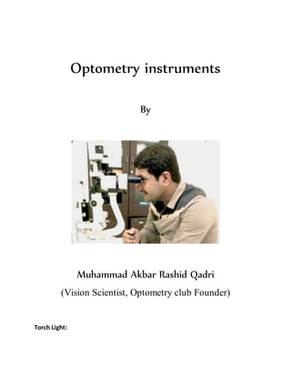
- 1. Optometry instruments By Muhammad Akbar Rashid Qadri (Vision Scientist, Optometry club Founder) Torch Light:
- 2. Torch light is used for: General eye examination To check pupil reactions 1) Hand HeldOccluder: Hand held occlude is used to block light rays to enter into the eye. Uses: Used to take monocular vision Used in cover/ uncover test Used in alternate occlusion method of binocular balancing
- 3. 2) Snellenchart: A Snellen chart is an eye chart that can be used to measure visual acuity. Snellen charts are named after the Dutch ophthalmologist Herman Snellen who developed the chart in 1862. Many ophthalmologists and vision scientists now use an improved chart known as the LogMAR chart but still mostly snellen chart is used in eye OPD’s. Snellen defined “standard vision” as the ability to recognize one of his optotypes when it subtended 5 minutes of arc. Thus the optotype can only be recognized if the person viewing it can discriminate a spatial pattern separated by a visual angle of one minute of arc. Outside of the United States, the standard chart distance is 6 metres (20 ft), and normal acuity is designated "6/6". Other acuities are expressed as ratios with a numerator of 6. Some clinics do not have 6 m eye lanes available, and either a half-size chart subtending the same angles at 3 metres (9.8 ft), or a reversed chart projected and viewed by a mirror is used to achieve the correct sized letters. In the most familiar acuity test, a Snellen chart is placed at a standard distance: 6 metres. At this distance, the symbols on the line representing "normal" acuity subtend an angle of five minutes of arc, and the thickness of the lines and of the spaces between the lines subtends one minute of arc. This line, designated 6/6 (or 20/20), is the smallest line that a person with normal acuity can read at a distance of 6 metres (20 ft). Three lines above, the letters have twice the height of those letters on the 6/6 (or 20/20 in the US) line. If this is the smallest line a person can read, the person's acuity is "6/12" ("20/40"), meaning that this person needs to approach to a distance of 6 metres (20 ft) to read letters that a person with normal acuity could read at 12m (40 feet). In an even more approximate manner, this person could be said to have "half" the normal acuity of 6/6. At exactly 6 meters distance from the patient, the letters on the 6/6 line shall subtend 5 minutes of arc (such that the individual limbs of the letters subtend 1 minute of arc), which means that the chart should be sized such that these letters are 8.73 mm tall and the topmost
- 4. (6/60) "E" should be 87.3 mm tall. Putting it another way, the eye should be at a distance 68.76 times the height of the top (6/60) letter. Available in different formats: English Urdu Pictures E charts for illiterate peoples All are based on snellen chart.
- 5. 3) Auto refractometer or Auto refractors: An autorefractor or automated refractor is a computer-controlled machine used during an eye examination to provide an objective measurement of a person's refractive error and prescription for glasses or contact lenses. This is achieved by measuring how light is changed as it enters a person's eye. Technique: The majority of autorefractors calculate the vision correction a patient needs (refraction) by using sensors that detect the reflections from a cone of infrared light. These reflections are used to determine the size and shape of a ring in the retina at the back of the eye. By measuring this zone, the autorefractor can determine when a patient’s eye properly focuses an image. Uses: In some offices, this process is used to provide the starting point for the ophthalmologist or optometrist in subjective refraction tests. Here, lenses are switched in and out of a phoropter and the patient is asked "which looks better" while looking at a chart. This feedback refines the prescription to one which provides the patient with the best vision. Automated refraction is particularly useful when dealing with non-communicative people such as young children or those with disabilities.
- 6. 4) Phoropter: A phoropter is an instrument used to test individual lenses on each eye during an exam. If, during an eye examination, your doctor has discovered a vision problem like nearsightedness, farsightedness or astigmatism, it's likely that one of the next steps you'll take will involve a phoropter. A phoropter is special machine used to switch multiple lenses in front of your eyes to correct your vision. Phoropters look imposing – like space-age visors – but are really an ingenious way to quickly determine the exact vision correction needed by your individual eyes. By having you look through the phoropter at a visual reference, image, or the “Big E” chart (the Snellen chart), your eye doctor will help guide you toward lenses that correct your vision impairment by switching lenses within the machine on the fly.
- 7. How does a phoropter work? he process of switching lenses in front of your eyes is less involved than it may look, given the imposing nature of the device. A phoropter is used to manually determine “refraction” – exactly how a lens must be shaped and curved to correct your vision to a normal state, nothing more. Phoropters are subjective however, based on your visual perception and response to your eye doctor's questions. Is your vision better, or worse? With this lens, or this lens? How about now? There are other procedures and technologies available that automatically measure the refraction needed within your eye and produce a "prescription" measurement without your input. These are called autorefractors and aberrometers.
- 8. 5) Trial Box: Trial Box is a box containing lenses, arranged in pairs, a trial spectacle frame, and other devices used in testing vision. It is also called as trial case. USES: OBJECTIVE REFRACTION SUBJECTIVE REFRACTION DIPLOPIA CHARTING DIAGNOSIS OF SQUINT ASSESS BINOCULAR VISION IT IS CONSIST OF TRIAL FRAME TRIAL LENS PRISM ACCESSORIES TRIAL FRAME: Trial frame an eyeglass frame designed to permit insertion of different lenses used in correcting refractive errors of vision. Features of Trial Frame: Light weight Adjustable It should have comfortable nose resting Readily adjustable and allow accurate centering vertically and horizontally for each eye.
- 9. Compartments of trial frame: 1st - High powered lens 2nd - spherical lens 3rd - cylindrical lens 4th - accessory lens & prisms Cylindrical lens compartment should be capable of smooth & accurate rotation • Trial frame should be easy to adjust of both PD & corneal alignment while providing a sure mounting for trial lens. Types of trial frame aperture: Full Aperture trial frame: Accommodates up to five 38mm lenses for each eye. Independent screw adjustments for PD of 48 to 80mm. Screw operated bridge height and projection. Sides adjustable for length and angle. Reduced aperture trial frame: Skeoch reduced aperture trial frame. A very durable lightweight drop cell trial frame. It holds up to four 38mm lenses for each eye. Accessories may be easily inserted and extracted.
- 10. Half eye Trial Frame: Half eye trial frame with nosepiece, child. As Half Eye Trial Frame with PD of 54 to 58mm. Also available with fixed bridge or adjustable nosepiece. Available for adults with PD 59 to 67 mm Trial lenses: During refraction the practitioner utilize a set of known lenses called as Trial lenses. Full Aperture Lens: Approximately 38 mm diameter Biconvex or biconcave form They do not confirm any of the additive lens principles Preferred by many practitioner Do not obscure patients face Disadvantage : heavier and thicker , large additive errors Reduced Aperture Lens: Lenses of 20 mm diameter mounted in the metal rim of 38 mm diameter. Plano convex & Plano concave Used for refraction and neutralization For Refraction, Curved surface should face the eye. For Neutralization, Curved surface of the trial lens is placed against to the curved surface of spectacle lens.
- 11. Spherical Lenses: All meridians have same power There are 32 pairs of spherical lens in plus & minus power Reduced & full aperture are available Power range of spherical Lenses: Pair of positive lenses ranging +0.12 - +20.00D Pair of negative lenses ranging -0.12 - - 20.00D 0.12 DS and 0.25 DS to 4.00 DS in 0.25 steps 0.50 DS to 8.00 DS in 0.50 steps 9.00 D to 14.00 DS in 1.00 steps 16.00 D to 20.00 D in 2.00 steps Uses of spherical lenses: For spherical ametropia For hand neutralization Checking the refracting error Cylindrical lenses: Power lies in one meridian Axis meridian is marked on rim of the lenses 19 pairs of cyl lenses
- 12. Power range in cylindricallenses: Pair of positive lenses ranging +0.12 - +6.00D Pair of negative lenses ranging -0.12 - - 6.00D 0.12 DC and 0.25 DC to 4.00 DC in 0.25 steps 4.50 DC to 6.00 DC in 0.50 steps Uses of cylindrical lenses: For correction of astigmatism For checking the refractive error Accessories of trial Box: Prism: Prism is a refractive medium having two plane surfaces inclined at an angle. And the principle of prism is “1 prism dioptres produces displacement of the image at 1 cm when the object is situated at the distance at 1m.” The uses of prism are to correct & measure Strabismus, Exercising prism, Measure the fusional range. For the measurement and correction of the angle of deviation, It is also used in instruments like Gonioscopy, Keratometer , slit lamp and applanation tonometer. Plano Lenses: zero power lenses, It is used for satisfy & identify the malingering patients.
- 13. Occluder: It is an opaque plastic disc to Occlude one eye To relax accommodation Used to dissociate fusion Used to close one eye while the other eye can be tested for visual acuity. Pin Hole Disc Opaque disc with pinhole of 1-2 mm diameter in its centre. Allows only a pencil of light pass through the corneas. Helps to determine whether eye has refractive or pathological errors. Pinhole of 1.32 mm is more effective. Usually available pinhole is 1mm in ordinary trial case. Principle of Pinhole: Pinhole creates a smaller blur circle on retina & thus improves the V/A It gives clue about potential visual acuity To find out if the loss of vision is due to an error of refraction or some organic lesion or a combination.
- 14. Maddox Rod: It is made up of several series of high plus Plano cylindrical lenses. Patient sees streak of light through this lens Available in red and white in colour. Used as single and double Maddox rod depends upon the test Uses of Maddox Rod: To detect heterophoria To detect cyclophoria To measure the squint deviation To detect orthophoria Stenopaeic Slit: It has slit of 1mm width & 25mm in length It allows strip of light to pass through the corneas Uses of Stenopaeic Slit: To find out axis of cylinder + or – Emsley fincham test To find out whether patient is having astigmatism Vertex distance also measure.
- 15. Red and Green Filters: Red in RE & Green in LE Used for color dissociation Used to find out suppression of eyes To find out diplopia To do worth 4 dot test & FRIEND test Used to measure the Fusion , squint 6) Near Vision Chart: It was introduced by Snellen. It is a photographic reduction of Snellen’s distant chart. Uses: Mainly used for visual acuity Also used to measure the near point of accommodation.
- 16. 7) Jackson Cross Cylinder: it’s naming derives from the fact that each of those cylinders can be considered as the combination of two equal yet opposite astigmatic lenses, placed vertically between them. The sphere in the cross-cylinder is double and of opposite power to the cylinder. The two most commonly used in everyday practice are the following. +0.25/ -0.50 (or -0.25/ +0.50) +0.50/ -1.0 (or -0.50/ +1.0). As the spherical equivalent of the cylinder is zero placing it in front of the patient doesn’t change the position of the Sturm cone, it can however reduce or increase the astigmatic error. We start by correcting the axis of the astigmatic error and continue by fine tuning the power. Correcting the astigmatic axis: We start the standard examination of visual acuity by placing the patient in front of a Snellen’s chart. We place the sphere and the cylinder as determined by another method on the test frame. We have the patient looking at smallest line he can see reasonably comfortably. The examiner
- 17. holds the instrument, with its handle being the projection of the astigmatic axis. In this way there is a positive and a negative correction on equal distance. By turning the cylinder we alter the angle of the astigmatism. This can help the patient lead us to the position where he sees a clearer image. When he does, we turn the cylinder towards the respective angle. If the patient wears positive cylinder, we turn the axis 5 degrees towards it and the opposite if the patient wears a negative one instead. We repeat the process until the patient doesn’t refer any difference in his vision. This is the correct axis. Correcting the power of astigmatism: With the axis in place, we can accurately tune the power of the cylinder. We do that by turning the axis of the cylinder parallel to that of the trial spectacles. By repeating the same process as previously we can increase or decrease the astigmatic lens in jumps equal to the power of the cylinder. When the patient perceives no difference the trial lens is correct. If the difference of the astigmatism found by the cross-cylinder method is more than 1.0D , we need to numerically subtract from the sphere half of the alteration found, in order to keep the spherical equivalent unaltered.
- 18. 7) Retinoscope: Retinoscopy is a technique to obtain an objective measurement of the refractive error of a patient's eyes. The examiner uses a retinoscope to shine light into the patient's eye and observes the reflection (reflex) off the patient's retina. While moving the streak or spot of light across the pupil the examiner observes the relative movement of the reflex or manually places lenses over the eye (using a trial frame and trial lenses) to "neutralize" the reflex. 8) Fan and Block: The Fan and Block test is used to determine the axis and magnitude of astigmatism. The fan is used to determine the presence of any astigmatismand its principal axes.
- 19. 9) Clock and Dial: The clock and dial test is used to determine the axis and magnitude of astigmatism. This is used to determine the presence of any astigmatismand its principal axes.
- 20. 10) Douchrome Test: A duochrome test is a test commonly used to refine the final sphere in refraction, which makes use of the chromatic aberration of the eye. Because of the chromatic aberration of the eye, the shorter wavelengths (green) are focused in front of the longer red wavelengths. 11) Slit Lamp: The slit lamp is an instrument consisting of a high-intensity light source that can be focused to shine a thin sheet of light into the eye. It is used in conjunction with a biomicroscope. The lamp facilitates an examination of the anterior segment and posterior segment of the human eye, which includes the eyelid, sclera, conjunctiva, iris, natural crystalline lens, and cornea. The binocular slit-lamp examination provides a stereoscopic magnified view of the eye structures in detail, enabling anatomical diagnoses to be made for a variety of eye conditions. A second, hand-held lens is used to examine the retina. Illumination: Diffuse illumination, Direct focal illumination, Specular reflection, Transillumination or retroillumination,
- 21. Indirect lateral illumination or Indirect proximal illumination and Sclerotic scatter. Accessoriesof Slit lamp: 90 D lens: For fundoscopy
- 22. Goldmann’s Applanation tonometer: for the measurement of IOP. 12) Ophthalmoscope: Ophthalmoscopy, also called funduscopy, is a test that allows a health professional to see inside the fundus of the eye and other structures using an ophthalmoscope (or funduscope). It is done as part of an eye examination and may be done as part of a routine physical examination. It is crucial in determining the health of the retina, optic disc, and vitreous humor. The pupil is a hole through which the eye's interior will be viewed. Opening the pupil wider (dilating it) is a simple and effective way to better see the structures behind it. Therefore, dilation of the pupil (mydriasis) is often accomplished with medicated eye dropsbefore funduscopy. However, although dilated fundus examination is ideal, undilated examination is
- 23. more convenient and is also helpful (albeit not as comprehensive), and it is the most common type in primary care. 13) B-scan Medical ultrasound (also known as diagnostic sonography or ultrasonography) is a diagnostic imaging technique based on the application of ultrasound. It is used to see internal body structures such as tendons, muscles, joints,blood vessels and internal organs. Its aim is often to find a source of a disease or to exclude any pathology. The practice of examining pregnant women using ultrasound is called obstetric ultrasound, and is widely used. B-scan ultrasonography, or B-scan, is a diagnostic test used in optometry and ophthalmology to produce a two-dimensional, cross-sectional view of the eye and the orbit. It is otherwise called brightness scan. It is commonly used to see inside the eye when media is hazy due to cataract or any corneal opacity.
- 24. 14) Biometry A scan Keraometry A scan: Used to measure axial length of the eye) Keratometer: Used to find out Dioptric power of the cornea and radius of curvature of the cornea
- 25. 15) Fundus camera: A fundus camera is a specialized low power microscope with an attached camera. Its optical design is based on the indirect ophthalmoscope. Fundus cameras are described by the angle of view - the optical angle of acceptance of the lens. It is used to take pictures of the retina. 16) OCT: Optical coherence tomography (OCT) is a non-invasive imaging test. OCT uses light waves to take cross-section pictures of your retina. With OCT, your ophthalmologist can see each of the retina's distinctive layers.
- 26. 17) Perimeter: Perimetry is the systematic measurement of visual field function. The two most commonly used types ofperimetry are Goldmann kinetic perimetry and threshold static automated perimetry. With Goldmann or "kinetic" perimetry, a trained perimetrist moves the stimulus; stimulus brightness is held constant.