Contrast media used with ct
•Als PPTX, PDF herunterladen•
59 gefällt mir•21,655 views

description of the IV and oral contrast used in the CT ( diagnostic imaging )

Empfohlen
Weitere ähnliche Inhalte
Was ist angesagt?
Was ist angesagt? (20)
Ähnlich wie Contrast media used with ct
Ähnlich wie Contrast media used with ct (20)
Mehr von DR Laith
Mehr von DR Laith (20)
Kürzlich hochgeladen
Kürzlich hochgeladen (20)
Contrast media used with ct
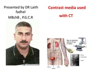
- 1. Presented by DR Laith fadhel MBchB , P.G.C.R Contrast media used with CT
- 2. Contrast media used with CT Presented by DR Laith fadhel
- 3. Information About Intravenous and Oral Contrast Used in CT • During many CT examinations, patients may be asked to take a special contrast agent (orally, rectally or via injection). Intravenous, oral , rectal and intra articular CT contrast are pharmaceutical agents (liquids) and are sometimes referred to as "dye". • CT contrast is used to make specific organs, blood vessels and/or tissue types "stand out" with more image contrast to better show the presence of disease or injury. Thus CT contrast highlights specific areas of the resultant CT image or "dyes" it. • In 1923 1st report of opacification of urinary tract after IV injection of Sodium iodide solution as treatment of syphilis by Osborne
- 4. Types of contrast There are four types of contrast agent used in CT 1. The type that is given via intravenous injection 2. The type that is given orally 3. The type that is given rectally 4. A much less common type of contrast used in CT as intra articular ( arthrography ) or inhaled as a gas and used for special lung and brain imaging. This technique (called Xenon CT) is only available at a small number of locations throughout the world and is only performed for rare cases.
- 5. How does CT Contrast Work? Iodide ( intravenous , other ) • Once the iodine contrast has been injected into the blood stream, it circulates through the heart and passes into the arteries, through the body's capillaries and then into the veins and back to the heart. As CT images are being acquired, the CT's x-ray beam is attenuated (weakened) as they pass through the blood vessels and organs flush with the contrast. This causes the blood vessels and organs filled with the contrast to "enhance" and show up as white areas on the x-ray or CT images. The kidneys and liver eliminate the contrast from the blood. Barium sulphate , Gastrografin (oral ) • Barium and Gastrografin are made up of substances which weaken (attenuate) x-rays. The oral contrast is swallowed and travels into the stomach and then into gastrointestinal tract. During the CT exam which follows, the CT x-ray beam is attenuated (weakened) as it passes through the organs containing the contrast, for example, the large intestine. The organs filled with the contrast are then "enhanced" and appear as highlighted white areas on the CT images.
- 6. Iodide compound contrast Its mainly classify according to the osmolarity , ionic or none ionic The osmolarity of blood about { 290 moSml/kg • Ionic – HOCM 1- Diatrizole ( urograffin hypaque ) 1500 moSml/kg 2- Metrizoate ( Isopaque ) 3- Iothalamate ( Conray ) 1500 1500 1- Ioxaglate ( hexabrix ) 490 1- HOCM (high osmolar contrast media ) • Ionic -- LOCM 1200-2000 moSml/kg 2- LOCM ( low osmolar contrast media ) • None Ionic – LOCM 350-500 moSml/kg 3- IOCM ( Iso osmolar contrast media ) 300 moSml/kg Note / Iso and Low osmolar are safer 5-10 than HOCM , and well tolerate 1- Iopamidole ( Niopam , Isovue ) 2- Iohexol ( Ominpaque ) 3- Iomeprol ( Iomeron ) 4- Ioversol ( Optiray ) 5- Iopromide ( Ultravist ) • None Ionic – ISO 1- Iotrolan ( Isovist ) 2- Iodixanol ( visipaque ) • Chapman / page 27 470 “ “ “ “ • Chapman / page 25 300 “
- 8. Dose of IV contrast and time of scan Using { 300 mg / ml } will depending of area examine -- ex • Head 150 ml • Chest or abdomen 100 ml • Children 2ml / kg • • • • • • • • • • •Chapman / page 12 , 85,106,109,140,269 In the scanning of abdomen the contrast inject 2ml/s Chest scanned after 20 s of start injection of contrast In abdomen or pelvis scanned after 30 s in arterial phase , 60 s in portal venous phase For CT angiography bolus tracking of contrast injected giving optimal acquisition timing In CT of pancreas , we use –Ve contrast like water , + Ve contrast ( iodinated ) , oral contrast / scan after 40 s In cholangiography , contrast infusion for 50 minute / scan after 35 minute In T-tube use( LOCM 150 , HOCM) 20ml during operation , and 10 day post op In CT urogram ( LOCM 300 ) 100-150 ml san with 3 mm slice thickness in 2 minute and 10 minute after contrast injection In CT arthrography ( LOCM ) 15 ml in shoulder , 6ml in elbow , 3 ml in wrist , double –contrast 4ml iodine + 40ml air CT contrast injectors
- 9. Adverse effect of IV contrast media • Adverse effect of none ionic iodinated CM are rare , occurring less than 1% • Sever or very sever adverse reaction occur in about 0.044% • The toxicity is due to function of osmolarity , and chemical structure of ionic CM •This reaction include 1- flushing , nausea , metal taste in moth , 2- peripheral burning , rigors 3- urticaria , warm , pain , abdominal pain 4- bronchospasm , none cardiac pulmonary edema 5- arrhythmias , hypotension 6- nephrotoxicity , CIN 7- hematological crisis in sickle cell patient 8- neurotoxicity , thyroid crisis in thyrotoxicosis patient 9- fetal reaction occurring in about 1.1-1.2 per million Identification of patient in high risk of anaphylactic reaction to IV CM • • • Previous reaction to CM Asthma previous allergic reaction • Special concern in patient complying from • • • • • renal insufficiency GFR < 30 ml/min , Serum creatinine > 130 µ mol/l DM , metformin drug Old age , Cardiovascular disease Thyrotoxicosis ,myasthenia gravis pheochromcytoma , sickle cell disease • Chapman / page 27-30
- 10. Barium compound contrast • Its made from barium sulphate with small particles size ( 0.1-3 mm ) none ionic suspension of 5.3 pH • barium carbonate is poisonous • mainly classify according to the density * E-Z Cat 1-2% is used in all GIT CT scan • Chapman / page 50 • Baritop 100 ( 100% all part of GIT ) • EPI -C ( 150% large bowel ) • E-Z HD ( 250 % esophagus , stomach and duodenum ) • E-Z paque ( 100% small bowel ) • Micropaque DC ( 100% eso , stoma , duo ) • Micropaque liquid ( 100% small and large bowel ) • Micropaque powder ( 76% small and large bowel ) • Polibar ( 115% large bowel ) • Polibar rapid ( 100% large bowel ) • Chapman / page 50
- 11. Dose of oral contrast and time of scan • Using of water -soluble 20 ml {Urograffin 150 , gastromiro } diluted in 1 litter orange squash • Barium suspension – low density ( 2% w/v ) • Adult dose for abdomen & pelvis ( 1000 ml ) gradually over 1 h before scan • upper abdomen , pancreas ( 500 ml ) gradually ½ h before scan • In Large bowel scan we must give the contrast before 4 h or before night / also use of CO2 for distention of bowel +20 mg buscopan + 1mg glucagon IV using IV contrast and low dose ( 80 mA ) CT technique scan at 70 s • in children over 10 years as adult • newborn 60-90 ml • below 1 year 120-240 ml • 1—5 years 240-360 ml • 5 –10 tears 360- 480 ml • Full dose 1 h before scan and ½ dose immediately prior scan •Chapman / page 13 ,86