
Recommended
More Related Content
What's hot
What's hot (20)
Similar to PRINCIPLES OF ACID BASE BALANCE
Similar to PRINCIPLES OF ACID BASE BALANCE (20)
More from Dr. Sindhu K., Asst. Prof., Dept. of VPT, VCG.
More from Dr. Sindhu K., Asst. Prof., Dept. of VPT, VCG. (20)
Recently uploaded
Recently uploaded (20)
PRINCIPLES OF ACID BASE BALANCE
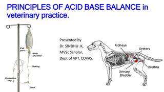
- 1. PRINCIPLES OF ACID BASE BALANCE in veterinary practice. Presented by Dr. SINDHU .K, MVSc Scholar, Dept of VPT, COVAS.
- 2. Water balance • Next to oxygen, water is most important for life. • Water & solutes (electrolytes & non-ionized organic molecules) constitutes body fluids, which are essential various physiological processes and maintenance of homeostasis. • Under normal steady state conditions, the fluid intake is carefully matched by equal output from the body to prevent body fluids volumes from increasing or decreasing. • Approximately 60% of a normal animal`s body weight is composed of water, with variations between and within species.
- 3. `• Electrolytes are compounds which exists as charged particles in aqueous solution and conduct a current in electricity. • The positively charged particles = Cations • The negatively charged particles = Anions • ECF contains mainly Na+, Cl-, HCO3- & HPO4`2- • ICF contains mainly K+, HCO3-, PO4`3-, SO4`2- & citrate. • These electrolytes dissolve in body fluids fulfil vital roles in virtually all of life process like nerve conduction, muscle contraction, metabolic reactions. • The balance between acidity & alkalinity in animal body is referred as acid base balance. & it is important homeostatic mechanism determined mainly by H+ ion concentration in various body fluids • Kidneys (excretory system) & circulating blood plays an important role in maintenance
- 4. `• Obese & older animals tends to have a smaller percentage of water, whereas neonates can have as much as 80% of their body weight as water. • Of 60% water, approximately 40% is with in the cells called ICF- Intra Cellular Fluids, and remaining 20% is outside the cells referred to as ECF- Extra Cellular Fluids. • The ECF may be further divided into intravascular fluids (blood plasma), extravascular fluids (interstitial fluid, lymph), inaccessible bone fluid(water trapped in deep layers of bone not readily exchangeable) & transcellular fluids(secretions of glandular tissues, GIT fluids, respiratory fluids, aqueous humour, peritoneal fluid, cerebrospinal fluid, etc) • Although ECF & ICF differ markedly in electrolyte composition, they are in osmotic equilibrium and water is freely diffusible between them.
- 5. Renal functions main function is excretion of waste products – urea, uric acid & creatinine acid-base balance regulation of salt & electrolyte content & volume of extracellular fluid production and release of hormones, autacoids, enzymes by kidney play a vital role in control of systemic blood pressure & RBC production
- 7. .Glomerular filtration – process by which water & small solutes are passed from the lumen of glomerular capillary to the space of Bowman`s capsule. force for glomerular filtration = hydrostatic pressure (60% of arterial pressure) 99% of glomerular filtrate is reabsorbed from tubular portion of Nephron. This mainly occurs to Reabsorb certain essential constituents like water, electrolytes & nutrients present in the glomerular filtrate, which have to be conserved. The Renin-Angiotensin-Aldosterone-System controls GFR & renal blood flow.
- 9. Tubular mechanism of renal epithelial transport simple diffusion Channel mediated diffusion (Na+, K+,Cl- solutes diffuses passively through ion channels/pores) Solvent drag/ convection solute flow (through aqueous pores by bulk flow) Facilitated transport/ uniport (solute binds to carrier protein & transported down electrochemical gradient without utilization of energy) Active transport/ primary active transport (carrier mediated process in which solute is transported against electrochemical gradient with ATP hydrolysis providing driving force) Secondary active transport – symport/ co-transport (Na+ -glucose, Na+ - Pi, Na+ - AA`s) -- antiport / counter-transport (Na+ -H+ counter transport)
- 11. Tubular transport Tubular cells perform the active secretory & resorptive functions, allowing passive diffusion of solute in direction appropriate to its electrochemical gradient. most important tubular mechanisms of electrolyte transport are = reabsorption of Na+, Cl- = secretion of H+, K+ separate mechanism for the transport of Ca++, Mg++, phosphate, Sulphate & organic acids and bases.
- 13. Approximate volumes of selected fluid compartments in DOGS COMPARTMENT % BODY WEIGHT METHOD Total Body Water TBW 60 Indicator substance Extra Cellular Fluid ECF 20-27 Indicator substance Red Blood Cells RBC 3 Counted + calculations Plasma Volume PV 5 Indicator substance Total blood volume BV 5.7-10 RBC volume + PV Interstitial lymph fluid 15 ECF – BV Transcellular fluid 1-6 Estimated Bone & dense connective tissue 5 Estimated ICF 33-40 TBW - ECF
- 15. Fluid therapy serves to correct – 1.Dehydration. 2.Acidosis and alkalosis. 3.Electrolyte deficiencies. 4.Nutrition and calorie.
- 16. Approximate values for blood volumes of various animals expressed as percentages of body weight SPECIES TOTAL BLOOD VOLUME PLASMA VOLUME RBC VOLUME Dogs 8.5 4.5 4.0 Cats 6.7 4.7 2.0 Chickens 6.5 4.5 2.0 Cattle 5.7 3.8 1.9 Goats 7.0 5.4 1.6 Horses Draft Through bred Saddle 7.0 10 7.5 4.0 6.0 5.2 3.0 4.0 2.5 Pigs 7.5 4.8 2.7 Sheep 6.5 4.5 2.0
- 17. WATER, SODIUM, & CHLORIDE. HOMEOSTASIS: Daily intake of water, nutrients, & minerals is normally balanced by daily excretion of these substances. water-turnover: term used to describe input/output of body water over a given period of time Value for water turnover in various domestic animals resting in cages /stalls range from about 40-132 mL/kg/day. Maintenance fluid needs: the volume of fluid required daily to maintain an animal in zero fluid balance, i.e., no net gain / loss of water. Normal water intake : occurs in response to thirst, which is stimulated by plasma hypertonicity &/ contracted ECF
- 18. Degrees of severity of dehydration and guidelines for assessment Body weight loss (%) Sunken eyes Shrunken face Skin fold test persists for (sec.) PCV (%) Fluid required to replace volume deficit (ml/kg bw) 4-6 Barely detectable -- 40-45 20-25 6-8 + + 2-4 50 30-50 8-10 + + + 6-10 55 50-80 10-12 + + + + 20-45 60 80-120
- 19. Dehydration : classification • Isotonic - mild dehydration with sodium loss. Observed in simple enteritis, copious sweating and nephrosis. • Hypertonic - mild dehydration without sodium loss. Observed in simple deprivation of water. • Hypotonic - severe dehydration with sodium loss. Observed in colibacillosis and salmonellosis.
- 20. Assessment of dehydration Mild Moderate Severe Skin Good elasticity Decreased elasticity No elasticity Eyes Slightly Sunken Bright Sunken Slightly Duller Sunken Deeply Dry Cornea Mouth Moist, Warm Sticky or Dry Dry , Cold Cyanotic Body weight decrease 4-6% 8% 10%
- 21. ,
- 22. Parameters to be monitored during fluid therapy • For the desired maximal response to occur with therapy the veterinarian must be familiar with following – 1. Cause and pathogenesis of dehydration. 2. Mechanism of fluid and electrolyte balance. 3. Composition and dosage of electrolyte solutions for treatment. 4. Blood parameters viz PCV & total proteins 5. Hemodynamics - Mean Arterial Pressure, Central Venous Pressure, Mean Pulmonary Arterial Pressure, Pulmonary Capillary Wedge Pressure 6. Urinary output. 7. Arterial pH & Arterial pCO2 8. Normal bronchovesicular lung sounds on auscultation
- 23. balanced electrolyte solutions are indicated for dehydration in large animals 1. Lactated Ringer ….. Commercially available. 2. Acidosis solution ….. NaCl – 21 g NaHCo3 – 18 g For 4 liters. KCL – 3 g 3. Ringer’s solution ….. NaCl – 3.4 g KCl – 1.2 g For 4 liters. CaCl2 – 1.3 g 4. Alkalosis solution ….. NaCl – 34 g KCL – 3 g MgSo4 – 1 g For 4 liters. Calcium gluconate – 4 g
- 24. Routes of administration Oral route : Always prefer isotonic fluid Intravenous route : 1. Always prefer isotonic solutions 2. Prefer in severe disturbances of fluid and electrolytes. 3. Do not use hypotonic solutions. Subcutaneous route: 1. only isotonic solutions are used. 2. If periphery is not cold, then fluid will be absorbed into the system in about 5-6 hours. 3. Contraindicated during oedematous conditions. Intraperitoneal route : 1. Asepsis is more important 2. Large quantities of fluid can be given. 3. Safest route for administration
- 25. Clinical observation during fluid therapy Until urine flow restored rate will be parallel to severity of dehydration, first rapidly then slowly. First hour ; 15 ml – 20 ml / kg body weight / hour. Second hour ; 10ml/kg body weight /hour continuously. If no urine is voided within 1 hour reduce to rate of fluid approximately half the quantity. • Clinical observation is very important during the administration of fluids.
- 26. Factors considered for fluid therapy 1.Types of illness and severity of conditions 2.Degree of dehydration 3.Conditions of patient 4.Organic functions of patient 5. Type of electrolyte imbalance
- 27. Drug contraindications Do not mix sulpha drugs with calcium and dextrose solutions. Do not mix oxytetracycline with calcium solutions. Try to avoid mixing of too many drugs in fluids. Do not mix chloramphenicol with vitamin B complex to the solutions.
- 28. Commercially available fluids 1. ELECTROLYTE SOLUTIONS: (ISOLYTE, PREMOLYTE, DEXTROSELYTE) designed for daily water and electrolyte maintenance and also for the replacement of loses. 2. DEXTROSE SOLUTIONS: It is used in prophylaxis and treatment of ketosis in starvation, diarrhea, vomiting or high fever. 3. PLASMA VOLUME EXPANDER: (DEXTRAN in 0.9% SODIUM CHLORIDE) . It is indicated in shock due to decreased effective blood volume, severe dehydration and surgical procedures and anesthesia. :
- 29. Clinical conditions I. Hyponatraemia: ( serum conc of Na+ < 140mEq/L in dogs) II. Hypernatraemia: ( dogs Na+ > 155mEq/L & cats > 160mEq/L ) III. Hypochloremia: ( excessive loss/sequestration of fluids) IV. Hyperchloremia: (hypernatremic animals due to loss of free H2O) V. Hypokalemia: ( serum K+ conc < 3.5mEq/L ) VI. Hyperkalemia: ( serum K+ conc > 6 mEq/L ) VII. Hypocalcemia: ( total serum Ca+ < 10-11mg/dL dogs ) VIII.Hypophosphatemia: ( post-parturient Haemoglobinuria ) IX. Hypomagnesaemia: (plasma level of Mg++ < 1.5-2.5 mEq/L )
- 30. Hyponatraemia • Etiology: • acute diarrhea in horses and calves, bladder rupture in new born foals, chronic wasting disease, intrinsic kidney disease and diuresis, gastrointestinal fistula, severe haemorrhage and excessive sweating. • In small animal’s hypoadrenocorticism, post obstructive diuresis, diuretic treatment, congestive heart failure, severe liver disease and nephritic syndrome. • Treatment: • 1) 5% sodium bicarbonate • 2) Lactated Ringers solution (precursor of bicarbonate). • 3) Normal saline • 4) 5% saline (avoided if acidosis is present) • Calcualtion Na+ requirement for replacement therapy in mEq • • = 140 – measured plasma sodium X weight in Kgs • 3 •
- 31. Hypernatraemia Etiology: In large animals - prolonged exposure to dry heat, respiratory loss with fever, low intake of water, excessive salt intake with adequate water. In small animals - pure water loss, diabetes insipidus, hypertonic NaHCO3 administration, cardiac arrest, feline urethral obstruction, acute renal failure. Types – hypervolaemic, hypovolaemic, isovolaemic Treatment : 1) Intake of fresh water in sufficient quantities. 2) 5% dextrose or maintenance fluid intravenously. 3) Salt poisoning – loop diurectics.
- 32. hypochloremia • occurs as a result of an increase in the net loss of electrolyte in the intestinal tract in acute intestinal obstruction, dilatation and impaction and torsion of the abomasum & enteritis. • Clinical findings include anorexia, weight loss, lethargy, mild polydipsia and polyurea. A marked metabolic alkalosis occurs with hypokalemia, hyponatremia, azotemia & death. • Meq of “chloride” required = body weight(kgs) X Plasma chloride deficit • 3 • • Litres of 0.9 % saline required = Meq of chloride required • 154
- 33. hypokalaemia • occurs as a result of decreased dietary intake, increased renal excretion, abomasal stasis, intestinal obstruction and enteritis, the prolonged use of potassium-free solutions in fluid therapy for diarrhoeic animals may result in excessive renal excretion of potassium. • Treatment : 1.Potassium chloride intravenously or orally potassium bicarbonate 2.potassium citrate orally • Meq of Kcl = Extra celluar deficit X Total body weight
- 34. hyperkalemia • occurs most commonly following severe metabolic acidosis. • In small animals rapid infusion of potassium salts, high dose of potassium, penicillin G, oliguric acute renal failure, Terminal stages of chronic renal failure, urethral obstruction, lower urinary tract rupture, metabolic acidosis and hypoadrenocorticism. • Hyperkalemia has a profound effect on cardiac function. There is usually marked bradycardia and arrhythmia and sudden cardiac arrest. • Treatment : 1) Administration of NaHCO3 intravenously to correct acidosis and shift K+ to the intracellular component. • 2) Administration of dextrose 0.5 gms per kg bw.t and Insulin 0.1 unit per kg B.wt to take up potassium for glycogen synthesis. • 3. Administration of calcium gluconate to temporarily allevate the effects of hyperkalemia on heart
- 35. hypocalcaemia • occurs immediately after parturition, hypoprotenemia, hypoparathyroid condition and in acute or chronic renal failure, puerperal tetany, ethylene glycol intoxication & inappropriate administration of a hypertonic phosphate enema. • Treatment: 1. Cattle 40% calcium borogluconate intravenously 2. calcium chloride / calcium gluconate iv 3. Maintainence dose = 10ml of 10% Ca gluconate added to 500ml of isotonic normal saline 0.9% Nacl solution.
- 36. Hypophosphatemia • occurs in cattle under conditions similar to those of hypocalcaemia, a decrease in feed intake or alimentary tract stasis will result in a decrease in serum inorganic phosphate. • Treatment: 1.sodium acid phosphate intravenously 2.parentral injection of phosphorous preparation 3.Dietary suppliments of phosphorus along with calcium in the ratio 1.5-2 parts Calcium : 1 part phosphrous.
- 37. Hypomagnesaemia • occurs due to inadequate energy intake while grazing lush pasture low in magnesium, starvation, anorexia, low dietary content of magnesium, diarrhea and hypothyroidism. • Treatment : 1.magnesium sulphate 10% intravenously. Usually concurrent administration of calcium is advisable. 2.Magnesium salts may be administered as a 20% solution in 5% dextrose.
- 38. Acid-base imbalance • Is abnormality in which the normal equilibrium between the acids & bases in body is disturbed and plasma pH is deviated out of the normal range (7.35- 7.45) • pH of arterial blood < 7.4 = acidosis • pH of arterial blood > 7.4= alkalosis • These changes are mainly the result of either respiratory or metabolic abnormalities, which disturb the normal ratio 20:1 HCO3- : H2CO3 ratio Respiratory acidosis/alkalosis=pH imbalance due to CO2 levels Metabolic acidosis/alkalosis=pH imbalance due to HCO3- level • In these abnormal conditions the body attempts to compensate for a pH imbalance by adjusting the activities of lungs/kidneys.
- 39. Horses • In case of severe diarrhea, shock & intestinal obstruction = predisposes to severe metabolic acidosis. • respiratory acidosis is very common sequel to closed circuit inhalation anesthesia in the horses. • Severe hypokalemia with blood K+ values < 2.5-3mEq/l. • Acidosis in foals = even dangerous than adults with blood K+ values >7mEq/L • Treatment: Prompt correction of the acidosis
- 41. Cattle • Abomasal disease coupled with an obvious fluid balance disorder, hypochloremia, hypokalemia, alkalosis. • Confirmed by appropriate lab tests for proper treatment. • Grain overloading = severe dehydration & metabolic acidosis. • Calf diarrhea = severe dehydration, metabolic acidosis with dangerous hyperkalemia • Treatment should focus on specific conditions & electrolyte balance.
- 43. Thank - you