
Empfohlen
Weitere ähnliche Inhalte
Was ist angesagt?
Was ist angesagt? (20)
Ähnlich wie Histology of cardiovascular system
Ähnlich wie Histology of cardiovascular system (20)
Mehr von RobbinsHobbin
Kürzlich hochgeladen
Kürzlich hochgeladen (20)
Histology of cardiovascular system
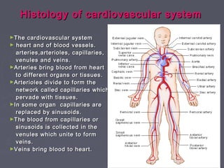
- 1. Histology of cardiovascular systemHistology of cardiovascular system ►The cardiovascular systemThe cardiovascular system ► heart and of blood vessels.heart and of blood vessels. arteries,arterioles, capillaries,arteries,arterioles, capillaries, venules and veins.venules and veins. ►Arteries bring blood from heartArteries bring blood from heart to different organs or tissues.to different organs or tissues. ►Arterioles divide to form theArterioles divide to form the network called capillaries whichnetwork called capillaries which pervade with tissues.pervade with tissues. ►In some organ capillaries areIn some organ capillaries are replaced by sinusoids.replaced by sinusoids. ►The blood from capillaries orThe blood from capillaries or sinusoids is collected in thesinusoids is collected in the venules which unite to formvenules which unite to form veins.veins. ►Veins bring blood to heart.Veins bring blood to heart.
- 2. Layers of ArteryLayers of Artery Fig: A medium sized muscular artery showing its layers.
- 3. Basic structure of ArteriesBasic structure of Arteries 1.The innermost layer, tunica1.The innermost layer, tunica intima ( tunica=coat).it consistsintima ( tunica=coat).it consists ofof a. An endothelial lininga. An endothelial lining b. A thin layer of glycoproteinb. A thin layer of glycoprotein called basal lamina.called basal lamina. c. A delicate layer ofc. A delicate layer of subendothelial connectivesubendothelial connective tissue.tissue. d. A membrane formed by thed. A membrane formed by the elastic fibres called theelastic fibres called the internalinternal elastic lamina.elastic lamina. 2.The tunica media.2.The tunica media. . Elastic tissue or smooth muscle. Elastic tissue or smooth muscle limited outside by a membranelimited outside by a membrane formed byformed by external elasticexternal elastic lamina.lamina. 3. The tunica adventitia3. The tunica adventitia consists of connective tissue inconsists of connective tissue in which collagen fibres arewhich collagen fibres are prominent.prominent.
- 4. Types of arteries:Types of arteries: ► On the basis of the kindOn the basis of the kind of tissue predominates inof tissue predominates in the tunica media arteriesthe tunica media arteries divide into elastic arterydivide into elastic artery and muscular.and muscular. ► Elastic arteriesElastic arteries :: ► The largest artery in theThe largest artery in the body.e.g. aorta,body.e.g. aorta, pulmonary trunk withpulmonary trunk with subdivisionsubdivision branchiocephalic ,branchiocephalic , common carotid,common carotid, subclavian ,vertebral,subclavian ,vertebral, pulmonary and commonpulmonary and common iliac.iliac. ► Tunica media is made upTunica media is made up of elastic tissueof elastic tissue ► Some smooth muscle areSome smooth muscle are also present in the media.also present in the media. ► The subendothelial tissueThe subendothelial tissue contains more elasticcontains more elastic tissues.tissues.
- 6. ►aa
- 8. ► Muscular Artery:Muscular Artery: ► They are theThey are the continuation of elasticcontinuation of elastic artery.artery. ► Predominant smoothPredominant smooth muscles in the tunicamuscles in the tunica media.media. ► Muscle fibres arrangedMuscle fibres arranged circularly.circularly. ► Connective tissues areConnective tissues are also present in betweenalso present in between the muscle fibres.the muscle fibres. Internal elastic lamina Tunica media Tunica adventitia
- 9. Muscular Artery (x4)Muscular Artery (x4)
- 10. Muscular Artery (L) vs Elastic ArteryMuscular Artery (L) vs Elastic Artery (R)(R)
- 11. Arterioles:Arterioles: ► The muscular arteriesThe muscular arteries having diameter lesshaving diameter less than 100 µm are calledthan 100 µm are called the arteriole.the arteriole. ► Muscular arterioles:Muscular arterioles: diameter 100-50 µm .diameter 100-50 µm . ► Lack of internal elasticLack of internal elastic lamina.lamina. ► Few layers of smoothFew layers of smooth muscles.muscles. ► Terminal arteriole:Terminal arteriole: diameter> 50diameter> 50 μμmm ► A thin layer of smoothA thin layer of smooth muscles fibres in themuscles fibres in the tunica media.tunica media. T.S. of arterioles
- 13. ►Capillaries :Capillaries : ► Terminal arterioles areTerminal arterioles are continued into capillary plexuscontinued into capillary plexus ► The arrangement of theThe arrangement of the capillary plexus and theircapillary plexus and their density varies.density varies. ► In the wall of capillaries thereIn the wall of capillaries there are the lining of endothelium.are the lining of endothelium. ► Endothelial cells are linedEndothelial cells are lined outside by a basal laminaoutside by a basal lamina ► Overlying the basal laminaOverlying the basal lamina there may be somethere may be some perivascular cells( pericyte)perivascular cells( pericyte) ► Capillaries are two typesCapillaries are two types ► ContinuousContinuous :: the edges ofthe edges of adjoining endothelial cells areadjoining endothelial cells are fuse .e g. in muscle, skin ,fuse .e g. in muscle, skin , lungs,brainlungs,brain ► Fenestrated:Fenestrated: apertures areapertures are present in endothelial cells.present in endothelial cells. E.g. small intestine, endocrineE.g. small intestine, endocrine glands and kidney.glands and kidney.
- 15. Sinusoids:Sinusoids: These are also network- differentThese are also network- different from the capillaries.from the capillaries. ► The wall of sinusoids containsThe wall of sinusoids contains only the endothelial cellsonly the endothelial cells covered by a layer ofcovered by a layer of connective tissue.connective tissue. ► In some places the wall isIn some places the wall is incomplete.incomplete. ► The lumen is broader butThe lumen is broader but irregular than the capillaries.irregular than the capillaries. ► The sinusoids are found in theThe sinusoids are found in the organ which is made up of theorgan which is made up of the cords of cells.e.g.cords of cells.e.g. liver,spleen ,suprarenal gland.liver,spleen ,suprarenal gland. and pitutary glandand pitutary gland
- 16. Fig : sinusoids in liver
- 17. veinsveins ► Basic structure is same asBasic structure is same as arteryartery ► The wall of vein is thinner.The wall of vein is thinner. ► Presence of abundantPresence of abundant collagen fibres in mediacollagen fibres in media layer.layer. ► The smooth muscle fibresThe smooth muscle fibres or elastic fibres reduced .or elastic fibres reduced . ► The tunica adventitia layerThe tunica adventitia layer is thicker.is thicker. ► Significant amount ofSignificant amount of elastic fibres and also theelastic fibres and also the muscle fibres in adventitia.muscle fibres in adventitia. ► There is no distinction of 3There is no distinction of 3 layers especially in smalllayers especially in small vein.vein. Fig: Medium sized vein. right (verhoff’s staining for elastic fibres the left (hematoxylin &Eosin staining)
- 18. Vein x10Vein x10
- 21. venulesvenules ► Smallest vein which drainSmallest vein which drain from capillaries are calledfrom capillaries are called the venule.the venule. ► Diameter 20-30Diameter 20-30 μμm.m. ► The wall consists ofThe wall consists of endothelial lining, basalendothelial lining, basal lamina and a thinlamina and a thin adventitia.adventitia. ► wall is very muchwall is very much permeable especiallypermeable especially lymphocytes and otherlymphocytes and other cells may pass out intocells may pass out into blood stream.blood stream. Fig : T.S. of venules
- 23. The heartThe heart ► Made up of 3 layersMade up of 3 layers ► Inner endocardiumInner endocardium ► A middle myocardiumA middle myocardium ► An outer epicardiumAn outer epicardium ► The endocardium: simple squmaous epitheliumThe endocardium: simple squmaous epithelium and a thin subendothelial connective tissue.and a thin subendothelial connective tissue. ► The myocardium: thickest layer and it consists ofThe myocardium: thickest layer and it consists of cardiac muscle fibres.cardiac muscle fibres. ► The epicardium consists of squamousThe epicardium consists of squamous mesothelium and an underlying subepicardialmesothelium and an underlying subepicardial layer of connective tissue.layer of connective tissue. ► The subepicaridal layer contains coronary bloodThe subepicaridal layer contains coronary blood vessels ,nerves and adipose tissue.vessels ,nerves and adipose tissue.
- 26. HEARTHEART
- 27. ►Purkinje fibers specialized for thePurkinje fibers specialized for the conduction of system of heart.conduction of system of heart. ►Short and larger fibers with central nucleus,Short and larger fibers with central nucleus, cytoplasm contains more glycogen.cytoplasm contains more glycogen.
- 28. .. ►..
- 29. Atherosclerotic LesionsAtherosclerotic Lesions ►Increase in thickening of intima.Increase in thickening of intima. ►Proliferation of smooth muscle cells,Proliferation of smooth muscle cells, increased deposition of CT fibers,increased deposition of CT fibers, lipoproteins.lipoproteins. ►Macrophages engulf lipoproteins.Macrophages engulf lipoproteins. ►Thickening occludes vessel.Thickening occludes vessel.
- 30. Atherosclerotic Plaque in CoronaryAtherosclerotic Plaque in Coronary ArteryArtery
- 31. THANK YOU!!!