SlideShare ist ein Scribd-Unternehmen logo
1 von 76
Downloaden Sie, um offline zu lesen
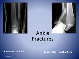
Presenter: Dr. Ravi Moderator – Dr. H.S. Selhi
5/12/2018
Ankle fractures – 10 % of all fractures
2nd most common lower limb fractures after hip
fractures
Mean age of injury 45 years
Low energy injury- simple fall/sports
5/12/2018
Ankle is a three bone joint
composed of the tibia ,
fibula and talus
Talus articulates with the
tibial plafond superiorly ,
posterior malleolus of the
tibia posteriorly and
medial malleolus medially
Lateral articulation is with
malleolus of fibula
5/12/2018
Ankle joint - saddle-shaped
Dome - wider anterior than posterior
Ankle dorsiflexes- external roatation fibula
to accommodate this widened anterior surface of
the talar dome
The tibiotalar articulation is considered to be highly
congruent such that 1 mm talar shift within the mortise
decreases the contact area by 42 %
5/12/2018
5/12/2018
3 groups of stabilizing
ligament complexes
• MEDIAL
• LATERAL
• SYNDESMOTIC
2 OUT OF 3 COMPLEXES SHOULD
BE INTACT FOR THEANKLE TO BE
STABLE
1) LATERAL
 anterior talofibular ligament
(ATFL)-WEAKEST
 calcaneofibular ligament
(CFL)
 posterior talofibular ligament
(PTFL).-STRONGEST
-limit ankleinversionand
prevent anterior and
lateral subluxationof the
talus
5/12/2018
2. MEDIAL LIGAMENT COMPLEX
SUPERFICIAL(little contribution to stability)
>Tibionavicular ligament
>Tibiocalcaneal ligament
>Superficial tibiotalar ligament
DEEP( primary medial stabilizer)
>Intraarticular
>Deep tibiotalar lig.
-stabilize the joint during eversion and
prevent talar subluxation
-20-50% stronger than lateral ligaments
5/12/2018
Medial talar tubercle
Navicular
tuberosity
Anterior colliculus
Sustentacular tali
5/12/2018
Medial
talusIntercollicular
groove
Posterior
colliculus
5/12/2018
• Axial, rotational & translational stability.
1- ANTERIORTIBIOFIBULARLIG.
2- POSTERIOR TIBIOFIBULARLIG.
3- TRANSVERSETIBIOFIBULARLIG.
4- INTEROSSEUS LIG.
3.SYNDESMOTIC LIGAMENT COMPLEX
5/12/2018
Chaput tubercle
Wagstaffe tubercle
Volkman
tubercle
5/12/2018
Swelling, echymosis, deformity
management of these fractures depends
upon careful identification of the extent of
bony injury as well as soft tissue and
ligamentous damage.
the key to successful outcome following
rotational ankle fractures is anatomic
restoration and healing of ankle mortise.
5/12/2018
Otawa ankle rules
X-raysare required
if there is bony painin the malleolar zone
AND anyone of the following:
• Age > 55 yrs
• Inability to bear weight
• Bone tenderness over the posterior edge or
tip of either malleolus.
validated and found to be both cost effective and reliable (up to
100% sensitivity)
5/12/2018
• Plain Films
AP & Lateral views
of the ankle
Mortise view - 15
degree internal rotation
Full length
radiograph of leg
when tenderness
of proximal fibula
Foot films when
tender to palpation
5/12/2018
An initial evaluation of the radiograph should
1st focus on
•Tibiotalar articulation and access for fibular
shortening
•Widening of joint space
•Malrotation of fibula
•Talar tilt
5/12/2018
Identifies
fractures of
◦ malleoli
◦ distal tibia/fibula
◦ plafond
◦ talar dome
◦ body and lateral
process of talus
◦ calcaneous
Ap view
5/12/2018
Tibiofibular clear space Tibiofibular overlap
5/12/2018
5/12/2018
On the anteroposterior view,
Important note
fibular (lateral) malleolus is
longer than the tibial (medial)
malleolus.
Even minimal displacement or shortening of the lateral
malleolus allows lateral talar shift to occur and may cause
incongruity in the ankle joint, possibly leading to
posttraumatic arthritis.
5/12/2018
Quantitative analysis
◦Tibiofibular overlap
◦< 1 0 m m is abnormal - implies
syndesmotic injury
◦Tibiofibular clear space
◦> 5 m m is abnormal - implies
syndesmotic injury
◦Talar tilt
◦> 2 m m is considered abnormal
Comparison with radiographs of the
normal side if there are unresolved
concerns of injury
5/12/2018
Taken with ankle in
15 degrees of
internal rotation
Useful in
evaluation of
articular surface
between talar
dome and mortise
5/12/2018
Medial clear space
◦ Between lateral border of
medial malleous and
medial talus
◦ <5mm is normal
◦ >5mm suggests lateral
shift of talus
◦ The joint spaces
medial and superior to
talus should be equal5/12/2018
Recess in distal fibula
lateral process
of talus
FIBULAR
LENGTH:
1.Shenton’s Line of the ankle
2.The dime test5/12/2018
5/12/2018
TALOCRURAL ANGLE : -
Approximately 83 degrees
and symetrical with
contalateral ankle
Assesment of fibular length
•Posterior mallelolar
fractures
•AP talar subluxation
•Distal fibular translation
&/or angulation
•Associated or occult
injuries
–Lateral process talus
–Posterior process talus
–Anterior process calcaneus
5/12/2018
The ankle is a ring
◦ Tibial plafond
◦ Medial malleolus
◦ Deltoid ligaments
◦ calcaneous
◦ Lateral collateral ligaments
◦ Lateral malleolus
◦ Syndesmosis
Fracture of single part
usually stable
Fracture > 1 part =
unstable
5/12/2018
• Stress Views
– Gravity stress view
– Manual stress views
5/12/2018
When radiographs of the ankle
are normal, stress views are
extremely important in evaluating
ligament injuries .
Inversion stress view. (A) For inversion
(adduction)-stress examination of the ankle, the
foot is fixed in the device while the patient is
supine. The pressure plate, positioned
approximately 2 cm above the ankle joint, applies
varus stress adducting the heel. (If the
examination is painful, 5 to 10 mL of 1%
Xylocaine or a similar local anesthetic is injected
at the site of maximum pain.) (B) On the
anteroposterior film, the degree of talar tilt is
measured by the angle formed by lines drawn
along the tibial plafond and the dome of the talus.
The contralateral ankle is subjected to the same
procedure for comparison.
This angle helps diagnose
tears of the lateral collateral
ligament5/12/2018
The anterior-draw stress film
for determining injury to the anterior talofibular
ligament
Values of up to 5 mm of
separation between the talus
and the distal tibia are
considered normal
between 5 and 10 mm may be
normal or abnormal, and the
opposite ankle should be
stressed for comparison.
Values above 10 mm always
indicate abnormality.
5/12/2018
5/12/2018
CT- Posterior malleolar fracture pattern
Joint invovement
Pre operative planning
MRI – ligament and tendon injuries
syndesmosis injury
• Classification of
ankle fractures
• Pott classification –
Unimalleolar
• bimalleolar
• timalleolar.
• Dennis-weber and AO/OTA
classifications
• Lauge-hansen
classification
5/12/2018
5/12/2018
Dennis –weber classification
describes the injury based on the
location of lateral malleolar fracture
A- below the level of syndesmosis
B- at the level of syndesmosis
C- above the level of syndesmosis
Does not predict the level or presence of syndesmotic injury
Does not address the presence of injury to medial side of ankle
Does not provide robust prognostic information
Good interobserver reliability
5/12/2018
AOclassification divides the three Danis Weber types further for
associated medial injuries
Infrasyndesmotic=44A
Transsyndesmotic=44B
Suprasyndesmotic=44C
v
5/12/2018
c
v
Infrasyndesmotic=44A
5/12/2018
Transsyndesmotic=44B
5/12/2018
Suprasyndesmotic=44C
Based on cadaveric study
it employs 2 words and a number
• First word: position of foot at time of injury
• Second word: force applied to foot relative to
tibia at time of injury
Number refers to the progression through
stages of bony and soft tissue injury
5/12/2018
5/12/2018
Types:
Supination External Rotation
Supination Adduction
Pronation External Rotation
Pronation Abduction
In each type there are several stages of injury
• Imperfect system:
– Not every fracture fits exactly into one category
– Even mechanismspecific pattern has been
–
questioned
–
Inter and intraobserver variation not ideal
Still useful and widely used
5/12/2018
Advantage
• useful for reconstructing the mechanism of
injury a guide for the closed reduction
• Sequential pattern –inference of ligament
injuries
complicated, variable inter observer reliability
doesn’t signify prognosis
doesn’t indicate stability
5/12/2018
Disadvantage
5/12/2018
1
3 2
4
Stage 1 Anterior
tibio- fibular
ligament
Stage 2 Fibula fx
Stage 3 Posterior
malleolus fx or
posterior tibio-
fibular ligament
Stage 4 Deltoid
ligament tear or
medial malleolus
fx5/12/2018
Lateral Injury: classic posterosuperioranteroinferior
fibula fracture
Medial: Stability maintained
Standard: Closed management5/12/2018
Lateral Injury: classic posterosuperioranteroinferior
fibula fracture
Medial Injury: medial malleolar fracture &*/or deltoid
ligament injury
Standard: Surgical management
5/12/2018
GOAL: TO EVALUATE DEEP DELTOID [i.e.
INSTABILITY]
Medial tenderness,
swelling, echymosis
STRESS VIEWS- GRAVITY OR
MANUAL
5/12/2018
SER-2
Negative Stress view
External rotation of
foot with ankle in
neutral flexion (00)
+ Stress View
Widened Medial Clear Space
SE-4
5/12/2018
• Stage 1: fibula
fracture is transverse
below mortise.
• Stage 2: medial
malleolus fracture is
classic vertical
pattern.
1
2
5/12/2018
Lateral Injury: transverse fibular fracture at/below
level of mortise
Medial injury: vertical shear type medial
malleolar fracture BEWARE OF
IMPACTION
5/12/2018
• Important to restore:
– Ankle stability
– Articular congruity- including medial
impaction
5/12/2018
5/12/2018
Stage 1 Deltoid
ligament tear or
medial malleolus
fx
Stage 2 Anterior
tibio-fibular
ligament and
interosseous
membrane
Stage 3 Spiral,
proximal fibula
fracture
Stage 4 Posterior
malleolus fx or
posterior tibio-
fibular ligament34
1 2
5/12/2018
Medial injury: deltoid ligament tear &/or transverse medial
malleolar fracture
Lateral Injury: spiral proximal lateral malleolar fracture
HIGHLY UNSTABLE…SYNDESMOTIC INJURY COMMON
5/12/2018
• Must x-ray knee to ankle to assess
injury
• Syndesmosis is disrupted in most cases
– Eponym: Maissoneuve Fracture
• Restore:
– Fibular length and rotation
– Ankle mortise
– Syndesmotic stability
5/12/2018
Stage 1 Transverse
medial malleolus fx
distal to mortise
Stage 2 avulsion fx of
tubercle of chaput
or tibio-fibular
ligament
Stage 3 Fibula fracture,
typically proximal to
mortise, often with a
butterfly fragment
1 2
3
5/12/2018
Medial injury: tranverse to short oblique medial malleolar
fracture
Lateral Injury: comminuted impaction type distal lateral
malleolar fracture
5/12/2018
Function:
Stability- prevents posterior
translation of talus & enhances
syndesmotic stability
Weight bearing- increases
surface area of ankle joint
5/12/2018
• Fracture pattern:
– Variable
– Difficult to assess on standard lateral
radiograph
• External rotation lateral view
• CT scan
5/12/2018
Type I- posterolateral
oblique type
Type II- medial extension
type
Type III- small shell
67
%
19
%
14
%
5/12/2018
FUNCTION:
Stability- resists external rotation, axial,
& lateral displacement of talus
Weight bearing- allows for standard
loading
5/12/2018
• Maisonneuve Fracture
– Fracture of proximal fibula
with syndesmotic disruption
5/12/2018
5/12/2018
• Volkmann Fracture
– Fracture of tibial attachment of PITFL
– Posterior malleolar fracture type
5/12/2018
• Tillaux-Chaput Fracture
– Fracture of tibial attachment of AITFL
In the Pott fracture, the fibula
is fractured above the intact
distal tibiofibular
syndesmosis, the deltoid
ligament is ruptured, and the
talus is subluxed laterally
5/12/2018
Pott fracture
Dupuytren
fracture.
(A) This fracture usually
occurs 2 to 7 cm above the
distal tibiofibular
syndesmosis, with disruption
of the medial collateral
ligament and, typically, tear
of the syndesmosis leading
to ankle instability.
(B) In the low variant, the
fracture occurs more distally
and the tibiofibular ligament
remains intact.
5/12/2018
Wagstaffe-LeFort
fracture.
In the Wagstaffe-LeFort
fracture, the medial portion
of the fibula is avulsed at the
insertion of the anterior
tibiofibular ligament. The
ligament, however, remains
intact.
5/12/2018
•Collicular
Fractures
–Avulsion fracture of distal
portion of medial malleolus
–Injury may continue and
rupture the deep deltoid
ligament
I
POSTERIOR
COLLICULUS
NTERCOLLICULAR GROOVE
ANTERIOR
COLLICULUS
5/12/2018
5/12/2018
•Bosworth fracture dislocation
–Fibular fracture with posterior dislocation
of proximal fibular segment behind tibia
5/12/2018
Initialmanagement
• Reducethe talus
• If joint unstable - slab
• other options -spanning
external fixator , calcaneal pin
traction.
• Rest,Ice, elevation
5/12/2018
Closed reduction and immobilization
• Stableankle fractures
• Usually with only fibulafractures
• Immobilization in castfor 4-6weeksis the preferred
treatment.
5/12/2018
Indications of surgery
 Inability to obtain or maintain an
anatomic mortise (unstable fracture
pattern)
 Open fractures
LateralMalleolarfractures
5/12/2018
• Avoidinjuring the superficial peronealnerve
• Makesurethat distal fibula isfully
out to length
• Laterallycomminuted pronation
abduction patterns aremostdifficult
• Formaximum stability placeplateposteriorly
Medialmalleolarfixation
5/12/2018
• 4.0mmpartially threaded screws
• Screwsshouldbe
perpendicular to the
fracture line andparallel
for maximal
compression.
• Spreadtwo screwsfor goodstability
• Usefluoroscopy to besurescrewsare
clearof the joint
Deltoidligamenttear
5/12/2018
• Thedeltoid ligament, especially
its deep branch isimportant to
the stability of the ankle
becauseit prevents lateral
displacement and external
rotation of thetalus
• Xray will show displacement and
tilting ofthe talus with
increased medial clearspace
• It isrepaired with
nonabsorbablesutures.
5/12/2018
SyndesmosisFixation
• Syndesmotic instability
after fixation of
malleolus
• Consider if fibula fracture
>4 cm above joint
line &
Maisonneuve’s
fracture
• Havebone hook
on back table to
checkstability
Complications following ankle
fractures
5/12/2018
EARLY :- wound infection/ dehiscence
loss of reduction
thromboembolism
LATE: - symptomatic hardware
osteoarthritis
nonunion
compartment syndrome
neuroma
5/12/2018

Weitere ähnliche Inhalte

Was ist angesagt?

Floating knee injuries
Floating knee injuriesFloating knee injuries
Floating knee injuriesKavin Khatri
 
Posterior malleolus fracture
Posterior malleolus fracturePosterior malleolus fracture
Posterior malleolus fractureAnuragSai7
 
Ankle & Foot Xray & Surgical Approaches
Ankle & Foot Xray & Surgical ApproachesAnkle & Foot Xray & Surgical Approaches
Ankle & Foot Xray & Surgical ApproachesMirant Dave
 
Distal Humerus Fracture Management- Rejul
 Distal Humerus Fracture Management- Rejul Distal Humerus Fracture Management- Rejul
Distal Humerus Fracture Management- RejulRejul Raj
 
Surgical approaches to hip joint
Surgical approaches to hip jointSurgical approaches to hip joint
Surgical approaches to hip jointadityachakri
 
Evolution of tunnel placement in ACL reconstruction
Evolution of tunnel placement in ACL reconstructionEvolution of tunnel placement in ACL reconstruction
Evolution of tunnel placement in ACL reconstructionDhananjaya Sabat
 
Bone defects in tkr
Bone defects in tkrBone defects in tkr
Bone defects in tkrankitjose
 
Capitellum fractures
Capitellum fracturesCapitellum fractures
Capitellum fracturesApoorv Jain
 
Scaphoid fracture and non union
Scaphoid fracture and non unionScaphoid fracture and non union
Scaphoid fracture and non unionratish mishra
 
Recurrent Dislocation of patella
Recurrent Dislocation of patellaRecurrent Dislocation of patella
Recurrent Dislocation of patellasabir khadka
 
Biomechanics and biology of relative stability
Biomechanics and biology of relative stabilityBiomechanics and biology of relative stability
Biomechanics and biology of relative stabilityOrthosurg2016
 
ACL RECONSTRUCTION FAILURE
ACL RECONSTRUCTION FAILUREACL RECONSTRUCTION FAILURE
ACL RECONSTRUCTION FAILUREGonzalo Samitier
 
distal end radius fracture
distal end radius fracturedistal end radius fracture
distal end radius fractureibsta
 

Was ist angesagt? (20)

Floating knee injuries
Floating knee injuriesFloating knee injuries
Floating knee injuries
 
Acute scaphoid fractures
Acute scaphoid fracturesAcute scaphoid fractures
Acute scaphoid fractures
 
Posterior malleolus fracture
Posterior malleolus fracturePosterior malleolus fracture
Posterior malleolus fracture
 
Ankle xrays
Ankle xraysAnkle xrays
Ankle xrays
 
Ankle & Foot Xray & Surgical Approaches
Ankle & Foot Xray & Surgical ApproachesAnkle & Foot Xray & Surgical Approaches
Ankle & Foot Xray & Surgical Approaches
 
Distal Humerus Fracture Management- Rejul
 Distal Humerus Fracture Management- Rejul Distal Humerus Fracture Management- Rejul
Distal Humerus Fracture Management- Rejul
 
Surgical approaches to hip joint
Surgical approaches to hip jointSurgical approaches to hip joint
Surgical approaches to hip joint
 
Evolution of tunnel placement in ACL reconstruction
Evolution of tunnel placement in ACL reconstructionEvolution of tunnel placement in ACL reconstruction
Evolution of tunnel placement in ACL reconstruction
 
Scaphoid fracture
Scaphoid fractureScaphoid fracture
Scaphoid fracture
 
Bone defects in tkr
Bone defects in tkrBone defects in tkr
Bone defects in tkr
 
Capitellum fractures
Capitellum fracturesCapitellum fractures
Capitellum fractures
 
Distal end radius fracture
Distal end radius fractureDistal end radius fracture
Distal end radius fracture
 
Scaphoid fracture and non union
Scaphoid fracture and non unionScaphoid fracture and non union
Scaphoid fracture and non union
 
Fracture Talus
Fracture TalusFracture Talus
Fracture Talus
 
THR
THRTHR
THR
 
Recurrent Dislocation of patella
Recurrent Dislocation of patellaRecurrent Dislocation of patella
Recurrent Dislocation of patella
 
Elbow instability
Elbow instabilityElbow instability
Elbow instability
 
Biomechanics and biology of relative stability
Biomechanics and biology of relative stabilityBiomechanics and biology of relative stability
Biomechanics and biology of relative stability
 
ACL RECONSTRUCTION FAILURE
ACL RECONSTRUCTION FAILUREACL RECONSTRUCTION FAILURE
ACL RECONSTRUCTION FAILURE
 
distal end radius fracture
distal end radius fracturedistal end radius fracture
distal end radius fracture
 

Ähnlich wie Ankle Fractures Classification and Management

Anklefracturesfinal 120314092959-phpapp01
Anklefracturesfinal 120314092959-phpapp01Anklefracturesfinal 120314092959-phpapp01
Anklefracturesfinal 120314092959-phpapp01Drpraveen Kumar
 
Anterior cruciate ligament-Injury & management
Anterior cruciate ligament-Injury & managementAnterior cruciate ligament-Injury & management
Anterior cruciate ligament-Injury & managementAnand Rao
 
floating knee on Lower Limb trauma orthopedics.pptx
floating knee on Lower Limb trauma orthopedics.pptxfloating knee on Lower Limb trauma orthopedics.pptx
floating knee on Lower Limb trauma orthopedics.pptxArya RaveendranNair
 
Ankle injuries & Trimallelor fracture classification
Ankle injuries & Trimallelor  fracture classification Ankle injuries & Trimallelor  fracture classification
Ankle injuries & Trimallelor fracture classification Ponnilavan Ponz
 
Ankle fractures management
Ankle fractures   managementAnkle fractures   management
Ankle fractures managementSunil Santhosh
 
Lis Franc Injury
Lis Franc InjuryLis Franc Injury
Lis Franc Injuryjfreshour
 
Intertrochentric femur fracture by DR.NAVEEN RATHOR
Intertrochentric femur fracture by DR.NAVEEN RATHORIntertrochentric femur fracture by DR.NAVEEN RATHOR
Intertrochentric femur fracture by DR.NAVEEN RATHORDR.Naveen Rathor
 
Ankle fractures final
Ankle fractures finalAnkle fractures final
Ankle fractures finalAnkur Mittal
 
Ant cruciate ligament injuries
Ant cruciate ligament injuriesAnt cruciate ligament injuries
Ant cruciate ligament injuriesGaurav Singh
 
Complications of total hip replacement final
Complications of total hip replacement finalComplications of total hip replacement final
Complications of total hip replacement finalHumayun Israr
 
The effect of intact fibula on functional outcome of reamed intramedullary in...
The effect of intact fibula on functional outcome of reamed intramedullary in...The effect of intact fibula on functional outcome of reamed intramedullary in...
The effect of intact fibula on functional outcome of reamed intramedullary in...Love2jaipal
 
Orthopedics 5th year, 7th/part two & 8th lectures (Dr. Ali A.Nabi)
Orthopedics 5th year, 7th/part two & 8th lectures (Dr. Ali A.Nabi)Orthopedics 5th year, 7th/part two & 8th lectures (Dr. Ali A.Nabi)
Orthopedics 5th year, 7th/part two & 8th lectures (Dr. Ali A.Nabi)College of Medicine, Sulaymaniyah
 
fracture shaft of femur.pptx
fracture shaft of femur.pptxfracture shaft of femur.pptx
fracture shaft of femur.pptxSaurabh Agrawal
 

Ähnlich wie Ankle Fractures Classification and Management (20)

Ankle injury
Ankle injuryAnkle injury
Ankle injury
 
Anklefracturesfinal 120314092959-phpapp01
Anklefracturesfinal 120314092959-phpapp01Anklefracturesfinal 120314092959-phpapp01
Anklefracturesfinal 120314092959-phpapp01
 
Anterior cruciate ligament-Injury & management
Anterior cruciate ligament-Injury & managementAnterior cruciate ligament-Injury & management
Anterior cruciate ligament-Injury & management
 
floating knee on Lower Limb trauma orthopedics.pptx
floating knee on Lower Limb trauma orthopedics.pptxfloating knee on Lower Limb trauma orthopedics.pptx
floating knee on Lower Limb trauma orthopedics.pptx
 
Intro case
Intro caseIntro case
Intro case
 
Ankle injuries & Trimallelor fracture classification
Ankle injuries & Trimallelor  fracture classification Ankle injuries & Trimallelor  fracture classification
Ankle injuries & Trimallelor fracture classification
 
Ankle fractures management
Ankle fractures   managementAnkle fractures   management
Ankle fractures management
 
Lis Franc Injury
Lis Franc InjuryLis Franc Injury
Lis Franc Injury
 
Intertrochentric femur fracture by DR.NAVEEN RATHOR
Intertrochentric femur fracture by DR.NAVEEN RATHORIntertrochentric femur fracture by DR.NAVEEN RATHOR
Intertrochentric femur fracture by DR.NAVEEN RATHOR
 
Ankle fractures final
Ankle fractures finalAnkle fractures final
Ankle fractures final
 
Ankle seminar
Ankle seminarAnkle seminar
Ankle seminar
 
distal femoral fx2.pptx
distal femoral fx2.pptxdistal femoral fx2.pptx
distal femoral fx2.pptx
 
Ant cruciate ligament injuries
Ant cruciate ligament injuriesAnt cruciate ligament injuries
Ant cruciate ligament injuries
 
Complications of total hip replacement final
Complications of total hip replacement finalComplications of total hip replacement final
Complications of total hip replacement final
 
L12 ankle fxs
L12 ankle fxsL12 ankle fxs
L12 ankle fxs
 
ACL Recon.pptx
ACL Recon.pptxACL Recon.pptx
ACL Recon.pptx
 
The effect of intact fibula on functional outcome of reamed intramedullary in...
The effect of intact fibula on functional outcome of reamed intramedullary in...The effect of intact fibula on functional outcome of reamed intramedullary in...
The effect of intact fibula on functional outcome of reamed intramedullary in...
 
Ankle fracture
Ankle fractureAnkle fracture
Ankle fracture
 
Orthopedics 5th year, 7th/part two & 8th lectures (Dr. Ali A.Nabi)
Orthopedics 5th year, 7th/part two & 8th lectures (Dr. Ali A.Nabi)Orthopedics 5th year, 7th/part two & 8th lectures (Dr. Ali A.Nabi)
Orthopedics 5th year, 7th/part two & 8th lectures (Dr. Ali A.Nabi)
 
fracture shaft of femur.pptx
fracture shaft of femur.pptxfracture shaft of femur.pptx
fracture shaft of femur.pptx
 

Kürzlich hochgeladen

Study on the Impact of FOCUS-PDCA Management Model on the Disinfection Qualit...
Study on the Impact of FOCUS-PDCA Management Model on the Disinfection Qualit...Study on the Impact of FOCUS-PDCA Management Model on the Disinfection Qualit...
Study on the Impact of FOCUS-PDCA Management Model on the Disinfection Qualit...MehranMouzam
 
World-Health-Day-2024-My-Health-My-Right.pptx
World-Health-Day-2024-My-Health-My-Right.pptxWorld-Health-Day-2024-My-Health-My-Right.pptx
World-Health-Day-2024-My-Health-My-Right.pptxEx WHO/USAID
 
Statistical modeling in pharmaceutical research and development.
Statistical modeling in pharmaceutical research and development.Statistical modeling in pharmaceutical research and development.
Statistical modeling in pharmaceutical research and development.ANJALI
 
Tans femoral Amputee : Prosthetics Knee Joints.pptx
Tans femoral Amputee : Prosthetics Knee Joints.pptxTans femoral Amputee : Prosthetics Knee Joints.pptx
Tans femoral Amputee : Prosthetics Knee Joints.pptxKezaiah S
 
Culture and Health Disorders Social change.pptx
Culture and Health Disorders Social change.pptxCulture and Health Disorders Social change.pptx
Culture and Health Disorders Social change.pptxDr. Dheeraj Kumar
 
LESSON PLAN ON fever.pdf child health nursing
LESSON PLAN ON fever.pdf child health nursingLESSON PLAN ON fever.pdf child health nursing
LESSON PLAN ON fever.pdf child health nursingSakthi Kathiravan
 
Big Data Analysis Suggests COVID Vaccination Increases Excess Mortality Of ...
Big Data Analysis Suggests COVID  Vaccination Increases Excess Mortality Of  ...Big Data Analysis Suggests COVID  Vaccination Increases Excess Mortality Of  ...
Big Data Analysis Suggests COVID Vaccination Increases Excess Mortality Of ...sdateam0
 
PNEUMOTHORAX AND ITS MANAGEMENTS.pdf
PNEUMOTHORAX   AND  ITS  MANAGEMENTS.pdfPNEUMOTHORAX   AND  ITS  MANAGEMENTS.pdf
PNEUMOTHORAX AND ITS MANAGEMENTS.pdfDolisha Warbi
 
PULMONARY EMBOLISM AND ITS MANAGEMENTS.pdf
PULMONARY EMBOLISM AND ITS MANAGEMENTS.pdfPULMONARY EMBOLISM AND ITS MANAGEMENTS.pdf
PULMONARY EMBOLISM AND ITS MANAGEMENTS.pdfDolisha Warbi
 
Presentació "Real-Life VR Integration for Mild Cognitive Impairment Rehabilit...
Presentació "Real-Life VR Integration for Mild Cognitive Impairment Rehabilit...Presentació "Real-Life VR Integration for Mild Cognitive Impairment Rehabilit...
Presentació "Real-Life VR Integration for Mild Cognitive Impairment Rehabilit...Badalona Serveis Assistencials
 
Presentation on Parasympathetic Nervous System
Presentation on Parasympathetic Nervous SystemPresentation on Parasympathetic Nervous System
Presentation on Parasympathetic Nervous SystemPrerana Jadhav
 
Informed Consent Empowering Healthcare Decision-Making.pptx
Informed Consent Empowering Healthcare Decision-Making.pptxInformed Consent Empowering Healthcare Decision-Making.pptx
Informed Consent Empowering Healthcare Decision-Making.pptxSasikiranMarri
 
Primary headache and facial pain. (2024)
Primary headache and facial pain. (2024)Primary headache and facial pain. (2024)
Primary headache and facial pain. (2024)Mohamed Rizk Khodair
 
METHODS OF ACQUIRING KNOWLEDGE IN NURSING.pptx by navdeep kaur
METHODS OF ACQUIRING KNOWLEDGE IN NURSING.pptx by navdeep kaurMETHODS OF ACQUIRING KNOWLEDGE IN NURSING.pptx by navdeep kaur
METHODS OF ACQUIRING KNOWLEDGE IN NURSING.pptx by navdeep kaurNavdeep Kaur
 
Giftedness: Understanding Everyday Neurobiology for Self-Knowledge
Giftedness: Understanding Everyday Neurobiology for Self-KnowledgeGiftedness: Understanding Everyday Neurobiology for Self-Knowledge
Giftedness: Understanding Everyday Neurobiology for Self-Knowledgeassessoriafabianodea
 
The next social challenge to public health: the information environment.pptx
The next social challenge to public health:  the information environment.pptxThe next social challenge to public health:  the information environment.pptx
The next social challenge to public health: the information environment.pptxTina Purnat
 
VarSeq 2.6.0: Advancing Pharmacogenomics and Genomic Analysis
VarSeq 2.6.0: Advancing Pharmacogenomics and Genomic AnalysisVarSeq 2.6.0: Advancing Pharmacogenomics and Genomic Analysis
VarSeq 2.6.0: Advancing Pharmacogenomics and Genomic AnalysisGolden Helix
 
LUNG TUMORS AND ITS CLASSIFICATIONS.pdf
LUNG TUMORS AND ITS  CLASSIFICATIONS.pdfLUNG TUMORS AND ITS  CLASSIFICATIONS.pdf
LUNG TUMORS AND ITS CLASSIFICATIONS.pdfDolisha Warbi
 
MedDRA-A-Comprehensive-Guide-to-Standardized-Medical-Terminology.pdf
MedDRA-A-Comprehensive-Guide-to-Standardized-Medical-Terminology.pdfMedDRA-A-Comprehensive-Guide-to-Standardized-Medical-Terminology.pdf
MedDRA-A-Comprehensive-Guide-to-Standardized-Medical-Terminology.pdfSasikiranMarri
 
systemic bacteriology (7)............pptx
systemic bacteriology (7)............pptxsystemic bacteriology (7)............pptx
systemic bacteriology (7)............pptxEyobAlemu11
 

Kürzlich hochgeladen (20)

Study on the Impact of FOCUS-PDCA Management Model on the Disinfection Qualit...
Study on the Impact of FOCUS-PDCA Management Model on the Disinfection Qualit...Study on the Impact of FOCUS-PDCA Management Model on the Disinfection Qualit...
Study on the Impact of FOCUS-PDCA Management Model on the Disinfection Qualit...
 
World-Health-Day-2024-My-Health-My-Right.pptx
World-Health-Day-2024-My-Health-My-Right.pptxWorld-Health-Day-2024-My-Health-My-Right.pptx
World-Health-Day-2024-My-Health-My-Right.pptx
 
Statistical modeling in pharmaceutical research and development.
Statistical modeling in pharmaceutical research and development.Statistical modeling in pharmaceutical research and development.
Statistical modeling in pharmaceutical research and development.
 
Tans femoral Amputee : Prosthetics Knee Joints.pptx
Tans femoral Amputee : Prosthetics Knee Joints.pptxTans femoral Amputee : Prosthetics Knee Joints.pptx
Tans femoral Amputee : Prosthetics Knee Joints.pptx
 
Culture and Health Disorders Social change.pptx
Culture and Health Disorders Social change.pptxCulture and Health Disorders Social change.pptx
Culture and Health Disorders Social change.pptx
 
LESSON PLAN ON fever.pdf child health nursing
LESSON PLAN ON fever.pdf child health nursingLESSON PLAN ON fever.pdf child health nursing
LESSON PLAN ON fever.pdf child health nursing
 
Big Data Analysis Suggests COVID Vaccination Increases Excess Mortality Of ...
Big Data Analysis Suggests COVID  Vaccination Increases Excess Mortality Of  ...Big Data Analysis Suggests COVID  Vaccination Increases Excess Mortality Of  ...
Big Data Analysis Suggests COVID Vaccination Increases Excess Mortality Of ...
 
PNEUMOTHORAX AND ITS MANAGEMENTS.pdf
PNEUMOTHORAX   AND  ITS  MANAGEMENTS.pdfPNEUMOTHORAX   AND  ITS  MANAGEMENTS.pdf
PNEUMOTHORAX AND ITS MANAGEMENTS.pdf
 
PULMONARY EMBOLISM AND ITS MANAGEMENTS.pdf
PULMONARY EMBOLISM AND ITS MANAGEMENTS.pdfPULMONARY EMBOLISM AND ITS MANAGEMENTS.pdf
PULMONARY EMBOLISM AND ITS MANAGEMENTS.pdf
 
Presentació "Real-Life VR Integration for Mild Cognitive Impairment Rehabilit...
Presentació "Real-Life VR Integration for Mild Cognitive Impairment Rehabilit...Presentació "Real-Life VR Integration for Mild Cognitive Impairment Rehabilit...
Presentació "Real-Life VR Integration for Mild Cognitive Impairment Rehabilit...
 
Presentation on Parasympathetic Nervous System
Presentation on Parasympathetic Nervous SystemPresentation on Parasympathetic Nervous System
Presentation on Parasympathetic Nervous System
 
Informed Consent Empowering Healthcare Decision-Making.pptx
Informed Consent Empowering Healthcare Decision-Making.pptxInformed Consent Empowering Healthcare Decision-Making.pptx
Informed Consent Empowering Healthcare Decision-Making.pptx
 
Primary headache and facial pain. (2024)
Primary headache and facial pain. (2024)Primary headache and facial pain. (2024)
Primary headache and facial pain. (2024)
 
METHODS OF ACQUIRING KNOWLEDGE IN NURSING.pptx by navdeep kaur
METHODS OF ACQUIRING KNOWLEDGE IN NURSING.pptx by navdeep kaurMETHODS OF ACQUIRING KNOWLEDGE IN NURSING.pptx by navdeep kaur
METHODS OF ACQUIRING KNOWLEDGE IN NURSING.pptx by navdeep kaur
 
Giftedness: Understanding Everyday Neurobiology for Self-Knowledge
Giftedness: Understanding Everyday Neurobiology for Self-KnowledgeGiftedness: Understanding Everyday Neurobiology for Self-Knowledge
Giftedness: Understanding Everyday Neurobiology for Self-Knowledge
 
The next social challenge to public health: the information environment.pptx
The next social challenge to public health:  the information environment.pptxThe next social challenge to public health:  the information environment.pptx
The next social challenge to public health: the information environment.pptx
 
VarSeq 2.6.0: Advancing Pharmacogenomics and Genomic Analysis
VarSeq 2.6.0: Advancing Pharmacogenomics and Genomic AnalysisVarSeq 2.6.0: Advancing Pharmacogenomics and Genomic Analysis
VarSeq 2.6.0: Advancing Pharmacogenomics and Genomic Analysis
 
LUNG TUMORS AND ITS CLASSIFICATIONS.pdf
LUNG TUMORS AND ITS  CLASSIFICATIONS.pdfLUNG TUMORS AND ITS  CLASSIFICATIONS.pdf
LUNG TUMORS AND ITS CLASSIFICATIONS.pdf
 
MedDRA-A-Comprehensive-Guide-to-Standardized-Medical-Terminology.pdf
MedDRA-A-Comprehensive-Guide-to-Standardized-Medical-Terminology.pdfMedDRA-A-Comprehensive-Guide-to-Standardized-Medical-Terminology.pdf
MedDRA-A-Comprehensive-Guide-to-Standardized-Medical-Terminology.pdf
 
systemic bacteriology (7)............pptx
systemic bacteriology (7)............pptxsystemic bacteriology (7)............pptx
systemic bacteriology (7)............pptx
 

Ankle Fractures Classification and Management

  • 1. Presenter: Dr. Ravi Moderator – Dr. H.S. Selhi 5/12/2018
  • 2. Ankle fractures – 10 % of all fractures 2nd most common lower limb fractures after hip fractures Mean age of injury 45 years Low energy injury- simple fall/sports 5/12/2018
  • 3. Ankle is a three bone joint composed of the tibia , fibula and talus Talus articulates with the tibial plafond superiorly , posterior malleolus of the tibia posteriorly and medial malleolus medially Lateral articulation is with malleolus of fibula 5/12/2018
  • 4. Ankle joint - saddle-shaped Dome - wider anterior than posterior Ankle dorsiflexes- external roatation fibula to accommodate this widened anterior surface of the talar dome The tibiotalar articulation is considered to be highly congruent such that 1 mm talar shift within the mortise decreases the contact area by 42 % 5/12/2018
  • 5. 5/12/2018 3 groups of stabilizing ligament complexes • MEDIAL • LATERAL • SYNDESMOTIC 2 OUT OF 3 COMPLEXES SHOULD BE INTACT FOR THEANKLE TO BE STABLE
  • 6. 1) LATERAL  anterior talofibular ligament (ATFL)-WEAKEST  calcaneofibular ligament (CFL)  posterior talofibular ligament (PTFL).-STRONGEST -limit ankleinversionand prevent anterior and lateral subluxationof the talus 5/12/2018
  • 7. 2. MEDIAL LIGAMENT COMPLEX SUPERFICIAL(little contribution to stability) >Tibionavicular ligament >Tibiocalcaneal ligament >Superficial tibiotalar ligament DEEP( primary medial stabilizer) >Intraarticular >Deep tibiotalar lig. -stabilize the joint during eversion and prevent talar subluxation -20-50% stronger than lateral ligaments 5/12/2018
  • 8. Medial talar tubercle Navicular tuberosity Anterior colliculus Sustentacular tali 5/12/2018
  • 10. • Axial, rotational & translational stability. 1- ANTERIORTIBIOFIBULARLIG. 2- POSTERIOR TIBIOFIBULARLIG. 3- TRANSVERSETIBIOFIBULARLIG. 4- INTEROSSEUS LIG. 3.SYNDESMOTIC LIGAMENT COMPLEX 5/12/2018
  • 12. Swelling, echymosis, deformity management of these fractures depends upon careful identification of the extent of bony injury as well as soft tissue and ligamentous damage. the key to successful outcome following rotational ankle fractures is anatomic restoration and healing of ankle mortise. 5/12/2018
  • 13. Otawa ankle rules X-raysare required if there is bony painin the malleolar zone AND anyone of the following: • Age > 55 yrs • Inability to bear weight • Bone tenderness over the posterior edge or tip of either malleolus. validated and found to be both cost effective and reliable (up to 100% sensitivity) 5/12/2018
  • 14. • Plain Films AP & Lateral views of the ankle Mortise view - 15 degree internal rotation Full length radiograph of leg when tenderness of proximal fibula Foot films when tender to palpation 5/12/2018
  • 15. An initial evaluation of the radiograph should 1st focus on •Tibiotalar articulation and access for fibular shortening •Widening of joint space •Malrotation of fibula •Talar tilt 5/12/2018
  • 16. Identifies fractures of ◦ malleoli ◦ distal tibia/fibula ◦ plafond ◦ talar dome ◦ body and lateral process of talus ◦ calcaneous Ap view 5/12/2018
  • 17. Tibiofibular clear space Tibiofibular overlap 5/12/2018
  • 19. On the anteroposterior view, Important note fibular (lateral) malleolus is longer than the tibial (medial) malleolus. Even minimal displacement or shortening of the lateral malleolus allows lateral talar shift to occur and may cause incongruity in the ankle joint, possibly leading to posttraumatic arthritis. 5/12/2018
  • 20. Quantitative analysis ◦Tibiofibular overlap ◦< 1 0 m m is abnormal - implies syndesmotic injury ◦Tibiofibular clear space ◦> 5 m m is abnormal - implies syndesmotic injury ◦Talar tilt ◦> 2 m m is considered abnormal Comparison with radiographs of the normal side if there are unresolved concerns of injury 5/12/2018
  • 21. Taken with ankle in 15 degrees of internal rotation Useful in evaluation of articular surface between talar dome and mortise 5/12/2018
  • 22. Medial clear space ◦ Between lateral border of medial malleous and medial talus ◦ <5mm is normal ◦ >5mm suggests lateral shift of talus ◦ The joint spaces medial and superior to talus should be equal5/12/2018
  • 23. Recess in distal fibula lateral process of talus FIBULAR LENGTH: 1.Shenton’s Line of the ankle 2.The dime test5/12/2018
  • 24. 5/12/2018 TALOCRURAL ANGLE : - Approximately 83 degrees and symetrical with contalateral ankle Assesment of fibular length
  • 25. •Posterior mallelolar fractures •AP talar subluxation •Distal fibular translation &/or angulation •Associated or occult injuries –Lateral process talus –Posterior process talus –Anterior process calcaneus 5/12/2018
  • 26. The ankle is a ring ◦ Tibial plafond ◦ Medial malleolus ◦ Deltoid ligaments ◦ calcaneous ◦ Lateral collateral ligaments ◦ Lateral malleolus ◦ Syndesmosis Fracture of single part usually stable Fracture > 1 part = unstable 5/12/2018
  • 27. • Stress Views – Gravity stress view – Manual stress views 5/12/2018 When radiographs of the ankle are normal, stress views are extremely important in evaluating ligament injuries .
  • 28. Inversion stress view. (A) For inversion (adduction)-stress examination of the ankle, the foot is fixed in the device while the patient is supine. The pressure plate, positioned approximately 2 cm above the ankle joint, applies varus stress adducting the heel. (If the examination is painful, 5 to 10 mL of 1% Xylocaine or a similar local anesthetic is injected at the site of maximum pain.) (B) On the anteroposterior film, the degree of talar tilt is measured by the angle formed by lines drawn along the tibial plafond and the dome of the talus. The contralateral ankle is subjected to the same procedure for comparison. This angle helps diagnose tears of the lateral collateral ligament5/12/2018
  • 29. The anterior-draw stress film for determining injury to the anterior talofibular ligament Values of up to 5 mm of separation between the talus and the distal tibia are considered normal between 5 and 10 mm may be normal or abnormal, and the opposite ankle should be stressed for comparison. Values above 10 mm always indicate abnormality. 5/12/2018
  • 30. 5/12/2018 CT- Posterior malleolar fracture pattern Joint invovement Pre operative planning MRI – ligament and tendon injuries syndesmosis injury
  • 31. • Classification of ankle fractures • Pott classification – Unimalleolar • bimalleolar • timalleolar. • Dennis-weber and AO/OTA classifications • Lauge-hansen classification 5/12/2018
  • 32. 5/12/2018 Dennis –weber classification describes the injury based on the location of lateral malleolar fracture A- below the level of syndesmosis B- at the level of syndesmosis C- above the level of syndesmosis Does not predict the level or presence of syndesmotic injury Does not address the presence of injury to medial side of ankle Does not provide robust prognostic information Good interobserver reliability
  • 33. 5/12/2018 AOclassification divides the three Danis Weber types further for associated medial injuries Infrasyndesmotic=44A Transsyndesmotic=44B Suprasyndesmotic=44C v
  • 37. Based on cadaveric study it employs 2 words and a number • First word: position of foot at time of injury • Second word: force applied to foot relative to tibia at time of injury Number refers to the progression through stages of bony and soft tissue injury 5/12/2018
  • 38. 5/12/2018 Types: Supination External Rotation Supination Adduction Pronation External Rotation Pronation Abduction
  • 39. In each type there are several stages of injury • Imperfect system: – Not every fracture fits exactly into one category – Even mechanismspecific pattern has been – questioned – Inter and intraobserver variation not ideal Still useful and widely used 5/12/2018
  • 40. Advantage • useful for reconstructing the mechanism of injury a guide for the closed reduction • Sequential pattern –inference of ligament injuries complicated, variable inter observer reliability doesn’t signify prognosis doesn’t indicate stability 5/12/2018 Disadvantage
  • 42. 1 3 2 4 Stage 1 Anterior tibio- fibular ligament Stage 2 Fibula fx Stage 3 Posterior malleolus fx or posterior tibio- fibular ligament Stage 4 Deltoid ligament tear or medial malleolus fx5/12/2018
  • 43. Lateral Injury: classic posterosuperioranteroinferior fibula fracture Medial: Stability maintained Standard: Closed management5/12/2018
  • 44. Lateral Injury: classic posterosuperioranteroinferior fibula fracture Medial Injury: medial malleolar fracture &*/or deltoid ligament injury Standard: Surgical management 5/12/2018
  • 45. GOAL: TO EVALUATE DEEP DELTOID [i.e. INSTABILITY] Medial tenderness, swelling, echymosis STRESS VIEWS- GRAVITY OR MANUAL 5/12/2018
  • 46. SER-2 Negative Stress view External rotation of foot with ankle in neutral flexion (00) + Stress View Widened Medial Clear Space SE-4 5/12/2018
  • 47. • Stage 1: fibula fracture is transverse below mortise. • Stage 2: medial malleolus fracture is classic vertical pattern. 1 2 5/12/2018
  • 48. Lateral Injury: transverse fibular fracture at/below level of mortise Medial injury: vertical shear type medial malleolar fracture BEWARE OF IMPACTION 5/12/2018
  • 49. • Important to restore: – Ankle stability – Articular congruity- including medial impaction 5/12/2018
  • 51. Stage 1 Deltoid ligament tear or medial malleolus fx Stage 2 Anterior tibio-fibular ligament and interosseous membrane Stage 3 Spiral, proximal fibula fracture Stage 4 Posterior malleolus fx or posterior tibio- fibular ligament34 1 2 5/12/2018
  • 52. Medial injury: deltoid ligament tear &/or transverse medial malleolar fracture Lateral Injury: spiral proximal lateral malleolar fracture HIGHLY UNSTABLE…SYNDESMOTIC INJURY COMMON 5/12/2018
  • 53. • Must x-ray knee to ankle to assess injury • Syndesmosis is disrupted in most cases – Eponym: Maissoneuve Fracture • Restore: – Fibular length and rotation – Ankle mortise – Syndesmotic stability 5/12/2018
  • 54. Stage 1 Transverse medial malleolus fx distal to mortise Stage 2 avulsion fx of tubercle of chaput or tibio-fibular ligament Stage 3 Fibula fracture, typically proximal to mortise, often with a butterfly fragment 1 2 3 5/12/2018
  • 55. Medial injury: tranverse to short oblique medial malleolar fracture Lateral Injury: comminuted impaction type distal lateral malleolar fracture 5/12/2018
  • 56. Function: Stability- prevents posterior translation of talus & enhances syndesmotic stability Weight bearing- increases surface area of ankle joint 5/12/2018
  • 57. • Fracture pattern: – Variable – Difficult to assess on standard lateral radiograph • External rotation lateral view • CT scan 5/12/2018
  • 58. Type I- posterolateral oblique type Type II- medial extension type Type III- small shell 67 % 19 % 14 % 5/12/2018
  • 59. FUNCTION: Stability- resists external rotation, axial, & lateral displacement of talus Weight bearing- allows for standard loading 5/12/2018
  • 60. • Maisonneuve Fracture – Fracture of proximal fibula with syndesmotic disruption 5/12/2018
  • 61. 5/12/2018 • Volkmann Fracture – Fracture of tibial attachment of PITFL – Posterior malleolar fracture type
  • 62. 5/12/2018 • Tillaux-Chaput Fracture – Fracture of tibial attachment of AITFL
  • 63. In the Pott fracture, the fibula is fractured above the intact distal tibiofibular syndesmosis, the deltoid ligament is ruptured, and the talus is subluxed laterally 5/12/2018 Pott fracture
  • 64. Dupuytren fracture. (A) This fracture usually occurs 2 to 7 cm above the distal tibiofibular syndesmosis, with disruption of the medial collateral ligament and, typically, tear of the syndesmosis leading to ankle instability. (B) In the low variant, the fracture occurs more distally and the tibiofibular ligament remains intact. 5/12/2018
  • 65. Wagstaffe-LeFort fracture. In the Wagstaffe-LeFort fracture, the medial portion of the fibula is avulsed at the insertion of the anterior tibiofibular ligament. The ligament, however, remains intact. 5/12/2018
  • 66. •Collicular Fractures –Avulsion fracture of distal portion of medial malleolus –Injury may continue and rupture the deep deltoid ligament I POSTERIOR COLLICULUS NTERCOLLICULAR GROOVE ANTERIOR COLLICULUS 5/12/2018
  • 67. 5/12/2018 •Bosworth fracture dislocation –Fibular fracture with posterior dislocation of proximal fibular segment behind tibia
  • 68. 5/12/2018 Initialmanagement • Reducethe talus • If joint unstable - slab • other options -spanning external fixator , calcaneal pin traction. • Rest,Ice, elevation
  • 69. 5/12/2018 Closed reduction and immobilization • Stableankle fractures • Usually with only fibulafractures • Immobilization in castfor 4-6weeksis the preferred treatment.
  • 70. 5/12/2018 Indications of surgery  Inability to obtain or maintain an anatomic mortise (unstable fracture pattern)  Open fractures
  • 71. LateralMalleolarfractures 5/12/2018 • Avoidinjuring the superficial peronealnerve • Makesurethat distal fibula isfully out to length • Laterallycomminuted pronation abduction patterns aremostdifficult • Formaximum stability placeplateposteriorly
  • 72. Medialmalleolarfixation 5/12/2018 • 4.0mmpartially threaded screws • Screwsshouldbe perpendicular to the fracture line andparallel for maximal compression. • Spreadtwo screwsfor goodstability • Usefluoroscopy to besurescrewsare clearof the joint
  • 73. Deltoidligamenttear 5/12/2018 • Thedeltoid ligament, especially its deep branch isimportant to the stability of the ankle becauseit prevents lateral displacement and external rotation of thetalus • Xray will show displacement and tilting ofthe talus with increased medial clearspace • It isrepaired with nonabsorbablesutures.
  • 74. 5/12/2018 SyndesmosisFixation • Syndesmotic instability after fixation of malleolus • Consider if fibula fracture >4 cm above joint line & Maisonneuve’s fracture • Havebone hook on back table to checkstability
  • 75. Complications following ankle fractures 5/12/2018 EARLY :- wound infection/ dehiscence loss of reduction thromboembolism LATE: - symptomatic hardware osteoarthritis nonunion compartment syndrome neuroma