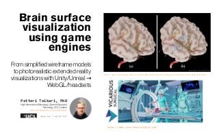
Brain surface visualization using game engines
•
1 gefällt mir•252 views

From simplified wireframe models to photorealistic extended reality visualizations with Unity/Unreal → WebGL/headsets 1) Deep learning brain segmentation for CT/MRI 2) Surface reconstruction from segmented brain 3) Surface visualization of regions of interest using game engines such as Unity or Unreal Slides are compiled mostly for people into 3D visualization, computer graphics, game developers who have not seen so many many brain visualizations necessarily. Overview of brains are currently being visualized and how could be upgraded Alternative download link: https://www.dropbox.com/s/69b176g8d8kuw1b/brain_viz.pdf?dl=0

Empfohlen
Empfohlen
Weitere ähnliche Inhalte
Mehr von PetteriTeikariPhD
Mehr von PetteriTeikariPhD (20)
Kürzlich hochgeladen
Kürzlich hochgeladen (20)
Brain surface visualization using game engines
- 1. Brain surface visualization using game engines Fromsimplifiedwireframemodels tophotorealisticextendedreality visualizationswithUnity/Unreal → WebGL/headsets Petteri Teikari, PhD High-dimensionalNeurology,Queen’sSquareof Neurology,UCL, London https://www.linkedin.com/in/petteriteikari/ Version “14/12/20“ https://www.unrealengine.com/en-US/spotlights/helping-brain-surgeons-practice-with-real-time-simulation https://www.vicarioussurgical.com/
- 2. GOAL Visualize simple “Tuftean” clinically- relevantvectorizedsurfacesfor clinicians, insteadof“messier”voxelbrainimages Slides are compiled mostly for people into 3D visualization, computer graphics, game developers who have not seen so many many brain visualizations necessarily. Overview of brains are currently being visualized and how could be upgraded
- 3. ExecutiveSummary “Rasterbrain” e.g. 1mm3 MNI CT Withlabel masksforhematoma andbeyond “Vectorizedbrain” e.g.mesh/NURBS Labelmaskskept for surface soyoucan easilye.g.“peel off” in XRbrain around hematoma Could start modularwith2 models,but thiscould be eventually end-to-end model of course “PerfectMesh” (well surface+uncertainty) visualizationdevcouldbe done separatelyfrommeshextraction Start with simplewireframes, advance to morephotorealistic renders, scalable to XR applications. Use industry-standardgameengines
- 4. Ideaillustrated: Real-time interactive 3DModel See the moodboardend of this slideshow for more visualizations Visualize the brain vasculature ifthatisthatinterestof the clinician (e.g.CT Angiography) Nowelletal. 2016http://doi.org/10.3791/53450
- 5. Ideaillustrated: What complexity level? Nowelletal. 2016 http://doi.org/10.3791/53450 https://iiif.wellcomecollection.org/image/B0007786.jpg/full/ 2048%2C/0/default.jpg https://www.unrealengine.com/en-US/spotlights/ helping-brain-surgeons-practice-with-real-time -simulation Tradeoffs: ● Looking pretty vsbeing understood by the end-users(e.g. datascientistsand clinicians). You need todo a usabilitystudyonthe user experience (UX) atsome pointif you startdeveloping newvisualization tools ● Looking pretty requiresalsoheavier hardware? Isthisalwaysfound in hospitals(no!), and same visualization pipeline could have variouscomplexitylevelsimplemented? Or write anew “wireframe”library if the current off-the-shelfphotorealisticrenderersyou are happywith?
- 6. Howlowpolyyoucango whilestillkeepingthevisualized braininterpretable? “Engineered” planar surfaces Easytosimplify this forvisualizations withhighfidelity Leaves,likehumanbrain,require morehighpolyrepresentation Easier tomodelbrainsof species withnogyrificationifyouareintomodeling forexamplemousebrain https://www.livescience.com/47421-human-brain-wrinkles.html lowpolygang Artby 🚲 Art by @andreykrygovhttps://www.instagram.com/p/CIqh_8JDV_Y/ https://www.blendswap.com/blend/6770
- 7. TECHBASICS
- 9. Mesh→ Unreal/Unity WebGL, etc. ifyou are into visualization→ Helpingbrainsurgeonspractice withreal-time simulationAugust30,2019bySébastien Lozé https://www.unrealengine.com/en-US/spotlights/helping-brai n-surgeons-practice-with-real-time-simulation In their 2018 paper Enhancement Techniquesfor Human AnatomyVisualization, Hirofumi Seo and Takeo Igarashi state that “Human anatomy is so complex that just visualizing it in traditional ways is insufficient for easy understanding…” To address this problem, Seo has proposed a practical approach to brain surgery using real-time rendering with UnrealEngine. Now Seo and his team have taken this concept a step further with their 2019 paper Real-Time Virtual Brain Aneurysm ClippingSurgery, where they demonstrate an application prototype for viewing and manipulating a CG representation of a patient’sbrain in real time. The software prototype, made possible with a grant (Grant Number JP18he1602001) from JapanAgencyforMedical Researchand Development(AMED), helps surgeons visualize a patient’s uniquebrainstructurebefore, during,and after anoperation. BrainBrowser isanopensourcefree3DbrainatlasbuiltonWebGLtechnologies, ituses Three.JStoprovide3D/layeredbrainvisualization. Reviewedin medevel.com Blender.blendfilebyplacedintheAssetsfolderofaUnityproject https://forum.unity.com/threads/holes-in-mesh-on-import-from-blender.248126/ Interaction betweenVolumeRendered3DTextureandMeshObjects https://forum.unity.com/threads/interaction-between-volume-rendered-3d-texture-and-mes h-objects.451345/
- 10. Easythentovisualizeon computer/VR/MR/AR OCTOBER14,2017 BY ANDIJAKL VisualizingMRI &CT Scans inMixedReality /VR/AR,Part 4: SegmentingtheBrain https://www.andreasjakl.com/visualizing-mri-ct-scans-in-mixed-reality-vr-ar-part-4-segmenting-the-brain/ Combining3DscansandMRIdata http://www.neuro-memento-mori.com/combining-3d-scans-and- mri-data/ VRsoftwaremaybring MRIsegmentationinto thefuture MattO'Connor July30, 2018 AdvancedVisualization https://www.healthimaging. com/topics/advanced-visu alization/vr-software-mri-s egmentation-future Nextmed:Automatic Imaging Segmentation,3D Reconstruction, and 3DModel VisualizationPlatform Using Augmentedand VirtualReality (2020) http://doi.org/10.3390/s2 0102962
- 11. PhysicalModels? Vascular models niceto be3D printed for surgery practice, but hematoma?
- 12. Makingthe Brains physicalwith 3D Printing Makingdatamatter:Voxelprintingforthe digital fabricationof data acrossscalesanddomains Christoph Bader et al. The Mediated Matter Group,Media Lab,Massachusetts Institute of Technology,Cambridg https://doi.org/10.1126/sciadv.aas8652 (30 May2018) We present a multimaterial voxel-printing method that enables the physical visualization of data sets commonly associated with scientific imaging. Leveraging voxel-based control of multimaterial three-dimensional (3D) printing, our method enables additive manufacturing of discontinuous data types such as point cloud data, curve and graph data, image- based data, and volumetric data. By converting data sets into dithered material deposition descriptions, through modifications to rasterization processes, we demonstrate that data sets frequently visualized on screen can be converted into physical, materiallyheterogeneousobjects. Representative 3D-printed models of image-based data. (A) In vitro reconstructed living human lung tissue on a microfluidic device, observed through confocal microscopy (29). The cilia, responsible for transporting airway secretions and mucus-trapped particles and pathogens, are colored orange. Goblet cells, responsible for mucus production, are colored cyan. (B) Biopsy from a mouse hippocampus, observed via confocal expansion microscopy(proExM) (30). The 3D print visualizesneuronal cell bodies, axons, and dendrites. (H) White matter tractography data of the human brain, created with the 3D Slicer medical image processing platform (37), visualizing bundles of axons, which connect different regions of the brain. The original data wereacquiredthroughdiffusion-weighted(DWI) MRI.
- 13. Voxel–SurfaceReconstruction Probablistic data-driven deeplearningprobably the best bet here already with current models
- 14. Surface (mesh orNURBS) fromvolumetricdata FastSurfer- Afastandaccurate deep learning basedneuroimagingpipeline Leonie Henschel et al. German Center for Neurodegenerative Diseases (DZNE),Bonn, Germany https://arxiv.org/abs/1910.03866 (9Oct 2019) Citedby17 To this end, we introduce an advanced deep learning architecture capable of whole brain segmentation into 95 classes in under 1 minute, mimicking FreeSurfer’s anatomical segmentation and cortical parcellation. The network architecture incorporates local and global competition via competitive dense blocks and competitive skip pathways, as well as multi-slice information aggregation that specifically tailor network performance towards accurate segmentation of both corticaland sub-corticalstructures. Further, we perform fast cortical surface reconstruction and thickness analysis by introducing a spectral spherical embedding and by directly mapping the cortical labels from the image to the surface. This approach provides a full FreeSurfer alternative for volumetric analysis (within 1 minute) and surface-based thickness analysis (within only around 1h run time). For sustainability of this approach we perform extensive validation: we assert high segmentation accuracy on several unseen datasets, measure generalizability and demonstrate increased test-retest reliability, and increased sensitivity to disease effectsrelative to traditional FreeSurfer.
- 15. Surface (mesh orNURBS) fromvolumetricdata#2 Surface-BasedConnectivity Integration Martin Cole et al. (2020) https://doi.org/10.1101/2020.07.01.183038 DeepCSR:A3D Deep LearningApproachforCorticalSurface Reconstruction RodrigoSantaCruz et al. (2020) https://arxiv.org/abs/2010.11423 Moreover, DeepCSR isasaccurate, more precise, and faster thanthe widelyused FreeSurfer toolboxand itsdeep learningpowered variant FastSurfer on reconstructing cortical surfacesfrom MRIwhichshould facilitatelarge-scale medical studiesand new healthcare applications.
- 16. Surface (mesh orNURBS) fromvolumetricdata#3 Probabilistic3DSurfaceReconstructionfromSparseMRIInformation KatarínaTóthová,SarahParisot,MatthewLee,EstherPuyol-Antón,AndrewKing,MarcPollefeys, EnderKonukoglu(Oct2020) https://arxiv.org/abs/2010.02041 Futureworkwillconcentrateon generalisationofthemethod tosurface reconstructioninthepresenceof pathologiesandreconstructionofother typesof organsofmorevariableshapes,whichmayleadtoadaptationoftheusedshapemodel.
- 17. Surface (mesh orNURBS) fromvolumetricdata#4 DeepSpline:Data-Drivenreconstructionof ParametricCurvesandSurfaces JunGao,Chengcheng Tang,VigneshGanapathi-Subramanian,Jiahui Huang, Hao Su, LeonidasJ. Guibas Universityof Toronto;VectorInstitute;Tsinghua University; Stanford University; UCSan Diego (Submittedon 12Jan2019) https://arxiv.org/abs/1901.03781 -Citedby14 https://github.com/SteveJunGao/deepspline See also orbingol/NURBS-Python Reconstruction ofgeometrybased ondifferentinputmodes,suchasimagesorpoint clouds,hasbeen instrumentalin thedevelopmentof computeraided design and computergraphics.Optimalimplementationsoftheseapplicationshavetraditionally involvedtheuseof spline-based representations attheircore. Mostsuchmethods attempttosolveoptimization problemsthatminimizean output-target mismatch. However,theseoptimization techniques requireaninitializationthatiscloseenough,as they arelocalmethods by nature.Weproposea deeplearningarchitecturethat adaptstoperform splinefittingtasks accordingly, providingcomplementaryresults to theaforementionedtraditionalmethods. To tacklechallengeswiththe 2Dcases suchas multiplesplineswithintersections, we use a hierarchical Recurrent Neural Network (RNN) Krauseetal.2017 trained with ground truth labels, to predict a variable number of spline curves, each with an undeterminednumberofcontrolpoints. In the 3D case, we reconstruct surfaces of revolution and extrusion without sel- fintersection through an unsupervised learning approach, that circumvents the requirement for ground truth labels. We use the Chamferdistance to measure the distance between the predicted point cloud and target point cloud. This architecture is generalizable, since predicting other kinds of surfaces (like surfaces of sweeping or NURBS), would require only a change of this individual layer, with the rest of the modelremainingthesame. Petteri: What would be the open-source workflow in production with NURBS? Harder to use Rhinoceros 3D in production (open question)
- 18. HaveCT-MRIpairsfortesting? #1: UseMRIsegmentationforCT Integratedanalysisofanatomicalandelectrophysiologicalhuman intracranialdataStolk etal.(2017) http://dx.doi.org/10.1038/s41596-018-0009-6
- 19. HaveCT-MRIpairsfortesting? #2: Use MRIsegmentation for CT https://doi.org/10.1111/epi.12827
- 20. Meshresampling? Already in surfacereconstruction net? Controlthe complexity ofthebrain mesh for the platform used to viewthe render How suitable aretriangular meshes actually? Howeasy to simplify e.g. comparedto NURBS? Do youhavea NURBSpresentation that youjustconvert to meshes if thevisualization library only support meshes?
- 22. “OldSchool”Simplification#2 Libigl https://libigl.github.io/tutorial/#subdivision-surfaces https://twitter.com/_AlecJacobson/status/12692953746588 67205 AlecJacobson@_AlecJacobson Manymeshsimplificationtoolsignore UVsor don'tdoagreat jobmaintainingexistingUVs. SongrunLiu's https://github.com/songrun/SeamAwareDecimater…built usinglibiglwilldecimatesurfacemesh*and*itsuvmap.Great formakingLoDsthatusethesametexture. https://rapidcompact.com/doc/cli/02-simplifying/index.html
- 23. Stillaresearchissuetosimplifymeshes#1 SpectralMeshSimplification https://hal.archives-ouvertes.fr/hal-02953334 Limitations. There are at least two main areas where our approach can further be developed, both related to time performance. First, the eigenvectors need to be computed at the beginning of the algorithm, while one may seek computing them at query time, when needed. Second, our cost evaluation involves fairly large matrices, at each evaluation which could be optimized using an approximation scheme
- 25. Coarse-to-fineupsampling aswell with deep learning topological updates of Loop Subdivision, but predicting vertex positions using a neural network conditioned on the local geometry of a patch. This approach enables us to learn complex non-linear subdivision schemes, beyond simple linear averaging used in classicaltechniques.Oneofour keycontributionsisa novel self-supervised training setup that only requires a set of high-resolution meshes for learning network weights. For any training shape, we stochastically generate diverse low-resolution discretizations of coarse counterparts, while maintaining a bijective mapping that prescribestheexacttargetpositionofeverynewvertexduringthesubdivisionprocess
- 26. Startdevelopingcomputationally lightvisualization withaframework thatscalesto fancierphotorealisticrendersaswell
- 27. ExtendedReality(VR/AR/MR/XR)inMedicine “VRx”: A MedgadgetBook Interview with AuthorDr. Brennan Spiegel DECEMBER8TH,2020 SCOTTJUNG https://www.medgadget.com/2020/12/vrx-a-medgadget-book-interview- with-author-dr-brennan-spiegel.html https://doi.org/10.1007/s11936-019-0722-7 The most readily available benefits of XR are in the form of visualizations of 3D anatomy and real-time display of anatomy and tooling. There are currently no published prospective clinical trials using AR or XR in human subjects. However, given the rapid development in the field, we expect to see human data in the near future. The XR hardware landscape is changing rapidly. Future hardware advances should improve visual realism and user comfort. Incorporation of haptic feedback into XR systems may be an important breakthrough for interventional procedures. Readers interested in more information about this field, particularly XR display hardware considerations, should consult our previous review [23]. The book Mixed and Augmented Reality in Medicine [24] will be of interest to readers looking for an in-depth resourceabout XRin medicine.
- 28. KeeptheExtendedReality(XR-VR/ AR)optionopen #1 Multi-ThreadedIntegrationofHTC-ViveandMeVisLab February2018 doi: 10.13140/RG.2.2.18864.05121 Conference: SPIE MedicalImaging2018,Project: VirtualRealityinMedicine Simon Gunacker,MarkusGall,DieterSchmalstieg,Jan Egger Virtualinteractionandvisualisationof 3Dmedicalimagingdata withVTK andUnity Gavin Wheeler; Shujie Deng; NicolasToussaint;Kuberan Pushparajah; JuliaA. Schnabel;John M. Simpson;Alberto Gomez Healthcare TechnologyLetters ( Volume:5, Issue:5, 10 2018) https://doi.org/10.1049/htl.2018.5064 - Cited by 17 Thesurfacerenderingtechniquesused in recentVR and AR medical visualisation systemsbuiltusing Unity requirea patient-specific polygonalmodel oftheanatomy ofinterest. Such surfacemodelsaretypicallyderived frommedical imagesthrough segmentation, using manual orsemi-automaticmethods.Inmostcases, thisinvolvesmanual effort,and thetimeand skill to do thismay besignificant. Moreover, the segmentationprocessinherently losesinformationpresentin theoriginal volumedata. Volumedata often do nothavepreciseboundaries, butvolumerendering allowstheuser to interactivelytunerendering parametersor applyfilters,to achievethedesired appearance. By integratingvolume rendering in VR, weremovethe potentially erroneoussegmentation stepsandgive theusermore flexibility andcontrol. In thiswork, weaimto integrateVTK intoUnity to bring themedical imaging visualisation featuresof VTKinto interactivevirtual environments developed using Unity. Particularly,wedescribea method to integrateVTKvolumerendering of 3Dmedical imagesinto a VR Unityscene,and combinetherendered volumewithopaquegeometry,e.g.spherelandmarks.Wefocuson creatingcore technology to enablethisandgivedevelopersandresearchersthe easeofuse andflexibility ofUnity combined with thevolumerendering featuresof VTK. High level workflowdiagramshowing thecommunication and interaction between MeVisLab and theHTCVivevia OpenVR capsuled byan ownthread.
- 29. KeeptheExtendedReality(XR-VR/ AR)optionopen #2 NextMed,AugmentedandVirtualRealityplatform for 3Dmedical imagingvisualization González-Izard,S.AlonsoPlaza,Ó,Sánchez Torres, R.Juanes-Méndez,J.A. García-Peñalvo, F.J.(2020)http://repositorio.grial.eu/handle/grial/1803 The Grid Factory, a U.K.-based provider of NVIDIA GPU-accelerated services, is partnering with telecommunications company Vodafone to showcase the potential of 5G technology with a network built at Coventry University. Operating NVIDIACloudXR on the private 5G network, student nurses and healthcare professionalscan experience lessonsand simulations invirtual realityenvironments. https://blogs.nvidia.com/blog/2020/11/17/coventry-university-cloudxr/
- 30. KeeptheExtendedReality(XR-VR/ AR)optionopen #3 Validation of virtualreality orbitometry bridgesdigitaland physical worlds PeterM.Maloca,BalázsFaludi,MarekZelechowski,ChristophJud,TheoVollmar,SibylleHug,PhilippL.Müller,EmanuelRamosdeCarvalho,JavierZarranz-Ventura, MichaelReich,ClemensLange,CatherineEgan,AdnanTufail,PascalW. Hasler,Hendrik P.N.Scholl&PhilippeC.Cattin ScientificReportsvolume10,Articlenumber: 11815(July2020) https://doi.org/10.1038/s41598-020-68867-6 In summary, the orbit and possiblyall other three- dimensionalspaces can be non-invasively and digitally visualized and measured in close-to-reality conditions and investigated with a high precision using aVR image display method that measuredwhat it purportsto measure. An objective diameter measurement can be attained to quantifythe dimensionsof the orbit and improve spatial awareness, diagnosis, monitoringand pre-surgical planning.
- 31. KeeptheExtendedReality(XR-VR/ AR)optionopen #4 Virtualreality in advanced medicalimmersive imaging: a workflow for introducing virtual reality asasupporting tool in medicalimaging MarkusM.Knodel,Babett Lemke,MichaelLampe,MichaelHoffer,ClarissaGillmann,MichaelUder,JensHillengaß, GabrielWittum&TobiasBäuerle Computing andVisualization in Sciencevolume18,pages203–212(2018) https://doi.org/10.1007/s00791-018-0292-3 Our approach is based on the use of the graphical surface package VRL [4] (Visual Reflection Library) which preserves various own packages and brings together third party single packages to enable within one program the extraction of 3D volume and surface rendered CT and MRI image stacks. Such an approach is based on a very intuitive and simple GUI (graphical user interface) to project them into the virtual reality space within the environment of a versatile and prominent 3D virtual reality project, namely COVISE / opencover (COllaborative VIsualization and Simulation Environment) [5] of the HLRS (Höchstleistungsrechenzentrum) Stuttgart which is used widely within automotive development processes of e.g. Mercedesand Porsche. See UnityFormafor similar functionalityfor the automotive industry and beyond https://blogs.unity3d.com/2020/12/09/introducing-unity-forma-reimagine-m arketing-with-real-time-3d/
- 32. KeeptheExtendedReality(XR-VR/ AR)optionopen #5 Modeling and VisualizationforVirtual Interaction withMedical Image Data Fredrik Nysjö,DepartmentofInformation Technology, Division ofVisualInformation andInteraction,Box337,UppsalaUniversity, SE-75105Uppsala, Sweden. https://uu.diva-portal.org/smash/get/diva2:1388179/FULLTEXT01.pdf Theuseofraytracingandpath tracingforrealisticrendering ofglobalilluminationis becomingmorecommonin visualizationandinteractive applications,whilepreviously mainlybeingusedinfilmrendering andotherofflinerendering. Combining thecachingofglobal illuminationinPaper Vwiththehybrid raytracingofPaper VIfor improving theinteractiveperformanceis somethingthatwouldalsobe interestingtoinvestigate.
- 33. KeeptheExtendedReality(XR-VR/ AR)optionopen #6 How Iused AR To Build 3D MRI Scans RiyaMehtaNov19,2019 https://medium.com/@riyamehta9001/how-i-used-ar-to-build-3d-mri-scans-5e0df497c594 Autodesk 3D Itwasawesometoplayaroundwiththisandbeableto implementmodelsIbuiltprogrammedintothe augmentedrealityplatformIusedcalledSketchfab, whichconverts/programs3DmodelsintoeitherVRor ARdemos.Here’saquickglanceatwhatIwasabletodo. Augmentedrealitywillcompletelychangethewaywediagnosepatients,view3D models&observethemedicalindustryasawhole.
- 34. KeeptheExtendedReality(XR-VR/ AR)optionopen #7 Visualizing MRI& CT Scans inMixed Reality / VR /AR,Part 2: 3D Volume Rendering AndreasJakl(October10, 2017) https://www.andreasjakl.com/visualizing-mri-ct-scans-mixed-reality-vr-ar-part-2-3d-volume-rendering/ DigitalHealthcare,Augmented Reality,MobileAppsand more!AndreasJaklisa professor @St.PöltenUniversityof Applied Sciences,MicrosoftMVP forWindowsDevelopmentandAmazonAWS EducateCloud Ambassador.
- 35. KeeptheExtendedReality(XR-VR/ AR)optionopen #8 https://www.cgtrader.com/low-poly-3d-models/brain 165 low poly 3D Brain models are available for download. These models contain a significantly smaller number of polygons and therefore requireless computing power to render. Models which have fewer polygons are best used in real time applications that require fast processing, like virtual reality (VR), augmented reality (AR), mixed reality (MR), cross reality (XR) and games, especially mobile games. Choose from our collection of rigged and animated models to easily use them in your real-time applications. Get 3D assets for environments, pick props, objects or buy complete 3D model collections, bundles and packs with everything your game might need. Save development time and costs, make prototype experiences or use 3Dmodelsas placeholdersinyour project.Tofindmodelsthatrenderpredictably in variousengines,usethePBRfilternexttothesearchbar. https://www.cgtrader.com/3d-models/science/medical/low-polygon-art-medical-brain-color
- 36. GameEngines(e.g. Unity orUnreal) fromsimple wireframestoXRvisualization StartwithlightweightWebGL / 3DPDF visualizationswithclinicianswithlittle hardware and/ortechnicalskills.Three.JSapproache.g. used in X Toolkit mightbe even simpler?
- 37. WebGL as your baseline? https://medevel.com/15-webgl-medical-visualization-projects/ AnatomyLearning is a free 3D anatomy atlas that exported to work with WebGL using Unity Game Engine & Unity Web Player. It provides 2 versions one for the modern web browser that is built on WebGL2.0 and other for Android mobile systems. This 3D heart anatomyVR(Virtual Reality) project is built by BabylonJS: a WebGL JavaScript Framework, It's published as a VR (Virtual Reality) Project at VESTA a WebVR social network that provides a sharing platform VRartists, developers, & normal users. TheOpen AnatomyProject is an open source 3D anatomy atlas that works directly from the web browser as it uses pure HTML technologies and WebGL rendering. The Open Anatomy project is carried out and developed by the Brigham and Women's Hospital in Boston aiming to deliver rich digital anatomy atlases to students, doctors, researchers, and thegeneral public. BrainBrowser isanopensourcefree3DbrainatlasbuiltonWebGLtechnologies,ituses Three.JSto provide3D/layeredbrainvisualization.WehavereviewedBrainBrowser andlistedallofitscurrent features,youmayreadaboutitathere: BrainBrowser:OpensourceWeb-basedBrainVisualizationwithVolume&Surfaceviewers
- 38. WebGL as your baseline? ModernScientificVisualizationson the Web LoraineFrankeand DanielHaehn InformaticsSeptember2020,7(4),37; https://doi.org/10.3390/informatics7040037 Modern scientific visualization is web-based and uses emerging technology such as WebGL (Web Graphics Library) and WebGPU for three-dimensional computer graphics and WebXR for augmented and virtual reality devices. These technologies, paired with the accessibility of websites, potentially offer a user experience beyond traditional standalonevisualizationsystems. We review the state-of-the-art of web- based scientific visualization and present an overview of existing methods categorized by application domain. As part of this analysis, we introduce the Scientific Visualization Future Readiness Score (SciVis FRS) to rank visualizations for a technology-driven disruptive tomorrow. We then summarize challenges, current state of the publication trend, future directions, and opportunities for thisexciting research field.
- 39. WebVR Virtual Reality Volume Rendering forreal-time VisualizationofRadiologicAnatomy https://tobias.rautenkranz.ch/blog/code/webvr-vr.html (2017) A new Web technology (WebVR) in combination with powerful smartphone graphic processors and inexpensive virtual reality viewers (Google Cardboard) have made virtual reality cheap and easily obtainable. These new developments and the before mentioned possibility of a more natural visualization have lead me to implementanexperimentalVRwebpageforanMRIscan. Implementation Details The existing volume renderer for WebGL was adapted to support the new WebVR standard to allow virtual reality rendering in the browser. Like for mostVRrenderers aforward renderer is used. Thedifference being, that this is normally done to be able to use MSAA (see Optimizing the Unreal Engine 4Renderer for VR, Pete Demoreuille); but this is of no use for volume rendering. Instead, the ability of the existing WebGL renderer to output an image of the volume and its segmentation simultaneously is not needed. On the contrary, they need to be combined in one image. Thus, the renderer was adapted to a single stage. As a side effect, color renderings are now possible without usingthe WEBGL_draw_buffers extension. Since multiple layers are not supported by WebVR implementations, some additional overlay renderers are injected before or after the volume rendering. Please note that, due to its experimental nature, the source code is sometimesconfusingor even confused.
- 40. Example of Unityuse Interactive heartwithUnity3D http://www.medicalgraphics.de/en/projects/making-ofs/interactive-heart-with-unity3d.html To useWebGL there areanumberof toolsandenginesavailable. Sometimeagowedealtwith the possibilitiesof Sketchfab,aweb servicethatallowsasimplewaytovisualize3DdatainWebGL. Sketchfabismainlya puredataviewer,interaktionissomehow limited.Also themodelsareattachedto aweb serviceandcannot me usedstand-aloneoroffline. With theliberalizationoftheirlicenseconditionsandthe integrationof WebGL/html5Unity3Dwasbecomingavery interesting tool.Unityisactuallyagameengine,adevelopment environmentfor games.Through it´spossibilitiestwo writeown codeandscriptsandthenexporttheapplicationfordifferent devices(PC, Mac,web,consoles,mobile)the possibilities are virtuallyunlimited.AlsoUnityisnotawebservice,therefore youhavefullcontroloverthecreatedapplication. Asasimpletestobjectfor aninteractive,medical application,wechose ahuman heart,which shouldbe freely explorableintheapp.Inaddition,webuiltasimplefunctionalityto emphasizeanatomicalstructures- inthisparticularcase there is anoptiontoremove the coronaryvesselsandtoopena partofthe hearttolookintothe ventricles.Thisisasimple example ofthepossibilitiesofinteractivity3DModels.
- 41. Example of Unityuse #2 HolographicReconstructionofAxonalPathwaysinthe HumanBrain Mikkel V. PetersenJeffrey Mlakar Suzanne N. Haber Martin Parent Yoland Smith Peter L. Strick Mark A. Griswold Cameron C. McIntyre Published:November 07, 2019DOI: https://doi.org/10.1016/j.neuron.2019.09.030
- 42. Example of Unityuse #3 ColorRenderinginMedical Extended-RealityApplications AndreaSeungKim, Wei-ChungCheng, RyanBeams& AldoBadanoJournal of Digital Imaging (November 2020) https://doi.org/10.1007/s10278-020-00392-4 RGBinputandoutputforfivedigitalmaterial,digitallighting,anddigitalcamera configurationswithintheUnityengineintherenderingofcross-platform applicationsfor selectedscenes:(a)adigitalpathologyimage[8],(b) adigital chestradiograph,and(c)afull-fielddigitalmammogram[9] When buildingan XRapplication fordifferent platforms,developersshouldconsiderthefilesize with associatedmemorysizerequirements,pixel dimensions,andresolution oftheimagetexturesfor each targetplatform[25],asthetypeoftexture compression isdependenton theintendedplatform. Forinstance,astandaloneXR HMD,Androidmobile device,andaPCwill each havetheir own unique compression formatsthatwork with their specific hardwareassomegraphicsdevicesonlyusecertain compressed formats.Developershavetheabilityto designatespecificcompressionsettingsforeach platformin theimportsettingsoftheinspector window.
- 43. Example of Unityuse #4 Virtual linearmeasurementsystemforaccuratequantificationof medicalimages Gavin Wheeler;ShujieDeng;Kuberan Pushparajah;JuliaA. Schnabel;John M. Simpson;AlbertoGomez School ofBiomedicalEngineering& ImagingSciences, King'sCollegeLondon,London,UK HealthcareTechnologyLetters ( Volume:6, Issue:6,122019)https://doi.org/10.1049/htl.2019.0074 Hierarchical structureof themeasurementprefab, asimplemented in Unity.Themeasurement objecthas fivechild objects, asillustrated. Objectsmarked with ‘I’havephysicsinteractors.Blueand purplearrowsindicatethelinking of theconnectorlinesto thestartpoint, end pointand label. Green arrowsindicatetheUnityscriptsgoverning thescaleof theobjects. Thered arrow indicatesthe redirection of editing (translate, rotate)fromtheconnectorto themeasurementparent. Shapes, coloursand label textarearepresentativeexample We proposed a 3D VR system to carry out linear measurements on volumetric images, and demonstrated it on echocardiographic images of a calibration phantom and of cardiac patients. All measurements were carried out with Philips QLAB (our baseline), Tomtec (its 3D measurement system only) and our proposed VR platform. Overall, this study showed that a VR system can have measurement tools that are comparable to clinically used commercial tools, while providing further insight and understanding into complex 3D anatomy.
- 44. Example of Unityuse #5 ApplicationsofVRmedicalimage visualizationtochordallengthmeasurementsforcardiacprocedures PatrickCarnahan, John Moore, Daniel BainbridgeM.D., Gavin Wheeler, ShujieDeng, Kuberan Pushparajah, ElvisC. S. Chen, John M. Simpson, TerryM. Peters ProceedingsVolume11315,MedicalImaging2020: Image-GuidedProcedures, RoboticInterventions,andModeling; 1131528(2020) https://doi.org/10.1117/12.2549597
- 45. Example of Unrealuse #1 Helpingbrainsurgeonspractice withreal-time simulation August30,2019bySébastienLozé https://www.unrealengine.com/en-US/spotlights/helping-brai n-surgeons-practice-with-real-time-simulation In their 2018 paper Enhancement Techniquesfor Human AnatomyVisualization, Hirofumi Seo and Takeo Igarashi state that “Human anatomy is so complex that just visualizing it in traditional ways is insufficient for easy understanding…” To address this problem, Seo has proposed a practical approach to brain surgery using real-time rendering with Unreal Engine. Now Seo and his team have taken this concept a step further with their 2019 paper Real-Time Virtual BrainAneurysm ClippingSurgery, where they demonstrate an application prototype for viewing and manipulating a CG representation of a patient’sbrain in realtime. In developing the application, Seo’s team chose Unreal Engine as the underlying real-time technology because of its graphics and programming tools. “Unreal Engine has powerful mathematical C++ APIs such as FVector, FMath, and UKismetMathLibrary, so we find it to be asuitable platform for research on3D CG geometry,” saysSeo.
- 46. Example of Unrealuse #2 VolumeRendering inUnrealEngine4. 08-04-2016,04:20PMTobehonest,Iamnotsurethisshouldbehere,butIfelttheother topicswereevenlessrelevantasIamtalkingaboutrendering.Justnotthestandard methodsinUE4.FeelfreetomoveifIplaceditinthewrongarea.Tostart,letmebetransparent. IamworkingonamastersthesisusingVRandscientific visualization.Isawpotentialinthemergingof UE4andscientificvisualizationforstudents,scientists,gamersandallgraphicalartistsalike. https://forums.unrealengine.com/development-discussion/rendering/91596-your-thoughts-on-and-comments-to-volume-rendering-in-unreal-engine-4 https://youtu.be/z34X_52O20U
- 47. Example of Unrealuse #3 VolumetricMedicalData Visualizationfor Collaborative VREnvironments 27 October 2020 RolandFischer,Kai-ChingChang, René Weller,Gabriel Zachmann https://doi.org/10.1007/978-3-030-62655-6_11 Wepresentaneasy-to-useandexpandable systemforvolumetricmedicalimage visualizationwithsupportformulti-userVR interactions.Themain ideaistocombine astate-of- the-artopen-sourcegameengine,theUnreal Engine4,withanewvolumerenderer forCT images. Theunderlyinggameenginebasis guaranteesthe extensibility andallowsforeasy adaptionofour systemtonewhardwareandsoftware developments.Inourexample application,remote userscanmeetinasharedvirtualenvironmentandview, manipulate anddiscussthe volume-rendereddatain real-time. OurnewvolumerendererfortheUnrealEngineis capableofreal-timeperformance,aswellas,high-quality visualization. For the future we plan to expand the interaction possibilities with the volume visualization,specifically,wearelooking atintegratingadynamicclippingplaneforabetterview of internal regions and a volumetric drawing tool allowing for quick sketches and annotationsinside the volume.Other improvementswouldbe adirectintegrationand parallelization of the preprocessing part to speed up the workflow and allowing for a dynamic adjustment of the transfer functions. To improve the visualization of complex structures and organs that involve multiple materials support for multi-dimensional transferfunctionscouldbeadded.
- 48. Example of Unrealuse #4 3D Kinematicsof UpperLimb FunctionalAssessment UsingHTCVive in UnrealEngine4 KaiLiang Lew,Kok SweeSim,ShingChiangTan, FazlySalleh Abas19November 2020 https://doi.org/10.1007/978-3-030-63119-2_22 The purpose of research in this paper is to quantify the accuracy and precision of HTC Vive by making upper limb assessment measurements and performing functional tasks in the Unreal Engine 4. Thirty healthy males performed daily aim functional tasks, and arm length measurement and assessment were made. Each participant attended two testing sessions and one arm length measurement session. The upper limblength wasmeasured using HTC Vive after making three types of hand posture exercises. The arm assessment included the minimum and maximum angle of shoulder adduction, abduction, flexionand extension. The experiment showed all the upper limb measurements collected from the functional tasks as well as the position and rotation of the upper limb could be estimated correctly. The proposed system is potentially useful for assessing stroke rehabilitationinthe hospital and rehabilitationcenter.
- 49. Example of Unrealuse #5 The Uterine Games:UsingaGame EnginetoDevelop a 3D DigitalFemale ReproductiveTracttoAidin AnatomyEducation YunaK. Park DanielleRoyer(18 April2020) https://doi.org/10.1096/fasebj.2020.34.s1.04584 The aim of this project was to iteratively design and develop a mobile application (app) depicting a 3D model of the plastinated female reproductive tract. A 3D surface model of the plastinate was digitally reconstructed using an Artec Space Spider 3D Scanner. Artifacts were smoothed and texture was refined in ZBrushCore 2018 and Autodesk Maya 2019. The model was packaged into a mobile app using a gameengine, UnrealEngine4 (UE4). Compared to other app development software, UE4 was chosen for its robust visualization of 3D models, cross‐ platform deployment, and zero upfront costs. With online tutorials, UE4’s Blueprints visual scripting system is relatively simple to grasp, and the node based interface is‐ a powerful approach for non programmers‐programmers , allowing extreme flexibilitywithout the need for coding.Utilizing this flexibility, the app was designed to promote self paced‐ independent learning of the female reproductive tract and associatedpelvicanatomy. https://youtu.be/EFXMW_UEDco https://www.unrealengine.com/en-US/spotlights/vr-med ical-simulation-from-precision-os-trains-surgeons-five-ti mes-faster
- 50. Unity (C#, simpler?) vsUnreal(C++, morephotorealistic)
- 53. Nice exampleof hownotto visualizebrain Veryhard toanalyze structureswiththesemanuallyset anatomicalopacitieswithout skull-stripping surface visualization→ http://doi.org/10.1007/978-3-540-30497-5_81
- 55. BlenderandCycles arebothopen-source,andsuitableforproduction (atleastfornon-realtimesolutions),checkforlicenses? Blender2.91.0Python APIDocumentation https://docs.blender.org/api/current/index.html BVTKNodes - photorealistic rendering ofVTK data in Blender https://discourse.vtk.org/t/bvtknodes-photorealistic-rendering-of-vtk- data-in-blender/3268/16 cycles volumerendering 3D image texture (CTorMR dataset) https://blender.stackexchange.com/questions/18418/cycles- volume-rendering-3d-image-texture-ct-or-mr-dataset https://blender.stackexchange.com/questions/62110/using-image-sequence-of-medical-scans-as-volume-data-in-cycles
- 56. Aninteractiveframeworkforwhole-brainmapsat cellular resolution Fürthet al.(2017) https://doi.org/10.1038/s41593-017-0027-7
- 57. Collection of lowpoly brain models clickimagesfor source In other words, why hand-model these, if you could create automatically low-poly brains from acquired CT/MRI images, and these low-poly models with ROI overlays of the structure of interest highlighted in them, in a interactive 3D model. And if nothing else, you can use this as an inspiration for your startup branding
- 59. VessMorphoVis implementsdifferentalgorithms forvisualizingvascular networks.Theoutlineofthe morphologyissketchedin(A)usingthinpolylines andtinyspherestorepresentthesectionsand samplesofthemorphology,respectively.In(B),the morphologyisillustratedbyalistofpointsshowing onlytheindividualsampleswithoutany connectivity.Themorphologyisvisualizedasa disconnectedsetofsegmentsandsectionsusing thesamecolorin(C)and(F),withalternating colors in(D) and(G)andalsousingtransparentshadersin (E)and(H),respectively Users can control the visual quality of the skeleton, choosing between highly optimized geometry (A and C) for global far views or high-quality reconstructions(B and D) for close up views. Morphologypolylinesare rendered using bevel objects with 4 and 16 sides in A and B, respectively. The piecewise segments of the polylines (C) might limit the visual quality in case of close ups; therefore, we added another parameter to use spline interpolation to smooth their curvature (D) A high-qualityrendering of a largevasculaturemesh reconstructed froma vasculargraphhaving 2.1 million∼2.1 million samplesbased on ourmetaballs implementation. The meshisrendered using theartistic glossyshaderwith Cycles Thesamemeshreconstructedwithmetaballsalgorithmisrendered withfourdifferentshaders: glossy, flat, artisticbumby and artistic glossy inA,B,CandD,respectively,using theWorkbenchand CyclesrenderesinBlender https://doi.org/10.1093/bioinformatics/btaa461
- 60. TheNIH/NIGMSCenterforIntegrativeBiomedicalComputing UncertaintyVisualization (youshouldhaveyouruncertaintiespropagated fromsegmentationandsurfacereconstructionalgorithmsforthis) https://www.sci.utah.edu/cibc-research/highlights/24-cibc-highlights/175-uncertainty-visualization.html AnIsosurfacevisualizationofamagnetic resonanceimagingdataset(inorange)surrounded byavolumerenderedregionoflowopacity(in green) toindicateuncertaintyinsurfaceposition. F.Jiao,J.M.Phillips,J.Stinstra,J.Kueger,R.Varma,E.Hsu,J.Korenberg,C.R.Johnson. "MetricsforUncertaintyAnalysisandVisualizationofDiffusionTensorImages," InProceedingsofthe5thinternationalconferenceonMedicalimaging andaugmentedreality (MIAR),Beijing,China,Springer-Verlag,Berlin,Heidelbergpp.179--190.September,2010 https://doi.org/10.1007/978-3-642-15699-1_19 - Citedby17
- 62. PhysicallyBasedShadingformedicalimagedata 17Feb2017 https://www.openinventor.com/en/news/detail/id/2359 Classicaltechniquesforrenderingmedicalimagedatain3Dhavebeenaroundsincethe1980s,including multi-planar reformatting(MPR),maximumintensityprojection(MIP)anddirectvolume rendering (DVR)withcolorandopacitymapping.Thesetechniquesarehighlyusefulbutbasedonverysimplemodelsofcolor,lighting,andtransparencythatdonotaccuratelyrepresentthe appearanceofmaterialsintherealworld. PhysicallybasedshadingcombinesavarietyofGPUacceleratedtechniquesincludingimage-basedlighting,complexsurfacereflectionmodeling,ray-tracedshadowcasting,ambientocclusion, highdynamicrangeanddepthoffield. Thesetechniquescanbeusedinteractivelyontypicaldesktopmachineswithstandardgraphicshardware.
- 64. https://brainder.org/research/brain-for-blender/ Imageacquisition and reconstruction The images were acquired at the Research Imaging Institute, University of Texas Health Science Center at San Antonio, in a Siemens magnetom Trio 3T system, in two sessions, each consisting of 6 acquisitions of T1-weighted images, using a mprage sequence, with voxel size of 0.8×0.8×0.8 milimeters. The images were registered and averaged to improve signal-to-noise ratio, as described here, and bias corrected using spm8 software. The already realigned, averagedand bias-corrected volume,in nifti format, isavailable here. The generation of the cortical meshes and subcortical segmentations used FreeSurfer 5.2.0. The splitting of the cortical meshes into independent objects was performed using a custom script that soon will be released at Brainder.org (update: they are now available here). The subcortical meshes were produced from the volumetric segmentations, as described here. Subcortical structures In addition to the above cortical meshes, surfacesforsubcortical structures are also available.These are not produced directly by the FreeSurfer pipeline. However, the segmented volumesthat are part of the subcortical stream can be used to generate surfacesforvisualisation purposes, asdescribed here. The meshesforthe same brain, in different formats,can be downloaded here: srf mz3 obj ply.
- 68. Hybridrenderingof explodedviewsformedicalimageatlasvisualization https://docs.lib.purdue.edu/cgi/viewcontent.cgi?article=1013&context=cgtpubs:“In thisworkwehavedescribedan interactiveatlasvisualization systemwhich iscapableofcreatingexplodedviewsbasedon the hierarchicalstructureofthedata. Ourhybridrenderingtechniqueiscapableofexplodinganatomic meshesinto slabstorevealan underlyingmedical image.Wedetailedan OpenGLimplementation oftherenderingprocess, and presentedresultsfromtheAALneuroanatomical atlas.Ourimplementation isabletomaintain interactiveframerates,even on tablethardware.” https://doi.org/10.1080/21681163.2017.1343686
- 72. https://www.shutterstock.com/video/clip-1011364865-plexus-brain-animation-background-you-can-use
- 73. DashEnterpriseAppGallery Thispublicinstanceofthe 👑 DashEnterprise 👑 appmanager runs>60Dashappsfor100sofconcurrentusersonAzureKubernetesService.ClickonaDashapp'snamebelowformore infoandlinksto Python&Rsourcecode onGitHub.https://dash-gallery.plotly.host/dash-brain-viewer/
- 77. The importance of visualization AustenLester https://slideplayer.com/slide/12732663/
- 78. Simulation offlow incerebral blood vessels Reference: Adaptive Surface Visualization of VesselswithAnimated Blood Flow, The AuthorsComputer GraphicsForum, author:K. Lawonn et al.; Otto-von-Gericke- UniversityMagdeburg, Dept. of Simulation and Graphics Implementation of azSpace control within MeVisLab Reference: zSpace, author:P. Saalfeld; Otto-von-Gericke- UniversityMagdeburg, Dept. ofSimulati onand Graphics MeVisLab https://www.mevislab.de/mevislab/screenshots
- 85. Assessing performance of augmented reality- based neurosurgicaltraining Wei-Xin Si, Xiang-Yun Liao, Yin-LingQian, Hai-TaoSun, Xiang-Dong Chen, QiongWang & PhengAnnHeng Visual Computingfor Industry, Biomedicine, and Art volume 2, Article number: 6 (2019) https://doi.org/10.1186/s42492-019-0015-8
- 89. AntoineRosset@rossetantoine Testing #Cinematic 3DVRRenderinginOsiriX.This enginewillrequirethe28-coresofthenew #MacPro 5:05PM·Dec14,2019 Petteri: TODO! Overlay segmented surfaces with the volumetric “baseline anatomy”
- 91. 18 Healthcare AugmentedReality andVirtualRealityCompanies toWatch https://hitconsultant.net/2020/06/29/augmented-reality-and-virtual-reality-companies-to-watch/#.X9Nn-3UzZKg
- 92. NVIDIA Healthcare2.0– Developingand deployingAIin healthcare https://tectales.com/ai/nvidia-healthcare-2-0-developing-deploying-ai-in-healthcare.html ImFusionusesdeeplearningtoturn2Dultrasounddatainto3Dimages. NVIDIA is already working with various partners in adopting AI for their products. For example, Siemens Healthineers is using a NVIDIA GPU-based supercomputing infrastructure to develop AI software to generate organ segmentations that enable precision radiation therapy. Furthermore, Siemens’ SherlockAI supercomputer whichisused to run more than 500 AI experimentsdaily,is also powered byNVIDIAtechnology. However, NVIDIA is not only working with the industry, but also with academic and research institutions. They are collaborating with the King’s College London (Jorge Cardoso et al.) to bring AI in medical imaging to the point of care. In another project, they are applying ‘federated learning’ to algorithm development, allowing algorithms to be developed on site, using data from the local institutions, without the need for data to travel outside of its own domain. The work could lead to breakthroughs in classifying stroke and neurological impairments, determining the underlying causes of cancers, as well as recommendingthe best treatment forpatients.
- 93. Renderinga3DBrain volumein Blender https://blender.stackexchange.com/questions/15010/rendering-a-3d-volume InoneofmypostsItalkedaboutusing renderingbrainvolumesin-browser usingXTK.The results,I’lladmit,weren’tspectacular.Thevolumerenderingdidn’treallygiveverydefined edges.ButnowI’llshowacouplemethodsofrenderingabrainusing Blender.Thefirst methodisusingvolumetricdatainBlender,andthesecondusessurfacesgeneratedby FreeSurfer.Ithink itgivesprettycoolresults,check itoutbelow.(FreeSurfer .ascto Wavefront.objscript) https://mollermara.com/blog/blender-brain/
- 96. https://youtu.be/DH34mASfbTo Learn toturn yourCAT(CT)orMRI scan intoa3Dmodel.
- 97. Aorticstentfractureinthree-dimensional volumerendering (3DVR).(A)3DVR imagesdemonstratecompletetransverseaorticstentfracture (green circle)with angulation and slightlateralstentdisplacement.(B)Theeliminationof thesurrounding softtissueand vesselsallows fora bettervisualizationof thehardware. Coronaryarteriesinthree-dimensionalvolumerendering(3DVR).(A)3DVR imagesdemonstratethecourseoftheleftmaincoronaryartery(redarrow) originating(redasterisk) fromtherightcoronaryartery(greenarrow).(B) Itis alsoimportanttocorrelatethiscoronaryarteryanatomywiththesurrounding softtissuetoassessifthereisanymyocardialbridging. Spinalfixation hardwareand disc spacerin three- dimensionalvolumerendering (3DVR).3DVRimages demonstratespinalfixation hardwareand adiscspaceron (A)anterior-posteriorand (B) lateralviews.Theelimination of thesurroundingsofttissueand bones allowsforthebetter visualization ofthespinal fixation hardware(green arrow) anddisc spacer(red arrow)in ordertoevaluateforpossible hardwarecomplicationson (C) anterior-posteriorand(D) lateralviews. TheAdditionalDiagnosticValueoftheThree-dimensionalVolumeRendering ImaginginRoutineRadiologyPractice https://www.cureus.com/articles/22358-the-additional-diagnostic-value-of-the-three-dimensional-volume-rendering-imaging-in-routine-radiology-practice TheAdditionalDiagnosticValueof theThree-dimensionalVolumeRendering Imagingin RoutineRadiologyPractice
- 100. Surface Renderingof aBrain Tumor https://doc.pmod.com/p3d/example1surfacerenderingofabraintumor3131.html