Sensory and motor pathways
•Als PPTX, PDF herunterladen•
44 gefällt mir•33,591 views

Sensory and motor pathways

Empfohlen
Weitere ähnliche Inhalte
Was ist angesagt?
Was ist angesagt? (20)
Ähnlich wie Sensory and motor pathways
Ähnlich wie Sensory and motor pathways (20)
Mehr von Subramani Parasuraman
Mehr von Subramani Parasuraman (20)
Kürzlich hochgeladen
Kürzlich hochgeladen (20)
Sensory and motor pathways
- 1. S. Parasuraman, M.Pharm., Ph.D., Associate Professor, Faculty of Pharmacy, AIMST University Sensory and motor pathway
- 2. Learning Outcomes • At the end of this session, the student would be able to: – List various general sensations and receptors responsible for it. – Describe sensory pathways in spinal cord and brain and the somatosensory cortex – Describe upper and lower motor neuron. – Describe primary motor area and name the motor tracts. – Describe the corticospinal or pyramidal tract the somatosensory cortex
- 3. Myelinated and Non-myelinated Nerve Fibers • A motor neuron is a nerve that carries an impulse for a response from the CNS to an effector. • A mylinated neuron is covered by a myelin sheaths. A myelin sheath is membrane of a Schwann cell- the cell wraps itself around the axon of a neuron many times creating the sheath.
- 4. Myelinated and Non-myelinated Nerve Fibers • Non-myelinated fibers comprise the smaller axons of the CNS, in addition to peripheral postganglionic autonomic fibers, several types of fine sensory fibers (C fibers of skin, muscle and viscera), olfactory nerves, etc.
- 5. Myelinated and Non-myelinated Nerve Fibers Nerve fibres: A. Myelinated. B. Non-myelinated.
- 6. Myelinated and Non-myelinated Nerve Fibers Saltatory conduction of an impulse in a myelinated nerve fibre. • In myelinated neurones, the insulating properties of the myelin sheath prevent the movement of ions. • Therefore electrical changes across the membrane can only occur at the gaps in the myelin sheath, i.e. at the nodes of Ranvier. • When an impulse occurs at one node, depolarisation passes along the myelin sheath to the next node so that the flow of current appears to 'leap' from one node to the next. • This is called saltatory conduction.
- 7. Myelinated and Non-myelinated Nerve Fibers Simple propagation of an impulse in a non- myelinated nerve fibre. • The speed of conduction depends on the diameter of the neurone: the larger the diameter, the faster the conduction. • Myelinated fibres conduct impulses faster than unmyelinated fibres because saltatory conduction is faster than the complete conduction, or simple propagation. • The fastest fibres can conduct impulses 130 metres/ sec. (skeletal muscles) while the slowest impulses travel at 0.5 metres/ sec.
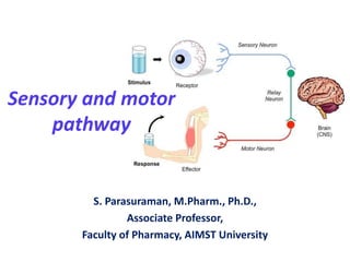
- 8. Sensory pathway (Transmit action potentials from periphery to brain)
- 10. Sensory nerve tracts (afferent or ascending) in the spinal cord • The white matter tracts in the spinal cord are highways for nerve impulse propagation. Sensory input travels along these tracts toward the brain, and motor output travels from the brain along these tracts toward skeletal muscles and other effector tissues. • The gray matter of the spinal cord receives and integrates incoming and outgoing information.
- 11. Sensory nerve tracts • There are two main sources of sensation transmitted to the brain via the spinal cord. – SKIN: Sensory receptors (nerve endings) in the skin, called cutaneous receptors, are stimulated by pain, heat, cold and touch, including pressure. Nerve impulses generated are conducted by three neurones to the sensory area in the opposite hemisphere of the cerebrum where the sensation.
- 12. Sensory nerve tracts • There are two main sources of sensation transmitted to the brain via the spinal cord. – Tendons, muscles and joints: Sensory receptors are nerve endings in these structures, called proprioceptors, and they are stimulated by stretch. Together with impulses from the eyes and the ears they are associated with the maintenance of balance and posture and with perception of the position of the body in space.
- 13. Sensory nerve tracts Receptor Route Pain, touch, temperature Neurone 1 -to spinal cord by posterior root Neurone 2 - decussation on entering spinal cord then in anterolateral spinothalamic tract to thalamus Neurone 3 - Touch, proprioceptors Neurone 1 -to medulla in posterior spinothalamic tract Neurone 2 - decussation in medulla, transmission to thalamus Neurone 3 - Proprioceptors Neurone 1 - to spinal cord Neurone 2 - Sensory nerve impulses: origins, routes, destination
- 14. Sensory and Motor Tracts Origin Name of tract Functions Midbrain and pons Rubrospinal tract decussates in brain stem Control of skilled muscle movement Reticular formation Reticulospinal tract does not decussate Coordination of muscle movement Maintenance of posture and balance Midbrain and pons Tectospinal tract decussates in midbrain Midbrain and pons Vestibulospinal tract, some fibres decussate in the cord Extrapyramidal upper motor neurones: origins and tracts
- 15. Types of sensory neuron
- 16. Types of sensory neuron Ref: https://d1yboe6750e2cu.cloudfront.net/i/b47dbd2f6420af8d66e52f3c3901790573ed4a86 [Last assessed on 06/11/2018]
- 17. Sensory receptors • In a sensory system, sensory receptors serve as the front-liners because they are in contact with the stimulus. • E.g.: Taste or gustatory receptors, odor or olfactory receptors have receptor molecules which undergo a process of binding to chemicals in the stimuli.
- 18. Sensory receptors Sensory Receptor Adequate Stimulus (sensory receptor responds to…) Ampullae of Lorenzini (electroreceptors) electric fields, salinity, temperature Baroreceptors pressure in blood vessels Chemoreceptors chemical stimuli Hydroreceptors humidity changes Mechanoreceptors mechanical stress or mechanical strain Nociceptors damage to body tissues (which leads to pain perception) Osmoreceptors osmolarity of fluids Photoreceptors light Proprioceptors sense of position Thermoreceptors temperature, heat, cold or both Electromagnetic receptors electromagnetic waves Pacinian Corpuscles pressure on skin; weight of an object Meissner’s Corpuscles fine touch Ref: https://explorable.com/sensory-receptors. Available in 06/11/2018
- 19. Functions of sensory neuron • External – Smell (olfactory receptors) – Taste (gustatory receptors) – Vision – Auditory – Temperature (Thermoreceptors ) – Mechanoreceptors (mechanoreceptors ) • Internal – Blood (peripheral chemoreceptors ) – Nociceptors
- 20. Motor pathway
- 21. Motor pathways • The motor system is the part of the central nervous system that is involved with movement. It consists of the pyramidal and extrapyramidal system. • Pyramidal tracts: – corticobulbar tract – corticospinal tract • Extrapyramidal tracts: – rubrospinal tract – pontine reticulospinal tract – medullary reticulospinal tract – lateral vestibulospinal tract – tectospinal tract
- 22. Motor pathways • Functions of pyramidal tracts: – nerves within the corticospinal tract are involved in movement of muscles of the body. – The nerves within the corticobulbar tract are involved in movement in muscles of the head. – The corticobulbar tract is also responsible for transmitting facial expression. • Functions of extrapyramidal tracts: – It causing involuntary actions – Extrapyramidal tracts involved in reflexes, locomotion, complex movements, and postural control.
- 23. Motor nerve tracts • Neurones which transmit nerve impulses away from the brain are motor (efferent or descending) neurones. • The motor pathways from the brain to the muscles are made up of two neurones [pyramidal (corticospinal), extrapyramidal]. • Motor neurone stimulation results in: – contraction of skeletal (striated, voluntary) muscle – contraction of smooth (involuntary) muscle, cardiac muscle and the secretion by glands controlled by nerves of the autonomic nervous system
- 24. Motor nerve tracts – Voluntary muscle movement The motor nerve pathways: upper and lower motor neurones.
- 25. Motor nerve tracts – Voluntary muscle movement • The stimulus to contract originates at the level of consciousness in the cerebrum. • The upper motor neurone: – This has its cell body (Betz's cell) in the precentral sulcus area of the cerebrum. – The axons pass through the internal capsule, pons and medulla. – The axons of these upper motor neurones make up the pyramidal tracts and decussate in the medulla oblongata, forming the pyramids. • The lower motor neurone: – This has its cell body in the anterior horn of grey matter in the spinal cord. – The motor end-plates of each nerve and the muscle fibres they supply form a motor unit.
- 26. Motor nerve tracts – Involuntary muscle movement • The upper motor neurone: • Spinal reflexes: – These consist of three elements (sensory neurones, connector neurones in the spinal cord and lower motor neurones). simple reflex arc
- 27. Motor nerve tracts – Involuntary muscle movement • Spinal reflexes (Cont.,): – It is an involuntary and nearly instantaneous movement in response to a stimulus. – In higher animals, most sensory neurons do not pass directly into the brain, but synapse in the spinal cord. This characteristic allows reflex actions to occur relatively quickly by activating spinal motor neurons without the delay of routing signals through the brain, although the brain will receive sensory input while the reflex action occurs.
- 28. Motor nerve tracts – Involuntary muscle movement • Stretch reflexes (Myotatic reflex): – The stretch reflex is a muscle contraction in response to stretching within the muscle. – This reflex has the shortest latency of all spinal reflexes. It is a monosynaptic reflex that provides automatic regulation of skeletal muscle length. • Autonomic reflexes: – Autonomic reflexes are also known as visceral reflexes because they often involve the internal organs of the body. – Eg: Urination and defecation, are spinal reflexes that can take place without input from the brain.
- 29. Motor nerve tracts – Involuntary muscle movement • Autonomic reflexes (Cont.,): – Autonomic reflexes are all polysynaptic, with at least one synapse in the CNS between the sensory neuron and the preganglionic autonomic neuron, and an additional synapse in the ganglion between the preganglionic and postganglionic neurons. Ref: https://ay12- 14.moodle.wisc.edu/prod/pluginfile.php/48623/mod_re source/content/2/autonomic_reflex.html [Last assessed on 06/11/2018]
- 30. Thank you
Hinweis der Redaktion
- Mechanoreceptors are sensory receptors which respond to mechanical forces, such as pressure or distortion