Pathophysiology inflammation and repair
•Als PPTX, PDF herunterladen•
242 gefällt mir•106,087 views

Pathophysiology inflammation and repair

Empfohlen
Weitere ähnliche Inhalte
Was ist angesagt?
Was ist angesagt? (20)
Ähnlich wie Pathophysiology inflammation and repair
Ähnlich wie Pathophysiology inflammation and repair (20)
Mehr von Subramani Parasuraman
Mehr von Subramani Parasuraman (20)
Kürzlich hochgeladen
Kürzlich hochgeladen (20)
Pathophysiology inflammation and repair
- 1. Inflammation and repair S. Parasuraman, M.Pharm., Ph.D., Associate Professor, Faculty of Pharmacy, AIMST University
- 2. Learning Outcomes • Upon successful completion of this topic, students should be able to: – Define various types of inflammation and highlight the various inflammation processes.(CLO1) – List the causes of acute and chronic inflammation. (CLO1) – Outline the role of biological mediators in the inflammation process. (CLO1) – Explain the process of healing and regeneration.(CLO1)
- 3. Inflammation
- 4. Causes of inflammation • Inflammation is defined as the local response of living mammalian tissues to injury due to any agent. • Causes of inflammation – Infective agents like bacteria, viruses and their toxins, fungi, parasites. – Immunological agents like cell-mediated and antigen-antibody reactions. – Physical agents like heat, cold, radiation, mechanical trauma. – Chemical agents like organic and inorganic poisons. – Inert materials such as foreign bodies.
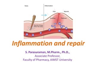
- 5. Signs of inflammation • Signs of inflammation Five cardinal signs of inflammation as • rubor (redness) • tumor (swelling) • calor (heat) • dolor (pain) • functio laesa (loss of function)
- 6. Types of inflammation • Depending upon the defense capacity of the host and duration of response, inflammation can be classified as – acute – chronic
- 7. Acute inflammation • Causes of acute inflammation: Infection, trauma, physical and chemical agents, necrosis, foreign bodies, and immune reactions. • Stages of acute inflammation: – Vasodilation – Increased vascular permeability – Movement of white blood cells from blood vessels into soft tissue at the site of inflammation
- 8. Acute inflammation • Stages of acute inflammation: – Vasodilation: Vasodilation occurs through release of mediators [include histamine, prostacyclin (PGI2), and nitric oxide (NO)] from cells. Vasodilation increases the hydrostatic pressure by causing slowing (sludging) of blood flow. Sludging of blood also causes margination of leukocytes along the wall of the blood vessel – Increased vascular permeability: Increased vascular permeability occurs through release of mediators [histamine, bradykinin, TNF, IL-1, leukotrienes C4, D4, and E4] from cells. – Movement of white blood cells from blood vessels into soft tissue at the site of inflammation: The steps required are rolling, pavementing, and transmigration.
- 9. Fate of acute inflammation • The acute inflammatory process can culminate in one of the following outcomes: – Resolution: complete return to normal tissue following acute inflammation. – Healing: Fibrosis takes place when the tissue destruction in acute inflammation is extensive so that there is no tissue regeneration. But when tissue loss is superficial, it is restored by regeneration. – Ulcer: Loss of the mucosa and deeper tissues.. – Fistula: Anomalous patent connection between two organs; most commonly organs with a lumen.
- 10. Fate of acute inflammation – Suppuration: When the pyogenic bacteria causing acute inflammation result in severe tissue necrosis, the process progresses to suppuration. Subsequently, mixture of neutrophils, bacteria, fragments of necrotic tissue, cell debris and fibrin comprise pus which is contained in a cavity to form an abscess. – Scar formation: Replacement of lost parenchyma with disorganized connective tissue (e.g., collagen). – Chronic inflammation
- 11. Chronic inflammation • Prolonged inflammation consisting of active inflammation and tissue destruction and repair, all occurring simultaneously. • Causes of Chronic inflammation: – Chronic inflammation following acute inflammation – Recurrent attacks of acute inflammation – Chronic inflammation starting de novo
- 12. Types of chronic inflammation • Based on histological features, chronic inflammations are classified as following 2 corresponding types – Chronic non-specific inflammation: It is characterised by non-specific inflammatory cell infiltration e.g. chronic osteomyelitis, lung abscess. e.g. actinomycosis. – Chronic granulomatous inflammation: It is characterised by formation of granulomas e.g. tuberculosis, leprosy, syphilis, actinomycosis, sarcoidosis etc. Actinomycosis Histology of lung Histology of lung with pulmonary tuberculosis
- 13. Cells involved in chronic inflammation • Macrophages: Activated macrophages produce proteases, IL-1, TNF, arachidonic acid metabolites, NO, angiogenesis and growth factors such as platelet- derived growth factor (PDGF) or fibroblast growth factor (FGF). • Lymphocytes : Activated lymphocytes produce – FGF stimulates fibroblasts to produce collagen, which results in scarring. – PDGF and transforming growth factor-β (TGF-β). – Interferon-γ (activates macrophages).
- 14. Systemic effects of chronic inflammation • Chronic inflammation is associated with following systemic features: – Fever – Anaemia – Leucocytosis – ESR (elevated in all cases of chronic inflammation} – Amyloidosis (long-term, chronic suppurative inflammation may develop secondary systemic (AA) amyloidosis)
- 15. Granulomatous inflammation • Granuloma is defined as a circumscribed, tiny lesion, about 1 mm in diameter, composed predominantly of collection of modified macrophages called epithelioid cells, and rimmed at the periphery by lymphoid cells.
- 16. Overview of inflammatory processes • Inflammation begins when a stimulus, such as infection, physical stress, or chemical stress, produces cellular damage.
- 17. Overview of inflammatory processes • The more important inflammatory mediators are the eicosanoids, biological oxidants, cytokines, adhesion factors, and digestive enzymes (proteases, hyaluronidase, collagenase, and elastase). • EICOSANOIDS: The pathway initiated by cyclooxygenase (COX) produces prostaglandins; the lipoxygenase pathway generates leukotrienes.
- 18. Overview of inflammatory processes • Biological Oxidants: The biologically derived oxidants are potent bacterial killers but are also a major contributing factor in tissue injury that results from the inflammatory response. These oxidants include the superoxide anion (∙O2-), hydrogen peroxide (H2O2), nitric oxide (∙NO), peroxynitrite (∙OONO∙), hypochlorous acid (HOCl), peroxidase-generated oxidants of undefined character, probably the hydroxyl radical (∙OH), and possibly singlet oxygen (1O2). • Cytokines: Tumor necrosis factor-α (TNF- α) and interleukin 1(IL-1) are produced primarily by cells of the monocyte–macrophage lineage.
- 19. Overview of inflammatory processes • Biological effects of eicosanoids
- 21. Healing of Tissues • Healing is the body response to injury in an attempt to restore normal structure and function. Healing involves 2 distinct processes: – Regeneration: healing takes place by proliferation of parenchymal cells and usually results in complete restoration of the original tissues – Repair: healing takes place by proliferation of connective tissue elements resulting in fibrosis and scarring. Both the processes take place simultaneously
- 22. Healing of Tissues – Regeneration: – Some parenchymal cells are short-lived while others have a longer lifespan. – In order to maintain proper structure of tissues, these cells are under the constant regulatory control of their cell cycle. These include growth factors such as Epidermal growth factor (EGF), Vascular endothelial growth factor (VEGF), Platelet-derived growth factor (PDGF), Fibroblast growth factor (FGF), Transforming growth factor-β (TGF-β).
- 23. Healing of Tissues – Repair: – Repair is the replacement of injured tissue by fibrous tissue. Two processes are involved in repair: • Granulation tissue formation • Contraction of wounds. – Repair response takes place by participation of mesenchymal cells, endothelial cells, macrophages, platelets, and the parenchymal cells of the injured organ.
- 24. Wound healing • Healing of skin wounds provides a classical example of combination of regeneration and repair. Wound healing can be accomplished in one of the following two ways: – Healing by first intention (primary union) • Characteristics: clean and uninfected, surgically incised, without much loss of cells and tissue and edges of wound are approximated by surgical sutures. – Healing by second intention (secondary union) • Characteristics: open with a large tissue defect (at times infected), having extensive loss of cells and tissues and the wound is not approximated by surgical sutures but is left open.
- 25. Wound healing – Healing by first intention (primary union) – Sequence of events in primary union: Initial haemorrhage, Acute inflammatory response, Epithelial changes, Organisation and Suture tracks.
- 26. Wound healing – Healing by second intention (secondary union) – Sequence of events in secondary union: Initial haemorrhage, Inflammatory phase, Epithelial changes, Granulation tissue, Wound contraction and Presence of infection.
- 27. Stages of wound healing • The stages of wound healing proceed in an organized way and follow FOUR processes: – Hemostasis – Inflammation – Proliferation – maturation • The stages of wound healing are linear, wounds can progress backward or forward depending on internal and external patient conditions.
- 28. Four stages of wound healing
- 29. Four stages of wound healing • Hemostasis Phase: Hemostasis starts when blood leaks Blood vessels constrict to restrict the blood flow Platelets stick together in order to seal the break in the wall of the blood vessel Coagulation occurs and reinforces the platelet plug with threads of fibrin Platelets adhere to the sub-endothelium surface within seconds of the rupture of a blood vessel's epithelial wall First fibrin strands begin to adhere (release of prothrombin)
- 30. Four stages of wound healing • Inflammatory Phase – Inflammation is the second stage of wound healing. – Inflammatory Phase is right after the injury when the injured blood vessels leak transudate (made of water, salt, and protein) causing localized swelling. – Inflammation both controls bleeding and prevents infection. – During the inflammatory phase, damaged cells, pathogens, and bacteria are removed from the wound area
- 31. Four stages of wound healing • Proliferative Phase – In Proliferative Phase, wound is rebuilt with new tissue made up of collagen and extracellular matrix. – In addition, a new network of blood vessels is constructed so that the granulation tissue can be healthy and receive sufficient oxygen and nutrients. – Myofibroblasts (cell that is in between a fibroblast and a smooth muscle cell) cause the wound to contract by gripping the wound edges and pulling them together using a mechanism similar to that of smooth muscle cells.
- 32. Four stages of wound healing • Maturation Phase – Also called the remodeling stage of wound healing – Maturation phase is when collagen is remodeled from type III to type I and the wound fully closes. – During the maturation phase, collagen fibers can lie closer together and cross-link. – Cross-linking of collagen reduces scar thickness and also makes the skin area of the wound stronger. *Type I is the predominant collagen type in a wound scar *The initial collagen in wounds is type III
- 33. Failure of wound healing • The stages of wound healing are a complex and fragile process. Failure to progress in the stages of wound healing can lead to chronic wounds. • Factors that lead up to chronic wounds are venous disease, infection, diabetes and metabolic deficiencies of the elderly. Ref: https://www.woundsource.com/blog/four-stages-wound-healing [Last assessed on 7/11/2018]
- 34. Thank you
Hinweis der Redaktion
- Ref: http://journal.frontiersin.org/article/10.3389/fcimb.2014.00069/full