Asthma Creation Mechanisms Bronchial Inflammation
•Als PPTX, PDF herunterladen•
6 gefällt mir•1,357 views

immunology/ bronchial asthma p.s. still it is pity that gifs don't open in this site.

Empfohlen
Weitere ähnliche Inhalte
Was ist angesagt?
Was ist angesagt? (20)
Ähnlich wie Asthma Creation Mechanisms Bronchial Inflammation
Ähnlich wie Asthma Creation Mechanisms Bronchial Inflammation (20)
Mehr von Nihal Yuzbasheva
Mehr von Nihal Yuzbasheva (20)
Kürzlich hochgeladen
Kürzlich hochgeladen (20)
Asthma Creation Mechanisms Bronchial Inflammation
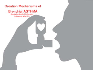
- 1. Azerbaijan Medical University Yuzbasheva Nihal 221 Creation Mechanisms of Bronchial ASTHMA
- 2. • Asthma is a type-I allergic airway disease characterized by Th2 cells and IgE. Episodes of bronchial inflammation, eosinophilic in nature and promoting bronchoconstriction, may become chronic and lead to persistent respiratory symptoms and irreversible structural airway changes. • Asthma is caused by a combination of complex and incompletely understood environmental and genetic interactions. These factors influence both its severity and its responsiveness to treatment. It is believed that the recent increased rates of asthma are due to changing epigenetics (heritable factors other than those related to the DNA sequence) and a changing living environment. Onset before age 12 is more likely due to genetic influence, while onset after 12 is more likely due to environmental influence.
- 3. • Peripheral blood cell generation of cytokines IFN-γ (TH1) and IL-5 (TH2) was used to evaluate the relationship of the balance of TH1/TH2 cytokines to asthma persistence and severity in a 42-year, longitudinal study. • Chemokines, including thymus and activation-regulated chemokine, are important to the regulation of inflammation and IgE synthesis. Their potential role in asthma has also been evaluated. • Finally, albuterol (R)- and (S)-enantiomers may have distinct effects on airway relaxation and regulation of inflammation, suggesting the possibility that monoisomeric therapy has therapeutic advantages. • The potential contribution of genetic factors and mechanisms to airway inflammation and remodeling also continues to be an area of intense investigation.
- 4. • Allergic asthma is triggered by an immune-inflammatory response driven by Th2 cells. This so-called “Th2-high” or “Th2-like” subphenotype of asthma originates from a complex interplay between the innate and adaptive branches of immune system • Differentiation of Th2 lymphocytes requires the cooperation of several promoting factors, including costimulatory molecules and cytokines expressed by dendritic cells and inflammatory cells.
- 5. • Two of the cytokines secreted by Th2 cells or by Tfh cells activated by the same antigen are IL-4 and IL-13. • These cytokines stimulate B lymphocytes to switch to IgE-producing plasma cells.
- 6. • IgE antibody produced in response to an allergen binds to high-affinity Fc receptors, specific for the ε heavy chain, that are expressed on mast cells. • The high-affinity receptor for IgE, called FcεRI ,consists of three polypeptide chains, one of which binds the Fc portion of the ε heavy chain very strongly, with a Kd of approximately 10−11 M. The other two chains of the receptor are signaling proteins.
- 7. • Mast cell activation results from binding of the allergen to two or more IgE antibodies on the cell. • When this happens, the FcεRI molecules that are carrying the IgE are cross-linked, triggering biochemical signals from the signal-transducing chains of FcεRI. • The signals lead to three types of responses in the mast cell: rapid release of granule contents (degranulation), synthesis and secretion of lipid mediators, and synthesis and secretion of cytokines.
- 8. • Bronchial asthma is form of respiratory allergy in which inhaled allergens (often undefined) stimulate bronchial mast cells to release mediators, including leuko-trienes, which cause repeated bouts of bronchial constriction and airway obstruction.
- 9. • In chronic asthma, large numbers of eosinophils accumulate in the bronchial mucosa, excessive secretion of mucus occurs in the airways, and the bronchial smooth muscle becomes hypertrophied and hyper-reactive to various stimuli. • Some cases of asthma are not associated with IgE production, although all are caused by mast cell activation.
- 10. 1. Development of mild asthma. A: On primary allergen exposure, DAMPs and/or PAMPs intrinsic to or accompanying the allergen activate DCs to become APCs biased toward the induction of Th2 cells. As a result, immune sensitization featuring allergen-specific IgE and Th2 cells is generated. B: Activation of IgE-bearing innate cells by inhaled allergen triggers the release of pro-inflammatory mediators such as histamine, leukotrienes, Th2-associated cytokines and chemokines, and reactive oxygen species. The resulting early-phase allergic reaction is manifested by smooth muscle contraction, mucus hypersecretion, and increased airway responsiveness. Inflammatory cell recruitment and activation leads to the production of a second wave of particularly Th2-associated inflammatory mediators such as IL-4, IL-5, IL-13, eotaxins, RANTES, and others. This late-phase reaction occurs 6 to 9 hours after allergen inhalation and usually lasts for not more than a day.
- 11. • In studies, the scientists aimed to define the mechanisms of what is referred to as immune tolerance and to detail the role that dendritic cells play in the process. • To model the events that occur when an asthma sufferer inhales a harmless allergen, the researchers exposed a number of mice to an egg allergen known as ovalbumin, or OVA. Within 24 hours of exposure to the allergen, dendritic cells migrated to the lymph nodes and induced the production of certain regulatory cells, as well as a specific type of cytokine called IL-10. The regulatory cells and IL-10 then worked to ensure that the innocent substance wasn't attacked.
- 12. • By displaying the critical role of dendritic cells in maintaining immune tolerance to certain antigens, the study points to the possibility that defective dendritic cells may contribute to asthma. If dendritic cells can't produce IL-10, for example, the word might not get out that the foreign visitor is friendly, and the immune system might launch an unwarranted attack.
- 13. Pathobiology of airway inflammation in asthma.
- 14. • Cellular inflammation of the airways with eosinophils and neutrophils is a characteristic feature of asthma and is considered relevant to the pathogenesis of the disease. • Eosinophilic asthma is a distinct phenotype of asthma that is associated pathologically by thickening of the basement membrane zone and pharmacologically by corticosteroid responsiveness. • In contrast, noneosinophilic asthma, a sizeable subgroup of asthma that includes patients with severe disease, is not characterized by thickening of the basement membrane zone, and it appears to be relatively corticosteroid resistant.
- 15. • Eosinophilic inflammation associated with poor asthma control, increased branchodilator response, lower lung function and exacerbations in ICS treated patients • Neutrophilic asthma seems to increase with increasing severity ICS use
- 16. Distribution of sputum cellular phenotypes in severe asthma (n = 88). Eosinophilic asthma (≥3% sputum eosinophils, <76% sputum neutrophils); Neutrophilic asthma (<3% sputum eosinophils, ≥76% sputum neutrophils); Paucigranulocytic asthma (<3% eosinophils and <76% neutrophils in induced sputum); Mixed granulocytic asthma (≥3% eosinophils and ≥76% neutrophils in induced sputum).
- 18. Clinical Syndrome •Bronchial asthma Clinical and pathological manifestations • Airway obstruction caused by bronchial smooth muscle hyperactivity; • inflammation and tissue injury caused by late- phase reaction
- 19. Syndrome Bronchial Asthma Therapy Corticosteroids Leukotriene antagonists Phosphodiesterase inhibitors Mechanism of action Reduce inflammation Relax bronchial smooth muscle and reduce inflammation Relax bronchial smooth muscles