
![Introduction ,[object Object],[object Object],[object Object],[object Object]](data:image/gif;base64,R0lGODlhAQABAIAAAAAAAP///yH5BAEAAAAALAAAAAABAAEAAAIBRAA7)
Empfohlen
Weitere ähnliche Inhalte
Was ist angesagt?
Was ist angesagt? (20)
Andere mochten auch
Andere mochten auch (20)
Ähnlich wie 2 pp vibrio
Ähnlich wie 2 pp vibrio (20)
Mehr von Bruno Mmassy
Mehr von Bruno Mmassy (20)
Kürzlich hochgeladen
Kürzlich hochgeladen (20)
2 pp vibrio
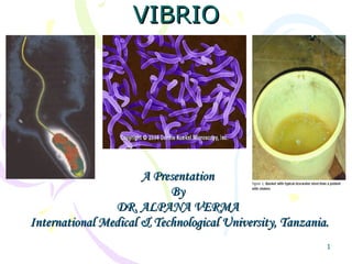
- 1. VIBRIO A Presentation By DR. ALPANA VERMA International Medical & Technological University, Tanzania.
- 4. The Vibrios Vibrios are among the most common bacteria in surface waters worldwide. They are curved aerobic rods and are motile, possessing a polar flagellum. V cholerae serogroups O1 and O139 cause cholera in humans, while other vibrios may cause sepsis or enteritis.
- 6. Growth Characteristics V cholerae regularly ferments sucrose and mannose but not arabinose. A positive oxidase test is a key step in the preliminary identification of V cholerae and other vibrios. Vibrio species are susceptible to the compound O/129 (2,4-diamino-6,7-diisopropylpteridine phosphate), which differentiates them from Aeromonas species, which are resistant to O/129. Most Vibrio species are halotolerant, and NaCl often stimulates their growth. Some vibrios are halophilic, requiring the presence of NaCl to grow. Another difference between vibrios and aeromonas is that vibrios grow on media containing 6% NaCl, whereas aeromonas does not.
- 7. Vibrio Species Associated With Human Disease Species Source of Infection Clinical Disease V. alginolyticus Seawater Wound infection, external otitis V. cholerae Water, food Gastroenteritis V. cincinnatiensis* Unknown Bacteremia, meningitis V. fluvialis * Seafood Gastroenteritis, wound infection, bacteremia V. furnissii * Seawater Gastroenteritis V. harveyi * Seawater Wound infection (shark bite) V. etschnikovii * Unknown Bacteremia V. mimicus * Fresh water Gastroenteritis, wound infection, bacteremia V. parahaemolyticus Shellfish, seawater Gastroenteritis, wound infection, bacteremia V. vulnificus Shellfish, seawater Bacteremia, wound infection, cellulitis
- 11. V. Cholerae Serological Classification Each O1 biotype can have 3 serotypes Classical El Tor Division into 2 epidemic serotypes Toxigenic V. cholerae O1 Division into 2 biotypes inaba ogawa hikojima A & B (A little C) Antigens A & C O139 A, B, C
- 13. V. cholerae Serological Classification I define Vibrios! I’m an O1 or O139 Strain NON-TOXIGENIC TOXIGENIC I may not be O1, Or O139! (but I can still stir up trouble)
- 22. Pathogenesis & Pathology Under natural conditions, V cholerae is pathogenic only for humans. A person with normal gastric acidity may have to ingest as many as 1010 or more V cholerae to become infected when the vehicle is water, because the organisms are susceptible to acid. When the vehicle is food, as few as 102–104 organisms are necessary because of the buffering capacity of food. Any medication or condition that decreases stomach acidity makes a person more susceptible to infection with V cholerae. Cholera is not an invasive infection. The organisms do not reach the bloodstream but remain within the intestinal tract. Virulent V cholerae organisms attach to the microvilli of the brush border of epithelial cells. There they multiply and liberate cholera toxin and perhaps mucinases and endotoxin.
- 23. Clinical Findings About 60% of infections with classic V cholerae are asymptomatic. The incubation period is 1–4 days,. There is a sudden onset of nausea ,vomiting and profuse diarrhea with abdominal cramps. Stools, which resemble "rice water," contain mucus, epithelial cells, and large numbers of vibrios. There is rapid loss of fluid and electrolytes, which leads to profound dehydration, circulatory collapse, and anuria.
- 24. Vibrio cholerae Enterotoxin V cholerae produce a heat-labile enterotoxin with a molecular weight of about 84,000, consisting of subunits A (MW 28,000) and B . ganglioside GM1 serves as the mucosal receptor for subunit B, which promotes entry of subunit A into the cell. Activation of subunit A1 yields increased levels of intracellular cAMP and results in prolonged hypersecretion of water and electrolytes. There is increased sodium-dependent chloride secretion, and absorption of sodium and chloride is inhibited. Diarrhea occurs—as much as 20–30 L/d—with resulting dehydration, shock, acidosis, and death. The genes for V cholerae enterotoxin are on the bacterial chromosome. Cholera enterotoxin is antigenically related to LT of Escherichia coli and can stimulate the production of neutralizing antibodies. However, the precise role of antitoxic and antibacterial antibodies in protection against cholera is still not clear.
- 30. African countries invaded and affected by cholera epidemics in the seventies.
- 37. Key Biochemical & Physiological Characteristics of Important Vibrios V. cholerae V. parahaemolyticus V. vulnificus Oxidase + + + D–Glucose (Gas) - - - Lactose - - V Sucrose + - V myo -inositol - - - Lysine decarboxylase + + + Arginine dihydrolase - - - Ornithine decarboxylase + + V Growth in 0% NaCl + - - Growth in 6% NaCl v + V TCBS growth Good Good Good Colony color on TCBS Yellow Green Green
- 39. Treating Cholera
- 42. The most important part of therapy consists of water and electrolyte replacement to correct the severe dehydration and salt depletion. Many antimicrobial agents are effective against V cholerae. Oral tetracycline tends to reduce stool output in cholera and shortens the period of excretion of vibrios. In some endemic areas, tetracycline resistance of V cholerae has emerged; the genes are carried by transmissible Plasmids. Treatment
- 43. Control rests on education and on improvement of sanitation, particularly of food and water. Patients should be isolated, their excreta disinfected, and contacts followed up. Chemoprophylaxis with antimicrobial drugs may have a place. Repeated injection of a vaccine containing either lipopolysaccharides extracted from vibrios or dense vibrio suspensions can confer limited protection to heavily exposed persons (eg, family contacts) but is not effective as an epidemic control measure.
- 44. THANK YOU
Hinweis der Redaktion
- Asporogenous - doesn’t sporulate. Two chromosomes, both have been completely sequenced. The following is the description of the circular representation of the V. cholerae genome given by Heidelberg et al. (2000). The two chromosomes, large and small, are depicted. From the outside inward: the first and second circles show predicted protein-coding regions on the plus and minus strand, by role (unknown and hypothetical proteins are in black). The third circle shows recently duplicated genes on the same chromosome (black) and on different chromosomes (green). The fourth circle shows transposon-related (black), phage-related (blue), VCRs (pink) and pathogenesis genes (red). The fifth circle shows regions with significant 2 values for trinucleotide composition in a 2,000-bp window. The sixth circle shows percentage G+C in relation to mean G+C for the chromosome.The seventh and eighth circles are tRNAs and rRNAs, respectively.
- Notes about this slide: Of the more than 200 strains that have been identified, only O1 and the newly emerged O139 have been associated with severe disease and cholera outbreaks (Weir, 2004). In any epidemic, one strain predominates. There is a complex classification system. V. cholerae is divided into two epidemic serotypes - O1 and O139 (there are many other environmental serotypes). All of the information presented below was derived from the review by (Crowcroft, 1994). The O1 strain predominated as the primary epidemic strain until 1992. O1 is further divided into two biotypes, Classical and El Tor. The Classical biotype was responsible for the first six pandemics until it was replaced by the El Tor biotype in 1961. The Classical and El Tor biotypes are further divided into three ribotypes based on the antigens they present: Inaba (A&C antigens); Ogawa (A & B antigens); and Hikojima (A&B&C antigens). The O139 serotype replaced the O1 serotype as the predominant pandemic strain in 1992 when it emerged in Southeast Asia and became the primary strain.
- Cholera classification is defined by the history of the disease. This information was from: http://www.who.int/mediacentre/factsheets/fs107/en/print.html When cholera appears in epidemic form in an unexposed population, it can affect all age groups since adults haven’t had the chance to acquire immunity. In contrast, in areas of endemic disease, most of the adult population has gained some degree of natural immunity because of illness or repeated asymptomatic infections. In endemic areas, usually children and the elderly are the most at risk for infection. The elderly are more at risk because they have lower gastric production and waning immunity. The poor are at a greater risk because they often lack safe water supplies, and may depend on street vendors or other unregulated sources of food and drink. For further reading, please refer to the handout that accompanies this presentation.
- Unless otherwise noted, the information presented in the notes for this slide was assimilated from the website: www.who.int/entity/water_sanitation_health/dwq/en/admicrob6.pdf After gaining entry into the host through ingestion, the organism colonizes the epithelial lining of the small intestine. The incubation period is one to five days, and patients are symptomatic for two to seven days. The production of Cholera Toxin, discussed in detail later, induces most of the symptoms associated with the disease cholera. For serious cases, death can occur as a result of hypovolemic shock (loss of vital organ function due to rapid fluid loss) within two to four hours of colonization. Two case types: Mild cases (90%) are difficult to distinguish from normal diarrheal diseases. Severe cases (10%) are associated with painless, watery diarrhea, with as much as 20 L day -1 fluid loss (Cotter, 2000) in as little as three to four hours, leading to hypovolemic shock. Severe dehyrdration results in muscle cramps, loss of skin turgor, scaphoid abdomen and weak pulse. (http://gsbs.utmb.edu/microbook.ch024.htm). 3. The onset of diarrhea in cholera allows for the rapid dissemination of copious quantities of this organism into the environment.
- This information was assimilated from (Sack et al., 2004). Microbiological methods of detection: Culture from fecal or water samples. Start culture from fecal matter in TCBS (thiosulphate citrate bile salts supports the growth of Vibrios but suppresses most other organisms) and allow it to grow for 18 hours. Start culture of fecal matter in peptone water, a high pH enrichment broth. After incubation in peptone water for 6-12 hours, inoculate a second TCBS plate and allow it to grow for 18 hours. V. cholerae appears as smooth yellow colonies with slightly raised centers. Appearance of these colonies gives a presumptive positive and should be reported to the government health department. Samples must be sent to the appropriate regional reference laboratory for confirmational testing. Rapid tests Dark field microscopy - inoculate a wet mount of the fecal specimen and examine for the appearance of darting microbes that are halted by the addition of O1 or O139 antiserum. Rapid immunoassays PCR and DNA probes If the reader wishes to know additional information about V. cholerae typing and microbiological methods of identification, the information presented below was derived from a publication of the Government of Canada on Laboratory Procedures for the Isolation and Identification of Vibrio cholerae O1 and Non-O1 from foods. 1995. Polyscience Publications . The procedures were written by S. Stavric and B. Buchanan. Biotyping (distinguishing between the Classical and El Tor biotypes as defined on slide 13) can be performed by the following tests: Polymyxin B sensitivity - Classical biotypes show a 12 to 15 mm zone of growth inhibition when subjected to polymixin B whereas El Tor biotypes show only a 1 to 2 mm zone. Hemolysin production - most El Tor biotypes will produce hemolysin and will lyse sheep red blood cells. Classical biotypes do not produce hemolysin and so will not lyse red blood cells. Phage sensitivity - El Tor biotypes are not sensitive to phage IV and will not be lysed. Classical biotypes are sensitive to phage IV and will be lysed. Agglutination with chicken red blood cells - El Tor strains will agglutinate while the Classical biotypes will not.
- Unless otherwise noted, the information in this notes section was assimilated from the website: http://www.who.int/mediacentre/factsheets/fs107/en/print.html Antibiotics are not necessary for most V. cholerae infections; however, they usually decrease the volume and duration of diarrhea and the period of Vibrio excretion. Antibiotics, when prescribed, should be ones to which the infective strain is susceptible because resistance is a growing problem. The susceptibility of infectious strains should be determined at the beginning of an epidemic using the standard disk diffusion test or by broth microdilution. For severe cases, tetracycline is the most-often prescribed antibiotic. Other antibiotics that are prescribed: cotrimoxazole, erythromycin, doxycycline, chloramphenicol, and furazolidone
- Sometimes the most effective measures at preventing the spread of disease are the simplest.
- Current cholera vaccines are effective in only 50% of recipients, and immunity lasts only 3 to 6 months (Crowcroft, 1994) Orochol is an attenuated live oral cholera vaccine, containing the genetically manipulated V. cholerae strain CVD 103-HgR. For more information, http://www.bernabiotech.com/products/orochol/infosheet/oro_2001_e.pdf/ Dukoral is a killed whole-cell V. cholerae O1 in combination with purified recombinant B subunit of cholera toxin. For more information, Dukoral - http://www.pharmeragroup.com/dukoralb.htm According to WHO (http://www.who.int/mediacentre/factsheets/fs107/en/print.html), both vaccines are suitable for use by travelers but not for use as a large scale public health measure: Use of these vaccines to prevent or control cholera outbreaks is not recommended because it gives, according to WHO, a false sense of security to vaccinated subjects and to health authorities who often neglect more effective measures. As of 1973, no country requires proof of cholera vaccination as a condition for entry. These vaccines only target O1 strains. Now, the appearance of the O139 strain has redirected efforts to develop an effective and practical cholera vaccine.