
Empfohlen
Weitere ähnliche Inhalte
Was ist angesagt?
Was ist angesagt? (20)
Ähnlich wie Osteoarthrosis,ppt
Ähnlich wie Osteoarthrosis,ppt (20)
Mehr von DrSiddique H. Ranna
Mehr von DrSiddique H. Ranna (19)
Kürzlich hochgeladen
Kürzlich hochgeladen (20)
Osteoarthrosis,ppt
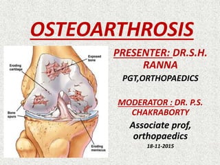
- 1. OSTEOARTHROSIS PRESENTER: DR.S.H. RANNA PGT,ORTHOPAEDICS MODERATOR : DR. P.S. CHAKRABORTY Associate prof, orthopaedics 18-11-2015
- 2. 0STEOARTH ROSIS/OSTE OARTHRITIS comes from three Greek words meaning bone, joint, and inflammation. refer to a group of non inflammatory arthritides. suffix,-itis means inflammation. it is generally agreed that the initial events are mechanical, not inflammatory.
- 3. Definition OA is a chronic non inflammatory, degenerative condition of joints characterized by progressive softening and disintegration of articular cartilage. new growth of cartilage and bone at the joint margins (osteophytes) and subchondral sclerosis, capsular fibrosis and subchondral cyst formations.
- 4. ACR definition heterogeneous group of condition leads to joint signs and symptoms’ which are associated with defective integrity of articular cartilage, in addition to related changes in the underline bone at the joint margins.
- 6. EPIDEMIOLOGY Most common arthritis & joint disease ¼OPD JAPANESE, 10 million AMERICAN OA KNEE BEFORE 45 YRS : M>F AFTER 45 YRS : F>M INCREASES WITH AGE
- 7. AETIO LOGY AGE GENETIC INHERIT ANCE CONGENITAL DEFECT OF JTS TRAUMA,INFLAM MATION, OVERUSE SPORTS/ LABOUR OBESITY MECHANICAL STRESS DM
- 8. ANATOMICAL DISTRIBUTION • Commonly affected joints:
- 9. LESS COMMONLY AFFECTED JOINTS:
- 11. PRIMARY GENERALISED OA INVOLVES 3 OR MORE JOINTS. USUALLY MIDDLE AGED WOMAN. PRESENTS WITH PAIN,SWEELING,STIFF NESS OF FINGER JTS, 1st CMC,BIG TOE MTP JTS,THEN KNEE,HIP FACET JTS.
- 12. PRIMARY LOCALISED OA HEBERDENS NODE : DIP JTS. BOUCHARDS NODE : PIP JTS without involvement of other jts. GENETIC FACTORS IMPORTANT. FEMALE 10 Times more than MALE.
- 13. SECONDARY OA
- 14. PATHOPHYSIOLOGY OF OSTEOARTHROSIS Intact cartilage morphology Increase hydration Failure of internal collagen that restrains the matrix gel. Breakdown of collagenous network Further hydration matrix Cartilage swelling occurs Repairmechanism EARLY STAGE
- 15. Mechanical overload persist Repair mech. overwhelmed Overall pgs degradation Further breakdown of collagen Chondrocytes starts secreting lysosomal enzymes Dissolves the matrix protein LATE CHANGES
- 16. RELEASE OF DEGRADATION PRODUCTS INTO JTS. SYNO VITIS LOSS OF SHOCK ABSORPTION PROPERTY Structural change in cartilage, BLISTERING FIBRILLATION,FISSURING LOSS OF INTEGRITY OF CARTILAGE
- 17. Mechanical forces increasingly concentrated in subchondral bone Increased vascularity in subchondral area Reactive sclerosis Focal trabecullar degeneration and subchondral cyst formation What cartilage remains still capable of regeneration, repair, remodeling
- 18. Cartilage at the ends undergo ENCHONDRAL OSSIFICATION Spur like bony outgrowth covered by hyaline cartilage develops at margins of joints OSTEOP HYTES Small bites of cartilage may actually break off into joints, leading to locking joints JOINT MICE
- 19. PATHOLOGY OF OSTEOARTHROSIS The cardinal pathological feature of osteoarthrosis are : Progressive cartilage destruction. Subarticular cyst formation. Sclerosis of surrounding bone. Osteophytes formation. Capsular fibrosis.
- 20. Eburnation: With progressive disintegration of cartilage the underlying bone becomes exposed and some areas may be polished or burnished to ivory- like smoothness called eburnation.
- 21. OSTEOPHYTES AND JOINT MICE Spur like bony outgrowths covered by hyaline cartilage, may develop at margins of joint & progressively enlarge Small bits of cartilage- covered bone, known as joint mice, may actually break off into the joint
- 22. SUBCHONDRAL CYST CYST MAY ARISE FROM INCREASE IN INTRASYNOVIAL PRESSURE.
- 23. SYNOVIAL MEMBRANE & CAPSULE Synovial membrane undergo hypertrophy and become edematous. CAPSULES undergoes fibrous degeneration and there are low-grade chronic inflammatory changes.
- 24. Histopathology of OA EARLY CHANGES Increased cellularity. Small irregularities/splits in the surface. Clusters of chondrocytes. Metachromasia due to depletion of PGS. LATE CHANGES Splits become more extentensive. Subchondral bone shows osteoblastic activity Increased vascularity. Cyst formation.
- 26. RADIOGRAPHIC GRADING OF OA STAGE -1 NILL OR MINIMAL CHANGES/ BONY SPUR ONLY STAGE -2 NARROWING OF JT. SPACE < ⅟2 OF NORMAL JT. SPACE STAGE -3 NARROWING OF JT. SPACE > ⅟2 OF NORMAL JT. SPACE STAGE -4 OBLITERATION OF JOINT SPACE STAGE -5 DERFORMITY OF JOINTS
- 28. PATHOLOGICAL GRADING OF OSTEOARTHROSIS GRADE-1 SOFTENING OR BLISTERING OF JOINT CARTILAGE GRADE -2 FRAGMENTATION OR FIBRILLATION AN AREA < 1cm. GRADE -3 FRAGMENTATION OR FISSURING AN AREA >1cm. GRADE -4 CARTILAGE FISSURING DOWN TO THE SUBCHONDRAL BONE.
- 29. SIGN AND SYMPTOMS OF OSTEOARTHROSIS 1. PAIN: Starts insidiously and increase slowly over months or years. It is aggravated by exertion and relieved by rest, although with time relief on rest is less. In late stage pain in the bed at night time may occur.
- 30. 2. STIFFNESS: Characteristically it occurs after periods of inactivity. With time it becomes constant and progressive. 3. SWELLING: May be intermittent suggesting of effusion. May be continuous suggesting capsular thickening or large osteophytes.
- 31. CREPITUS: Crunching sound or feelings during movements of joins. LOSS OF FUNCTION: May be most distressing symptoms. Locking joints due to joint mice. Difficulty in climbing stairs, restriction of walking distance or progressive inability to perform everyday tasks. 6. DEFORMITY: It results from capsular contracture or joint instability but be aware that the deformity may actually have preceded and contributed to the onset of osteoarthrosis.
- 32. INVESTIGATIONS OF OSTEOARTHROSIS BLOOD TEST THERE IS NO DIAGNOSTIC LAB. TEST FOR OA. Osteoarthrosis is not a systemic disease, therefore ESR, TC, DLC are within normal limit. Blood tests are done for differential diagnosis. SYNOVIAL FLUID
- 33. X-RAYS The cardinal x-ray features Narrowing of joint space. Sclerosis subchondral bone. Marginal osteophytes. Subchondral cyst.
- 35. CONSERVATIVE 1.PATIENT EDUCATION Patient education about the non systemic nature of disease. Patient should understand the condition and know what they themselves can do help. 2. LIFE STYLE MODIFICATION Avoid sitting on ground with cross leg position and sits up. Avoid squatting. Frequent shoe modification due to lateral wedging of shoe sole. Avoid use of high heel and up stairs. Walking instead of running on soft earth. Walking within the limit of pain.
- 36. 3. REDUCTION OF OVERLOADING OF JOINTS Slow down the rate of cartilage loss. Weight reduction for obese patient. Wearing of shock absorbing shoes. Using walking stick.
- 37. PHYSIOTHERAPY Physiotherapy should be directed to maintaining joint mobility and improving muscle strength. Range of motion /flexibility exercises. Zero gravity/ swimming is a good exercise Static muscle exercises
- 38. PAIN MANAGEMENT THERAPY Hot and cold therapy USG therapy IFT, SWD therapy etc. aerobic exercise but care should be taken to avoid activities which increases impact loading.
- 39. Analgesic medication: Patient with symptomatic OA can receive analgesic for short period of time unless there are contraindications ton this treatment. Paracetamole as analgesic doses. Other NSAIDS for acute pain for short periods. Oral corticosteroids usually have no role.
- 40. NUTRICEUTICALS GLUCOSEAMINE & CHONDROITIN SULPHATE longer period 3- 6 months. Stimulate proteoglycan synthesis by chondrocytes. increases the rate of formation of new cartilage mild anti-inflammatory properties.
- 41. DIACEREIN It is an IL-1 blocker. Diacerein actively inhibited the production of proteases . Stimulate the anabolic process of cartilage. Administered orally 50 mg 12 hrly for 3-6 months.
- 42. Intra-articular injections Hyal uron ic acid Stimulation of de novo synthesis of HA. Inhibition of arachidonic acid release and inhibition of IL-1 beta induced PGE2 synthesis by synoviocytes. It influences leucocytes adherence, proliferation, migration and phagocytosis. It protects cellular damage caused by reactive oxygen species. STE ROI D
- 43. ARTHROSCOPIC TREATMENT 1. JOINT DEBRIDGMENT & LAVAGE 2. REMOVAL OF LOOSE BODIES. 3. ARTHROSCOPIC CHONDROPLASTY .
- 45. Transplantation of multiple osteocartilagenous plugs or pegs. Auto grafts of upto 10 mm of diameter can be transferred into prepared DEFECTS IN THE FEMORAL CONDYLES. Reported clinical results with this technique are limited. MOSAICPLASTY
- 46. MOS AICPL ASTY
- 47. CORRECTIVE OSTEOTOMY High Tibial Osteotomy Realignment osteotomy is an option in active patients with symptomatic unicompartmental OA of the knee with mal-alignment.
- 49. REPLACEMENT SURGERY UNICONDYLAR REPLACEMENT TOTAL JOINT REPLACEMENT