
Empfohlen
Weitere ähnliche Inhalte
Was ist angesagt?
Was ist angesagt? (20)
Ähnlich wie Intervertebral disc prolapese
Ähnlich wie Intervertebral disc prolapese (20)
Mehr von DrSiddique H. Ranna
Mehr von DrSiddique H. Ranna (20)
Kürzlich hochgeladen
Kürzlich hochgeladen (20)
Intervertebral disc prolapese
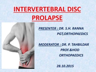
- 1. INTERVERTEBRAL DISC PROLAPSE PRESENTER : DR. S.H. RANNA PGT,ORTHOPAEDICS MODERATOR : DR. P. TAHBILDAR PROF.&HOD ORTHOPAEDICS 28.10.2015
- 2. HISTORY Aurelianus (5th century) clearly described the symptoms of SCIATICA. Andreas Vesalius (1543) first described the intervertebral disc. Middleton & Teacher (1911) described a case of paraplegia following attempting to lift heavy weight from floor on postmortem they found fibrocartilage in extradural space.
- 3. CONT.. • Mixter and Barr (1934) described disc herniation as the cause of Sciatica. Lindblom(1948) first described DISCOGRAPHY. Lyman Smith (1963) described CHEMONUCLEOLYSIS. Kambin & Gellman (1983) reported percutaneous approach for lumbar discectomy.
- 4. ANATOMY OF LUMBAR SPINE
- 5. Cont..
- 8. EFFECT OF AXIAL LOADING
- 10. DISC & NERVE ROOT RELATION L5 is TRAVERSING NERVE ROOT L5 is EXITING NERVE ROOT
- 11. RELATION OF INTRADISCAL PRESSURE AND POSTURE
- 13. IN RELATION TO MANUAL MATERIALS HANDLING
- 14. LUMBAR DISC PROLAPSE DEFINITION It is condition in which there is outpouching of the disc Nucleus pulposus along with few annular fibres and end plate cartilage through the tears in annulus fibrosus into the extradural space.
- 15. EPIDEMIOLOGY • AGE: 30 – 40 years. • SEX: Male affected more than female. • MOST COMMON LEVEL: L4-L5 (next common level is L5-S1). • MOST COMMON TYPE: Postero-lateral type.
- 16. ETIOLOGY
- 17. MICROTROUMA
- 18. EFFECT OF SMOKING Blood vessel get constricted Transport of nutrients & disposal of waste products decreased Disc cells get deficient nutrition or die Disc degenerates & results in DISC INSTABILITY
- 19. STAGES OF DISC DEGENERATION Stage of dysfunction Stage of instability Stage of stabilization
- 20. STAGE OF DYSFUNCTION Episode of rotational or compressive trauma (uncoordinated muscle contraction) Posterior facet joint & annular strain Small capsular & annular tear occurs Small subluxation of posterior joint Posterior joint synovium injured & result in SYNOVITIS Posterior segment muscle protect joint by sustained hypertonic contraction Muscle become ischemic & metabolites get accumulated cause pain Muscle splints the posterior joint subluxation &maintained
- 21. STAGE OF INSTABILITY Increased dysfunction FACET JOINT Degenerati on of cartilage Attenuation of capsule Laxity of capsule DISC Coalescenc e of tears Loss of nucleus internal disruption Bulging of annulus INCREASED ABNORMAL MOVEMENT
- 22. STAGE OF STABILIZATION FACET JOINT Destruction of cartilage Fibrosis in joint Enlargement of facets Locking facets Fibrosis around joint INCREASED STIFFNESS STABILIZ ATION Fibrosis in disc & osteophytes Destructio n of plates Approximati on of bodies Loss of nucleus DISC
- 23. PATHOPHYSIOLOGY OF PIVD With aging, vascular channels start to fail and vascular diffusion of nutrients decrease thus number of viable chondrocytes in the nucleus pulposus diminishes Synthesis rate & concentration of proteoglycans decreases & proportion of collagen increase in nucleus pulposus Water binding capacity of the nucleus decreases Nucleus becomes more fibrous & stiffer
- 24. CONT.. Nucleus is less able to bear & disburse load, transferring load to the posterior annulus ANNULU S IN TACT Facet joints share even more of the axial load ANNULU S FAIL Facet joints undergo degenerative changes & develop osteophytes FACET JOINT SYNDROME
- 25. CONT… ANNULUS FAIL Fissures develop across annular lamellae may extend upto disc periphery Internal disc disruption cause AXIAL PAIN Expression of this degraded nuclear material through these radial fissures DISC HARNIATION
- 26. FATE OF DISC HARNIATION Extrude disc & degraded nuclear material impinge on the nerve roots Nucleus pulposus is an immunogenic which induces an inflammatory response mediated by TNF alpha, IL, Phospholipase A2, Nitric oxide. Produces radicular pain syndrome & RADICULOPATHY
- 28. AXIAL LOCATION • Central (R/L) • Subarticular (R/L) • Foraminal (R/L) • Extra foraminal (R/L)
- 29. SAGITAL LOCATION • DISCAL. • PEDICULA —R. • SUPRAPED -ICULAR. • INFRAPEDI -CULAR.
- 30. CLINICAL FEATURE Level of prolapsed T12-L1 L1—L2 L2—L3 L3—L4 L4—L5 L5—S1 Nerve root compres sed L1 L2 L3 L4 L5 S1 pain Thorac o lumbar junctio n, groin, proxim al part of thigh Thorac o lumbar junctio n, groin, proxim al part of thigh Upper lumbar spine, anterio r aspect of proxim al thigh Lower back, hip, postero lateral thigh, anterio r leg Sacroili ac joint, hip, lateral thigh & lateral leg Sacroiliac joint, hip, postero lateral thigh & postero lateral leg to heel
- 31. Cont.. Level of prolapsed T12-L1 L1-L2 L2-L3 L3-L4 L4-L5 L5-S1 Nerve root compress ed L1 L2 L3 L4 L5 S1 Paresthesi a/Sensory loss Oblique band proximal 3rd of thigh anteriorly just below inguinal lig. Oblique band mid 3rd of thigh anteriorly Oblique band lower part of thigh anteriorly just above the knee Medial to shin of tibia, medial aspect of the foot Lateral leg, dorsum of foot, 1st web space Posterior aspect of thigh, back of calf, lateral side and sole of foot
- 32. C0NT… Level of prolapsed T12- L1 L1-L2 L2-L3 L3-L4 L4-L5 L5-S1 Nerve root compresse d L1 L2 L3 L4 L5 S1 reflexes Knee jerk slightly diminishe d Knee jerk slightly diminishe d Knee jerk diminishe d or absent Changes uncommo n (Posterior tibial reflex diminishe d or absent. Ankle jerk absent or diminishe d
- 33. CONT.. STAGE OF DEGENERATIVE DISEASE OF DISC STAGE OF DYSFUNCTION STAGE OF INSTABILITY STAGE OF STABILIZATION SYMPTOMS Low back pain often localized or referred to groin/ greater trochanter/ posterior thigh - Aggravated on movement - Relieved on rest Catch in back on movement. - Pain on coming to standing position after flexion. Low back pain decrease in severity SIGNS Local tenderness on one side & at one level -Hypo mobility - Extension painful - Neurological examination normal Abnormal movement of spine - Observation of catch Sway or shift when coming erect after flexion. Muscle tenderness - Stiffness - Reduced movements - Scoliosis
- 34. PHYSICAL EXAMINATION • ATTITUDE: The lumbar spine is flattened and slightly flexed, hip and knee slightly flexed on the affected side and hip rotates forward to relax Piriformis
- 35. GAIT • Slow and deliberate walk holding their loins with the hands. • TIP-TOE WALK due to not able to put the heel to the floor.
- 36. SIATIC SCOLIOSIS • Deviation of spine to one side to take the nerve away from the prolapsed disc is called SCIATIC SCOLIOSIS which becomes more obvious on bending forwards. • Trunk deviated to opposite side – SHOULDER TYPE (lateral) • Trunk deviated to same side – AXILLARY TYPE (medial)
- 37. CONT..
- 38. CLINICAL EXAMINATION • SLRT– positive < 40 degree. • Cross SLRT – May or may not +ve.
- 39. LASEGUE SIGN
- 40. BOWSTRING TEST
- 41. FEMORAL NERVE STRETCH TEST
- 43. DIFFERENTIAL DIAGNOSIS INTRASPINAL CAUSES Proximal to disc: Conus and Cauda equine lesions (eg. Neurofibroma, ependymoma) Disc level • Herniated nucleus pulposus • Stenosis (Canal or recess) • Infection: Osteomyelitis or discitis ( with nerve root pressure) • Inflammation: Arachnoiditis • Neoplasm: Benign or malignant with nerve root pressure
- 44. CONT.. EXTRASPINAL CAUSES Pelvis • Gynaecological conditions • Orthopaedic conditions ( osteoarthritis of hip, Muscle related disease, Facet joint arthropathy) • Sacroiliac joint disease • Neoplasm Peripheral nerve lesions • Neuropathy (Diabetic, tumour, alcohol) • Local sciatic nerve conditions (Trauma, tumour) • Inflammation (herpes zoster)
- 45. KEY DIAGNOSTIC POINTS LUMBAR DISC PROLAPSE Leg pain greater than back pain Neurological deficit present ANNULAR TEARS Back pain greater than leg pain Bilateral SLRT positive FACET JOINT ARTHROPATHY Localized tenderness present unilaterally over joint Pain occurs immediately on spinal extension Pain exacerbated with ipsilateral side bending
- 46. CONT.. SPINAL STENOSIS Back and/or leg pain develops after walks a limited distance. Flexion relieves symptoms MYOGENIC OR MUSCLE RELATED Pain localised to affected muscle Pain increases on prolonged muscle use Pain reproduced with sustained muscle contraction against resistance Contralateral pain with side bending
- 47. INVESTIGATIONS •THE CORNERSTONE OF DIAGNOSIS OF LUMBAR DISC DISEASE IS THE HISTORY AND PHYSICAL EXAMINATION NOT THE INVESTIGTION.
- 48. PLAIN RADIOGRAPHY • Narrowing of disc space • Osteophytes formation along the peripheries of the adjacent vertebral bodies • Sclerosis or condensation of subchondral bone of the adjacent vertebral bodies above and below the affected disc • Loss of lumbar lordosis • Translation of vertebral bodies.
- 49. MYELOGRAPHY
- 50. CT SCAN • ADVANTAGES • highly accurate & noninvasive tool. • superior imaging of cortical and trabecular bone. • identify root compressive lesions such as disc herniation. • differentiate between bony osteophyte from soft disc. • to diagnose foraminal encroachment of disc material
- 51. LIMITATION OF CT SCAN • It cannot differentiate between scar tissue and new disc herniation • It does not have sufficient soft tissue resolution to allow differentiation between annulus and nucleus.
- 52. MRI OF SPINE • It allows direct visualization of herniated disc material and its relationship to neural tissue including intrathecal contents.
- 53. CONT..
- 55. CONSERVATIVE • Majority of disc prolapse respond well to conservative therapy. Resolution of first disc prolapse takes place approximately 95% of patients over a period of 3 months.
- 56. BED REST
- 57. DRUGS • NSAIDS • SOME MUSCLE RELAXANTS • MOOD ELEVATORS & ANTI ANXIETY DRUGS • ETC…
- 58. PHYSIOTHERAPY • TENS &IFT THERAPY
- 60. Surgical treatment GOAL- To relive neural compression . and hence radiculopathy while minimizing complications.
- 61. INDICATIONS ABSOLUTE • Bladder and bowel involvement: The cauda equine syndrome • Increasing neurological deficit RELATIVE • Failure of conservative treatment • Recurrent sciatica • Significant neurological deficit with significant SLR reduction • Disc rupture into a stenotic canal • Recurrent neurological deficit
- 62. CONTRAINDICATIONS FOR SURGERY • Wrong patient ( poor potency for recovery) • Wrong diagnosis • Wrong level • Painless HNP (do not operate for primary complaint of weakness or paresthesia, in the absence of pain) • Inexperienced surgeon applying poor technical skills • Lack of adequate instruments
- 63. SOME OPERATIONS.. • HEMI OR PARTIAL LAMINECTOMY • FENESTRATION • TOTAL LAMINECTOMY • LAMINOTOMY & DISCECTOMY
- 64. FAILED BACK SYNDROME It is a condition characterized by persistent postoperative backache and sciatica. VERY COMMON CAUSES • Recurrent/ Persistent disc material at operated site • Herniated Nucleus Pulposus at other site • Epidural scar / Fibrosis • Facet arthrosis / Spinal stenosis
- 65. THANK YOU…