Exophthalmometer
•
57 likes•19,408 views

To know about Exophthalmos & use of Exophthalmometer

Recommended
More Related Content
What's hot
What's hot (20)
Similar to Exophthalmometer
Similar to Exophthalmometer (20)
More from Azizul Islam
More from Azizul Islam (20)
Recently uploaded
Recently uploaded (20)
Exophthalmometer
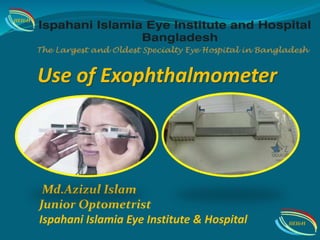
- 1. Use of Exophthalmometer Md.Azizul Islam Junior Optometrist Ispahani Islamia Eye Institute & Hospital IIEI&H IIEI&H
- 2. Declaration I have no financial interest by presenting this presentation IIEI&H
- 3. Aims of today,s topic Will be able…. • General idea @ Measurement Exophthalmos • To know about various type of Exophthalmometer • To know the normal value of Exophthalmometer & • How to documentation Exophthalmos finding IIEI&H
- 4. Exophthalmometer is an instrument used for measuring the degree of forward displacement of the eye in exophthalmos. The device allows measurement of the forward distance of the lateral orbital rim to the front of the cornea.Exophthalmometers can also identify Enophthalmos (retraction of the eye into the orbit), a sign of blow-out fracture or certain neoplasms. An Exophthalmometer IIEI&H
- 5. Exophthalmos : Abnormal protrusion of eye balls or Forward bulging of the globe in endocrine disorders specially thyroid dysfunction. Displacement of globe relative to orbital rims . ENOPHTHALMOS: Defined as retrodisplacement of eye into orbit.Inferior and posterior displacement of globe and intraorbital soft tissue . IIEI&H
- 6. Causes Of Exop:/Proptosis UNILATERAL BILATERAL CONGENITAL - DERMOID DEVELOPMENTAL ANOMALIES - OXYCEPHALY TRAUMATIC- ORBITAL HAGE/IOFB OSTEOPATHIES- RICKETS / ACROMEGALY INFLAMMATION – ORBITAL CELLULITIS/ABSCESS / CAVERNOUS SINUS /PSEUDOTUMOR/ TUBERCULOSIS TUMORS-LYMPHOMA / LEUKEMIA / SARCOMA / NEUROBLASTOMA VASCULAR-ORBITAL VARIX ENDOCRINAL - THYROID EYE DISEASE CYSTS-PARASITIC INFLAMMATORY - HISTIOCYTOSIS/FUNGAL GRANULOMA TUMOR- PRIMARY OR SECONDARY MUCOCOELE OF PARANASAL SINUSES IIEI&H
- 8. HISTORY TAKING Protrusion of eyeball: Age of Onset , duration , progression, Constant or intermittent, Variation with posture / strain Decreased vision : preceded, Stationary, Associated field defects H/O: Pain H/O :Double vision H/O :Trauma H/O: fever , systemic symptoms H/O :Cancer H/O :Thyroid disease H/O:Weight loss IIEI&H
- 9. LOCAL EXAMINATION HB Reflex : Central or Deviation Globe Displacement: Downward,Upward or Other Lid Retruction: +(ve) or – (ve) Lid lag/Lagophtalmos: ..? mm PALPATION : Size , shape, surface, margins consistency , tenderness , thyroid glangds, orbital rims , regional lymph nodes IIEI&H
- 10. EOM MOTILITY : Decreased in thyroid orbitopathy , extensive tum or growths and neurological defect VISUAL ACUITY : Maybe decreased due to Refractive changes due to pressure on eyeball, Optic nerve compresssion, Exposure keratopathy PUPIL REACTION : RAPD suggests optic nerve compression IOP : Maybe raised Rular Test: Horizontal with vertical measurement LOCAL EXAMINATION IIEI&H
- 11. SYSTEMIC EXAM The basis of the measurement for exophthalmos determination using the Hertel version is the outer orbital rim (orbital wall) and the apex of the cornea. All instruments are meant to measure the distance b2w the apex of cornea and lateral wall of orbit OPTICAL: Light Weight Hertels exophthalmometer Heavy Duty Hertel Exophthalmometer Naugle Exophthalmometer Luedde Exophthalmometer MECHANICAL : Gormaz Exophthalmometer IIEI&H
- 12. Hertel exophthalmometer is used for precise exophthalmos measurements (e.g. Exophthalmic goiter). Sturdy construction for many years of use. Overall dimensions are 25 x 7 x 2 cm. Weight is 117 grams . Scale for orbital wall goes from 75 mm to 121 mm. The scale to measure the proptosis ranges from 0 to 35 mm. L.W.Hertel exophthalmometer IIEI&H
- 13. Lowest price! Heavy Duty Exophthalmometer has metal construction for many years of use. Overall dimensions are 25 x 7 x 2 cm. Weight is 216 grams . Scale for orbital wall goes from 75 mm to 121 mm. The scale to measure the proptosis ranges from 0 to 35 mm. Heavy Duty Exophthalmometer IIEI&H
- 14. Naugle exophthalmometer is superior and inferior orbital rim- based instrument. This superior and inferior orbital rim-based instrument measures the position of the globe accurately even after lateral orbitotomy e.g. post tumor removal or thyroid decompression. It delivers fast reproducible results of highest precision, provides best patient comfort, thus increasing patient cooperation. Naugle exophthalmometer IIEI&H
- 15. Fastest, easiest and least expensive method for measuring exophthalmos. By sighting at right angles, this device eliminates parallax and allows the clinician to measure the amount of protrusion or bulging eye from its orbit. Luedde Exophthalmometer IIEI&H
- 16. Gormaz Exophthalmometer Measure distance between two lateral orbital margins Require topical anaesthesia IIEI&H
- 17. The basis of the measurement for exophthalmos determination using the Hertel version is the outer orbital rim (orbital wall) and the apex of the cornea. The internal refraction mirrors with millimeter scales for the left-hand and right-hand measuring halves of the exophthalmometer are calibrated so that the zero mark on the scale will be located in the plane of the resting points of the supports. For exophthalmus measurements the supports are placed against the two temporal orbital walls so that the orbital rim contacts the deepest point of the supports. Prisms are clear plastic with easy to read millimeter lines on both sides. Eye is seen through lower half of scale and the scale is seen through the upper half, eliminating parallax and aiding rapid determination of protrusion. The Measurement Procedure IIEI&H
- 18. The Measurement Procedure The examiner sits opposite the patient at eye level. The exophthalmometer is then positioned with the index points at the temporal lateral orbital walls. The instrument hold with both hands and firmly propped first against the patient’s right orbital wall on the temporal side(which should be felt against the lowest part of the support point). The moveable part on the right side is then set in such as way that the patient’s left orbital wall lies against the lowest part of the arched support. The distance between the lateral orbits (on scale 2) should be noted for future correlation.The examiner asks the patient to look at right ahead with eyelids wide open. The examiner measures for proptosis in each eye. IIEI&H
- 19. The normal range is 12–21 mm. Upper normal limit for people of African origin is a little higher, about 23– 24 mm. A difference greater than 2 mm between the eyes is significant. In children and teenagers mean exophthalmometric measurements increase with age: Less than 4 years old (13.2 mm), 5–8 years old (14.4 mm), 9–12 years old (15.2 mm) and 13–17 years old (16.2 mm). Depending upon the configuration of the osseous orbit, a value of 15mm might be pathological whereas 21mm might be normal. Axial Length of the eye affects exophthalmometer reading. Pseudoproptosis may be seen in severe myopia. Normal values Of Exophthalmometer IIEI&H
- 20. Work up of Exophthalmos Pt. IIEI&H
- 21. Work up of Exophthalmos Pt. IIEI&H
- 22. IMAGING USG/ CT-Scan/ MRI/ X-Ray INVESTIGATION Incisional biopsy Excisional biopsy Core biopsy Hematological : CBC , ESR, VDRL Thyroid function test: T3,T4,TSH Serum ANA , c- ANCA , ACE BUN , Creatinine FNAC IIEI&H
- 23. Management of Exop: Local management: Sun glasses,Sleep in supine position with head elevated,Taping of lids at night,Prisms in diplopia. Medical therapy: Topical tear substitutes,Systemic diuretics - minimal role,Parenteral antibiotics,Pain killers. Radiation: Radio Therapy & Chemo Therapy (Pseudotumour, Lymphoma, Rhabdomyosarcoma , Meningioma,Thyroid orbitopathy ). Surgical options: Orbitotomy, Orbital decompression, Combined ethmoidectomy Etc. IIEI&H
- 24. Take Home Messages An Exophthalmos is an important manifestation of a large number of orbital diseases. Thorough clinical examination coupled with appropriate investigations clinches the diagnosis and helps in management. A difference greater than 2 mm between the eyes is significant. IIEI&H
- 26. References The Orbit and Oculoplasty ,Dr.Agarwal www.ophthalmoloyweb.com Exophthalmometer in TheFreeDictionary“.Farlex. Retrieved 13 April 2013.” Picture: Me ,Google. IIEI&H IIEI&H
- 27. IIEI&H IIEI&H